

Loading, please wait...
Loading, please wait...

Revascularization of the cerebral circulation demands meticulous technical precision and rigorous preoperative preparation. Modern EC-IC bypass planning represents a critical milestone in cerebrovascular neurosurgery, especially when treating complex occlusive arteriopathies and intracranial giant aneurysms. Traditionally, vascular surgeons relied solely on two-dimensional cross-sectional imaging, including computed tomography angiography and digital subtraction angiography. Although these modalities provide high spatial resolution, they require the surgeon to mentally reconstruct intricate three-dimensional anatomical relationships. Consequently, neurosurgeons frequently experienced cognitive fatigue and variability during surgical trajectory selection. In recent years, mixed reality technologies have bridged this gap by projecting patient-specific anatomical holograms directly into a three-dimensional field. Therefore, clinicians can now explore superficial and deep intracranial vascular networks prior to skin incision. Furthermore, this advanced visualization allows multidisciplinary teams to select optimal donor vessels and recipient cortical branches with heightened confidence. In clinical practice, integrating holographic environments minimizes guesswork during craniotomy placement. As a result, surgical teams optimize operative workflows and enhance safety margins before entering the operating theatre.
The practical execution of mixed reality begins with the seamless segmentation of standard imaging data. Initially, neurosurgeons extract stereotactic computed tomography angiograms to delineate the skull, brain surface, and superficial temporal artery. Next, sophisticated rendering algorithms generate high-fidelity, interactive three-dimensional polygonal meshes. Clinicians subsequently upload these digital assets into head-mounted displays, such as optical see-through holographic visors. Because these headsets project interactive digital holograms onto the physical environment, surgeons can manipulate anatomical models in real space. For instance, the operator can rotate the virtual head, adjust tissue opacity, and inspect vascular bifurcations from unlimited vantage points. In addition, the holographic platform allows real-time depth perception that flat monitors cannot reproduce. Recent clinical data demonstrate that the mean preparation time for creating these patient-specific holograms is approximately 36 minutes. Thus, the pipeline integrates smoothly into routine elective neurosurgical workflows without causing excessive institutional delays. Moreover, the interactive software promotes collaborative case discussions among surgical teams.
Successful revascularization hinges entirely upon selecting an appropriate donor artery and a receptive cortical target. Traditionally, surgeons mapped the superficial temporal artery using manual palpation and transcutaneous acoustic Doppler ultrasound. However, transcutaneous Doppler tracing can become inaccurate in patients with deep branches or overlying tissue edema. In contrast, mixed reality allows direct visualization of the entire course of the superficial temporal artery and its frontal and parietal branches. A recent comparative technical investigation revealed that mixed reality delineates donor vessels significantly faster than conventional manual palpation or Doppler localization. Specifically, mixed reality reduced localization time by a mean difference of five minutes compared to manual methods, and four minutes compared to Doppler examination. Furthermore, holographic planning enables accurate assessment of vessel caliber, tortuosity, and branching anatomy along the planned incision line. Similarly, identifying the optimal recipient branch of the middle cerebral artery becomes substantially easier. As a consequence, neurosurgeons avoid unnecessary scalp dissection, preserve vital collateral vessels, and tailor the craniotomy directly over the target branch.
Skeptics of novel surgical technology often question whether virtual tools truly translate into measurable clinical benefits. Importantly, recent comparative findings confirm that mixed reality planning achieves excellent procedural safety without compromising operative efficacy. When matched against conventional planning cohorts, patients undergoing holographic preparation showed comparable intraoperative bypass flow rates. This equivalence proves that holographic pre-planning maintains the gold standard of vascular patency while refining technical steps. Additionally, the average craniotomy diameter did not differ significantly between cohorts, confirming that surgeons did not require oversized exposures to compensate for orientation uncertainties. On the contrary, mixed reality offered precise boundary definitions that prevented unnecessary bone removal. Postoperative imaging confirmed robust graft patency across the planned cohorts, demonstrating reproducible reconstructive success. Therefore, holographic assistance streamlines the initial operative phases while fully safeguarding microvascular flow parameters. By eliminating tactile ambiguity during donor harvesting, the surgical team can concentrate their efforts on the delicate microsurgical anastomosis. Consequently, the technology elevates procedural confidence without introducing unforeseen hemodynamic liabilities.
Integrating advanced mixed reality into daily hospital routines requires careful consideration of departmental costs, learning curves, and technological infrastructure. In evolving health systems, neurosurgical departments handle high volumes of complex vascular pathology, including Moyamoya disease and intracranial atherosclerotic stenosis. Fortunately, modern head-mounted displays and segmentation applications rely increasingly on cloud-based processing and consumer-grade hardware. Thus, institutions do not necessarily require multi-million-dollar capital investments to launch a mixed reality surgical planning program. Furthermore, junior residents and surgical fellows experience accelerated spatial learning curves when interacting with three-dimensional anatomical holograms. Instead of relying solely on mental extrapolation, trainees visualize complex skull base geometries in an intuitive, risk-free setting. However, hospital administrators must establish standardized protocols to ensure patient data security and rapid segmentation turnaround. In addition, neurosurgeons must remember that holograms serve as an adjunct rather than a complete replacement for fundamental microneurosurgical skills. Ultimately, adopting cost-effective holographic planning can enhance patient counseling, refine surgical precision, and optimize operating room efficiency across busy referral hospitals.
Although current mixed reality systems demonstrate remarkable planning utility, ongoing technological enhancements promise even broader clinical impact. Presently, investigators are integrating artificial intelligence algorithms to automate vessel segmentation, which could reduce preparation time from thirty minutes to mere seconds. Moreover, intraoperative dynamic navigation represents the next major milestone in augmented cerebrovascular surgery. By overlaying real-time holographic vascular anatomy directly onto the operative microscope oculars, surgeons could track deep recipient vessels beneath opaque arachnoid membranes. Additionally, integrating intraoperative indocyanine green angiography into holographic displays could deliver simultaneous functional and structural data. While multicenter randomized clinical trials are still necessary to evaluate long-term neurologic outcomes, existing technical evidence demonstrates undeniable feasibility. As these technologies mature, three-dimensional mixed reality will likely transition from an innovative adjunct into standard operative practice. Consequently, neurovascular surgery continues its rapid trajectory toward greater visual precision, tailored microdissection, and enhanced patient safety.
Mixed reality projects a patient-specific three-dimensional hologram directly onto the scalp surface, clearly displaying the anatomical course and bifurcations of the superficial temporal artery. This intuitive visual guidance allows surgeons to trace the vessel path rapidly, saving significant time compared to manual anatomical palpation or transcutaneous acoustic Doppler localization.
Recent comparative technical studies demonstrate that mixed reality planning does not significantly alter mean craniotomy diameter or intraoperative bypass blood flow rates. Instead, the technology ensures precise bone flap placement over recipient branches and preserves graft flow equivalency, maintaining standard microsurgical safety margins while optimizing preoperative anatomical orientation.
Generating a high-fidelity patient-specific vascular hologram requires an average preparation time of approximately thirty-six minutes. Trained clinical personnel can easily complete this image segmentation and rendering process prior to elective revascularization procedures, allowing hospitals to integrate holographic planning seamlessly without creating procedural delays in busy operating room suites.
Disclaimer: This content is for informational and educational purposes only, and does not constitute medical advice. It is not intended to be a substitute for professional medical advice, diagnosis, or treatment. Always seek the advice of your physician or other qualified health provider with any questions you may have regarding a medical condition. Refer to the latest local and national guidelines for clinical practice.
References

Read summarized clinical updates, watch expert medical content, and earn CME certifications right from your smartphone.
Open your smartphone camera to scan and install.





This technical note evaluates the role of three-dimensional mixed reality holograms in planning flow-augmentation EC-IC bypass surgery. Holographic planning significantly speeds up superficial temporal artery delineation while maintaining equivalent intraoperative bypass blood flow and craniotomy dimensions.
Today

Ambient digital scribes promise effortless medical documentation, but a critical sociotechnical commentary reveals that AI alters rather than eliminates charting work. Discover how shifting cognitive burdens, oversight risks, and workflow redesign impact clinical safety and healthcare practice.
Today

A new comparative study demonstrates that intraoperative imprint cytology achieves 96.6% diagnostic accuracy in ovarian tumours, outperforming frozen section in sensitivity for borderline lesions and offering an accessible, rapid alternative for resource-constrained surgical centres.
Today
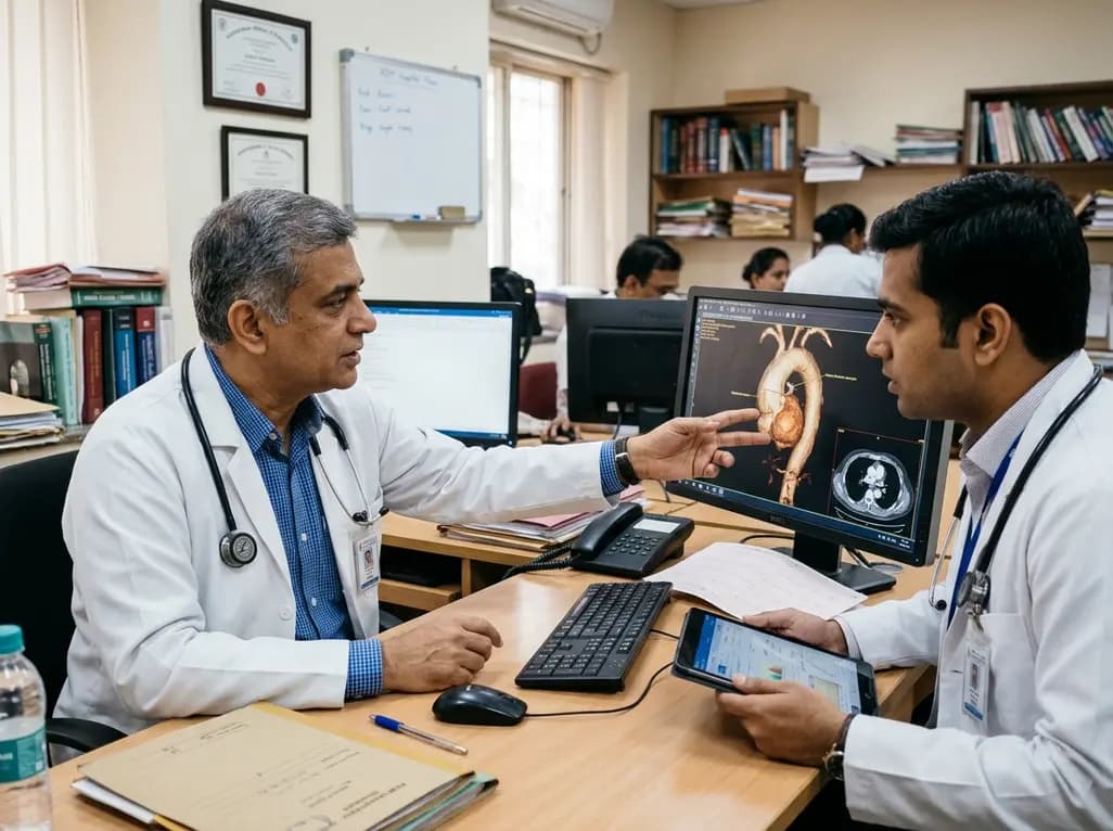
Thoracic aortic aneurysm lacks etiologic medical therapy. A new study reveals that targeting specific αV integrins with Cilengitide attenuates TGF-β and mechanotransduction pathways, effectively limiting pro-fibrotic remodeling and aortic bulb dilation in both syndromic and sporadic disease models.
Today
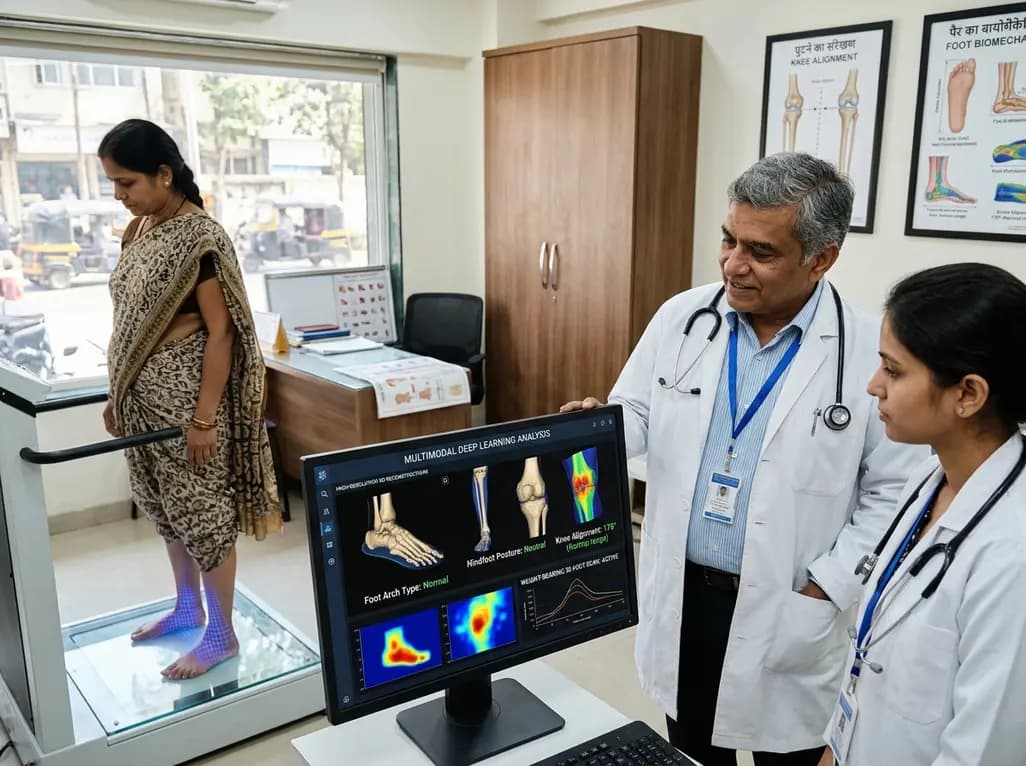
A multimodal deep learning framework utilizing weight-bearing 3D foot scans accurately screens lower-limb alignment, including arch type, hindfoot posture, and knee alignment, offering an accessible, radiation-free alternative to radiography for early biomechanical triage.
Today
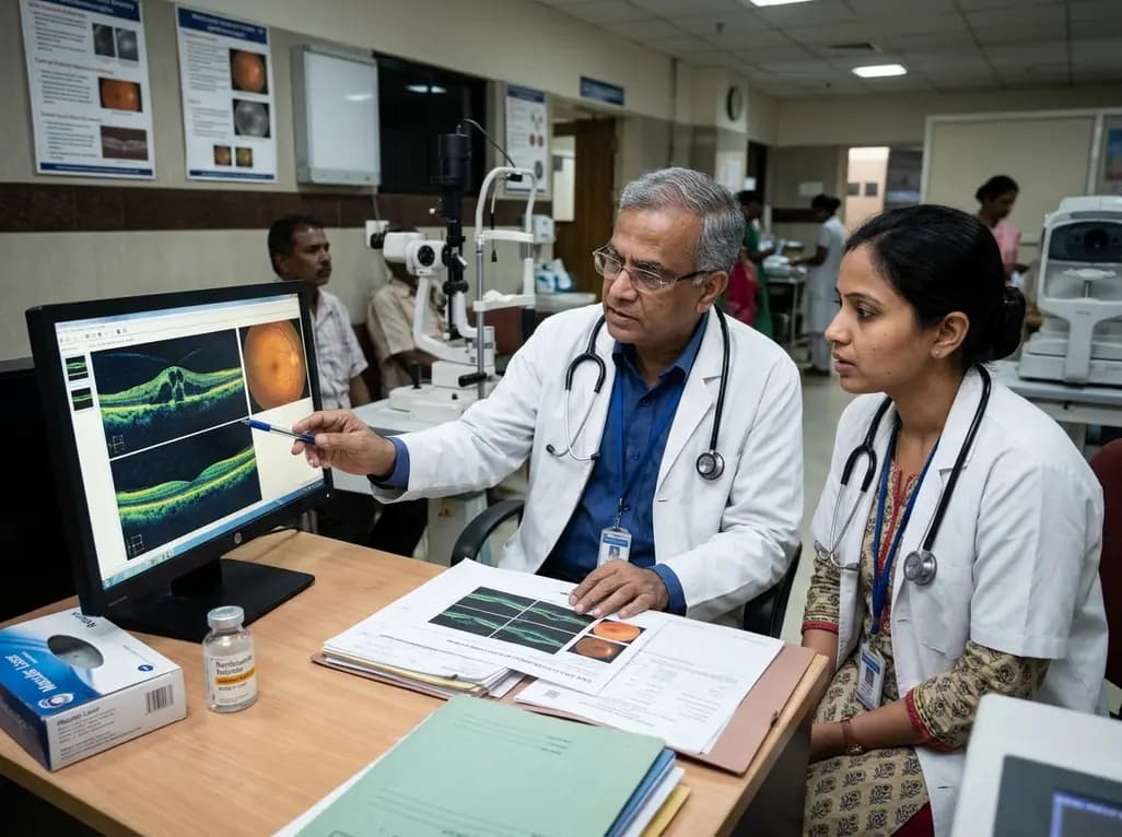
A real-world retrospective study demonstrates that combining intravitreal ranibizumab with focal/grid macular laser significantly improves visual acuity and central macular thickness compared to laser monotherapy in patients with center-involved diabetic macular edema.
Today
