

Loading, please wait...
Loading, please wait...
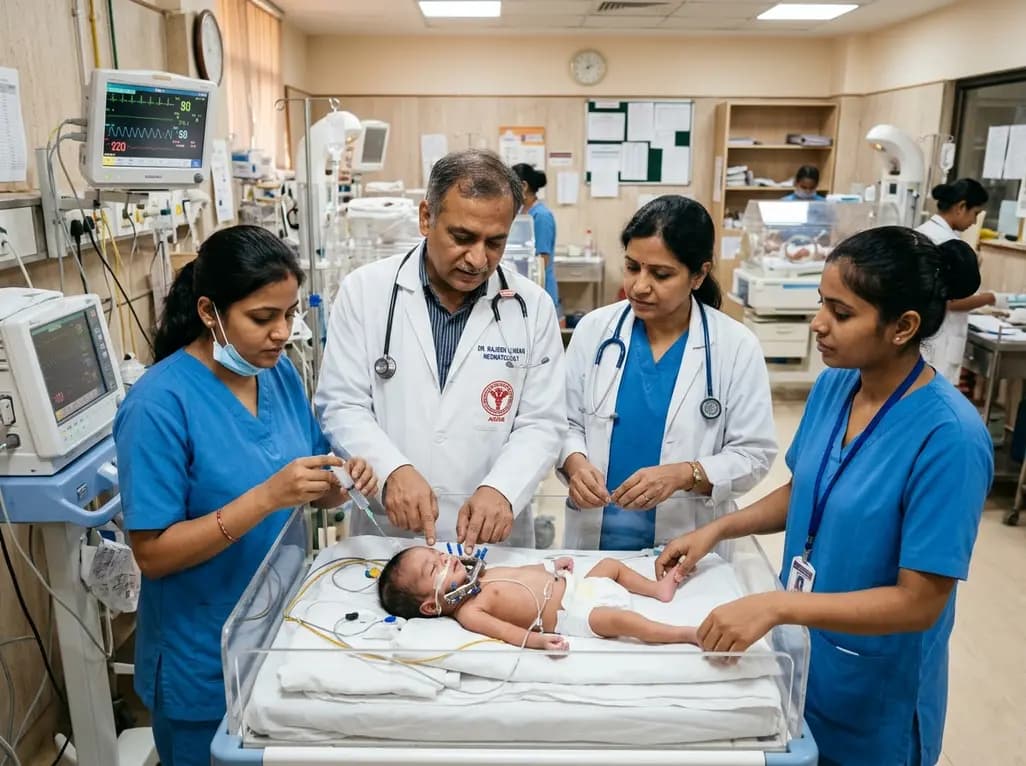
Neonates born with severe Pierre Robin sequence present significant clinical challenges characterized by micrognathia, glossoptosis, and life-threatening upper airway obstruction. In refractory cases failing positional therapy or non-invasive airway adjuncts, mandibular distraction osteogenesis serves as a definitive surgical intervention to prevent tracheostomy. However, successful surgical advancement of the hypoplastic mandible demands structured coordination across multiple pediatric subspecialties. Clinical teams must manage anatomical airway expansion alongside complex post-surgical device protocols and delayed neuromuscular feeding mechanisms.
Severe micrognathia forces the base of the tongue backward into the pharyngeal space, causing severe glossoptosis and upper airway compromise. Consequently, affected neonates experience severe obstructive sleep apnea, recurrent desaturations, and chronic respiratory fatigue. Although non-invasive strategies like prone positioning and nasopharyngeal stenting resolve mild obstruction, severe deformities demand surgical intervention. Therefore, pediatric surgeons perform mandibular distraction osteogenesis to advance the mandible forward incrementally. This mechanical bone lengthening draws the tongue base anteriorly, which significantly enlarges the retroglossal pharyngeal diameter. As a result, neonates experience rapid relief from upper airway obstruction without enduring the long-term morbidity of a tracheostomy. In addition, preoperative evaluation requires detailed computed tomography and dynamic endoscopic airway assessment. These diagnostic imaging modalities confirm that tongue-base obstruction represents the primary pathology rather than multilevel collapse or coexisting tracheobronchomalacia. Furthermore, precise preoperative planning helps clinicians select appropriate internal or external distraction vectors tailored to neonatal anatomy.
Managing the neonatal airway during distraction surgery demands exceptional expertise from pediatric anesthesiologists and critical care specialists. Intubation of neonates with severe retrognathia often presents extreme difficulty; therefore, anesthesiologists routinely employ video laryngoscopy or flexible fiberoptic bronchoscopy to secure the airway safely. Following bilateral osteotomy and distractor placement, clinicians frequently maintain mechanical ventilation during the initial latency phase. This temporary protective sedation prevents accidental tube displacement while perioperative soft-tissue edema gradually subsides. Consequently, extubation protocols require careful risk stratification and continuous monitoring in the neonatal intensive care unit. Once surgeons activate the distractor pins and achieve sufficient anterior displacement, the clinical team executes a controlled extubation trial. In recent institutional protocols, infants routinely extubate successfully around postoperative day 10 while maintaining oxygen saturation levels above 95% on room air. Additionally, bedside providers monitor closely for signs of post-extubation subglottic injury, stridor, or aspiration during spontaneous breathing.
Surgeons initiate distraction activation after a brief latency period, typically turning the distraction pins at an incremental rate of one millimeter daily. Because hardware manipulation occurs over several weeks, specialized nursing care plays an indispensable role in safeguarding device integrity. Nurses inspect the external pin sites daily, applying meticulous antiseptic protocols to prevent local cellulitis and pin-track infections. Furthermore, nursing teams regularly assess facial skin tension around the transcutaneous distraction arms to prevent pressure necrosis and localized ulceration. Meticulous device maintenance also demands that caregivers prevent inadvertent hardware trauma or accidental reverse-rotation of the distractors during routine infant handling. Consequently, clear bedside visual instructions and standardized documentation of pin rotations ensure accurate bilateral bone lengthening. Moreover, serial radiographic imaging confirms adequate bone gap expansion and symmetrical vector orientation before the infant enters the prolonged consolidation phase.
Although mandibular distraction osteogenesis rapidly resolves structural upper airway obstruction, it does not guarantee immediate oral feeding autonomy. Clinicians frequently observe an important clinical dissociation between anatomical airway stability and functional deglutition. Specifically, neonates who breathe effortlessly post-extubation often continue to demonstrate severe pharyngeal dysphagia, disorganized suckle patterns, and uncoordinated swallow-breathe mechanisms. This functional delay occurs because chronic anatomical deformation compromises the maturation of complex sensory-motor neural networks responsible for normal deglutition. Furthermore, surgical advancement alters the spatial alignment of the suprahyoid musculature, requiring time for neuromuscular recalibration. Consequently, many infants rely on nasogastric tube feeding for several months despite displaying robust somatic growth and normal respiratory parameters. Clinicians must recognize that anatomical restoration precedes functional neurodevelopment. Therefore, care teams should never equate successful airway extubation with immediate readiness for full oral feeding.
Because full oral competence requires prolonged functional rehabilitation, longitudinal multidisciplinary surveillance remains vital after hospital discharge. Speech-language pathologists, pediatric feeding therapists, and clinical dietitians formulate individualized rehabilitation pathways tailored to each infant’s developmental pace. Additionally, feeding therapists utilize specialized pacing techniques, modified nipples, and therapeutic positioning to foster safe, stress-free oral motor experiences. Meanwhile, dietitians monitor caloric density and daily enteral volumes through nasogastric tubes to safeguard continuous somatic weight gain. Periodic clinical follow-up at scheduled intervals—such as one, three, six, and seven months postoperatively—allows clinicians to adjust caloric targets and evaluate hardware consolidation. Eventually, surgical teams remove the distraction hardware once adequate cortical bone mineralization occurs across the distraction gap. Through sustained multidisciplinary collaboration, infants gradually overcome persistent oral aversion and dysphagia, ultimately achieving successful oral weaning alongside stable, long-term craniofacial development.
Mandibular distraction osteogenesis gradually lengthens the hypoplastic mandibular corpus through controlled mechanical bone separation. This forward displacement carries the attached anterior suprahyoid musculature and tongue base anteriorly away from the posterior pharyngeal wall. Consequently, the retroglossal and hypopharyngeal airway spaces expand significantly, relieving anatomical glossoptosis and eliminating life-threatening upper airway obstruction in neonates with severe micrognathia.
Airway patency relies on immediate structural enlargement of the retroglossal pharynx, whereas successful feeding requires sophisticated neuromuscular coordination among sucking, swallowing, and breathing. Chronic anatomical distortion, altered muscular vectors, and underlying sensory deficits impair deglutition reflexes. Consequently, infants require extended periods of neurological maturation and targeted feeding therapy before achieving safe, independent oral intake.
Hardware-related complications include localized pin-track infections, distractor pin loosening, cutaneous pressure necrosis, and facial nerve injury. Additionally, inappropriate pin turning can cause asymmetrical mandibular advancement or premature hardware failure. Meticulous pin-site hygiene, careful skin surveillance, daily standardized turning protocols, and routine radiographic tracking significantly reduce these postoperative hardware risks.
Disclaimer: This content is for informational and educational purposes only... Refer to the latest local and national guidelines for clinical practice.
References
Zhong J et al. Multidisciplinary Perioperative Management and Follow-Up After Mandibular Distraction Osteogenesis in a Neonate With Severe Pierre Robin Sequence: A Report. J Craniofac Surg. 2026 Oct 26. doi: 10.1097/SCS.0000000000013516. PMID: 42930455.
Flores RL, et al. Mandibular distraction osteogenesis for the treatment of neonatal tongue-based airway obstruction. J Craniofac Surg. 2014;25(4):1187-1191.
Breik O, et al. Pierre Robin sequence: A review of respiratory and feeding complications and recommendations for management. Int J Pediatr Otorhinolaryngol. 2016;89:229-242.
OrphanAnesthesia. Anaesthesia recommendations for Pierre Robin sequence. Orphanet Report Series. 2019.

Read summarized clinical updates, watch expert medical content, and earn CME certifications right from your smartphone.
Open your smartphone camera to scan and install.





Mandibular distraction osteogenesis reliably relieves severe airway obstruction in neonates with Pierre Robin sequence. However, functional oral feeding recovery frequently lags behind anatomical airway stabilization, underscoring the critical need for prolonged multidisciplinary nutritional rehabilitation.
Today

A nationwide UK Cystic Fibrosis Registry study demonstrates that higher updated mean HbA1c levels correlate with a graded, significant increase in all-cause mortality among individuals with cystic fibrosis-related diabetes, reinforcing the critical need for proactive glycemic monitoring and intervention.
Today

A KGMU study reveals over half of women with breast abnormalities fail to undergo diagnostic confirmation, leading to advanced-stage detection. Discover the socio-cultural barriers, clinical risks of incomplete resection, and community-level solutions needed to bridge the critical screening-to-diagnosis gap in India.
Today

A comparative non-contact study reveals that high-power radiofrequency delivery with the QDOT MICRO catheter induces an artifactual contact force rise. Electrophysiologists must understand this power-dependent thermal and electrical phenomenon to avoid inadvertent catheter withdrawal and ineffective ablation.
Yesterday

Researchers have engineered a self-powered Zn/C micro-galvanic electrospun membrane that couples sustained zinc release with endogenous microcurrents. This bioelectric scaffold stimulates M2 macrophage polarization and accelerates bone deposition to achieve robust cranial defect regeneration.
Today

Preclinical research demonstrates that BMI1 overexpression alleviates metabolic syndrome-related liver injury. By triggering ubiquitin-mediated METTL3 degradation, BMI1 prevents AHNAK m6A methylation and suppresses fibrogenic TGF-β/SMAD signaling, significantly improving hepatic steatosis and insulin resistance.
Today
