

Loading, please wait...
Loading, please wait...

Reconstructing large calvarial voids presents a severe challenge in reconstructive surgery and neurosurgical practice. Standard guided bone regeneration relies predominantly on static barrier membranes. However, these inert barriers lack the active biochemical and bioelectrical signals necessary to drive rapid osteogenesis. Recently, bioengineers engineered an innovative biomimetic electrospun scaffold that generates intrinsic microcurrents while steadily releasing bioactive zinc ions. This breakthrough provides dual bioelectric and biochemical stimulation to achieve efficient cranial defect regeneration.
Surgeons frequently encounter critical-sized calvarial defects resulting from severe traumatic brain injury, decompressive craniectomies, oncologic resections, or congenital malformations. In these demanding clinical scenarios, spontaneous bone healing fails completely. Consequently, persistent osseous non-unions leave vulnerable intracranial structures without adequate mechanical protection. Conventional therapeutic interventions typically utilize autologous bone grafts, titanium meshes, or passive polymeric barrier membranes. Although autografts remain the historical gold standard, they carry considerable donor-site morbidity, limited graft volume, and variable graft resorption rates. In contrast, inert barrier membranes effectively block fibrous tissue invasion into the bony space. Nevertheless, traditional membranes lack osteoinductive capability and cannot reproduce the native electrophysiological microenvironment of healing bone. Bone tissue naturally generates endogenous stress-generated potentials and electrical gradients during repair. Because passive scaffolds cannot supply bioelectrical signals, cellular migration and osteoblast differentiation often stall. Therefore, reconstructive specialists require advanced multifunctional membranes. Ideal modern scaffolds must actively modulate local inflammatory responses, stimulate osteogenic progenitor recruitment, and supply sustained biophysical cues directly to the injury site.
To resolve the limitations of passive implants, investigators designed a spatially patterned electrospun scaffold. The team fabricated a unique micro-galvanic membrane designated as P/C-Zn/C. This biomaterial incorporates a central zinc-containing core surrounded by a peripheral conductive carbon ring. By pairing metallic zinc with carbon nanoparticles across an electrospun polymeric matrix, the construct establishes miniature galvanic couples throughout its microarchitecture. Electrospinning yields an extracellular matrix-mimicking fibrous topography with interconnected microporosity. As a result, the scaffold promotes cell adhesion and supports adequate nutrient exchange. Furthermore, the deliberate spatial patterning creates an internal electrochemical gradient across the defect interface. The system harnesses the standard electrochemical potential difference between the zinc anode and the carbon cathode. Thus, the membrane generates continuous, low-level electrical fields upon contact with physiological fluids without requiring external battery packs or bulky transcutaneous wiring. Additionally, the composite polymer provides necessary mechanical resilience, which prevents membrane collapse into the calvarial defect during soft-tissue closure.
During extended in vitro immersion assessments lasting 28 days, the P/C-Zn/C scaffold demonstrated exceptional physicochemical stability and functional longevity. Notably, the membrane exhibited a sustained, time-dependent release of zinc ions without causing toxic concentration spikes. Simultaneously, the surrounding physiological medium maintained a stable physiological pH, avoiding cytotoxic local acidification or alkaline accumulation. Zinc serves as an essential trace element that stimulates alkaline phosphatase activity and promotes collagen synthesis in osteoprogenitor cells. Moreover, electrophysiological profiling confirmed that the micro-galvanic couples maintained stable voltage generation throughout the entire testing window. To isolate individual therapeutic mechanisms, researchers compared the dual-action membrane against control scaffolds with exogenous zinc alone or external electrical stimulation alone. Interestingly, the combined micro-galvanic membrane elicited far greater osteogenic differentiation than either single-modality group. These comparative findings verify that simultaneous ionic release and self-powered bioelectrical stimulation act synergistically. Together, they activate intracellular signaling cascades that strongly upregulate runt-related transcription factor 2, osteocalcin, and bone sialoprotein.
Successful bone regeneration relies heavily on favorable early osteoimmunomodulation. Prolonged M1 pro-inflammatory macrophage activation causes chronic tissue destruction and promotes fibrous scar encapsulation. Conversely, shifting macrophage phenotypes toward an anti-inflammatory, pro-regenerative M2 state accelerates bone deposition. In cellular assays, the P/C-Zn/C electrospun membrane triggered a profound phenotypic transition toward reparative M2 macrophages. Flow cytometry and immunofluorescence staining revealed marked upregulation of classic M2 surface markers, including CD206 and arginase-1. Furthermore, gene expression assays confirmed that the micro-galvanic environment significantly suppressed pro-inflammatory cytokines such as tumor necrosis factor-alpha and interleukin-6. Simultaneously, the membrane enriched anti-inflammatory and pro-osteogenic mediators, particularly transforming growth factor-beta and vascular endothelial growth factor. This favorable immune shift directly conditioned the local defect microenvironment. As a result, cultured human bone marrow-derived mesenchymal stem cells exhibited elevated mineralized nodule formation and enhanced alkaline phosphatase activity. Thus, the micro-galvanic scaffold couples electrophysical cues with ionic release to reprogram the local immune niche into a pro-osteogenic environment.
To evaluate translational efficacy, researchers implanted the P/C-Zn/C membrane into a standardized rat critical-sized calvarial defect model. Micro-computed tomography demonstrated dramatic architectural repair within the treatment group compared to passive controls. Specifically, defects treated with the micro-galvanic membrane achieved superior defect bridging, greater total bone volume, and significantly elevated bone volume fraction. Quantitative morphometric analysis confirmed dense, mature trabecular bone formation throughout the defect center rather than merely along the defect margins. Histological evaluations corroborating the radiographic findings revealed thick, organized lamellar bone sheets and extensive collagen deposition. Furthermore, in vivo immunohistochemical staining confirmed an abundant accumulation of F4/80 and CD206 double-positive M2 macrophages across the regenerating osteoid tissue. The scaffold maintained its structural integrity while progressively integrating with newly formed host bone. Consequently, the combination of intrinsic microcurrents and sustained zinc delivery prevented soft-tissue prolapse, suppressed osteoclastic resorption, and stimulated robust vascularized bone formation across the critical-sized cranial void.
These compelling experimental findings carry substantial clinical relevance for cranial and maxillofacial reconstructive surgery. Autologous bone harvesting often leads to infection, chronic pain, and extended surgical duration. In contrast, self-powered bioelectric scaffolds represent a transformative off-the-shelf alternative for complex osseous restoration. By eliminating external power supplies and fragile percutaneous leads, micro-galvanic membranes mitigate infection risks and simplify postoperative management. In clinical neurosurgery, these active barriers could serve as multifunctional cranioplasty covers that prevent fibrous adhesion while accelerating cranial flap integration. Similarly, periodontists and oral maxillofacial surgeons could deploy such membranes during vertical ridge augmentation and sinus floor elevation. However, several translational questions warrant rigorous future investigation. Researchers must perform direct in vivo bioelectrical measurements to quantify the exact voltage distribution within vascularized human tissues. Furthermore, long-term degradation kinetics, chronic immune profiling, and biomechanical testing in large animal models are vital next steps. Establishing these physiological parameters will accelerate the translation of smart bioelectric implants from preclinical laboratories into clinical practice.
The scaffold incorporates microscopic galvanic couples formed by pairing zinc and carbon within an electrospun polymer matrix. When physiological fluids contact the membrane, an electrochemical potential difference develops between the zinc anode and carbon cathode. This spontaneous oxidation-reduction reaction produces sustained, low-level microcurrents across the defect site. Consequently, the self-powered membrane delivers therapeutic electrical stimulation continuously without external power supplies or wires.
Zinc serves as an essential trace mineral that regulates cellular metabolism, enzyme function, and skeletal mineralization. During controlled degradation, the membrane releases zinc ions that activate alkaline phosphatase and upregulate osteogenic transcription factors like Runx2. Furthermore, zinc exhibits moderate antimicrobial properties and acts synergistically with endogenous microcurrents to suppress pro-inflammatory cytokines. As a result, zinc ion delivery promotes stem cell osteogenesis and accelerates osseous defect healing.
Macrophage polarization strongly governs the osteoimmune microenvironment during bone healing. While pro-inflammatory M1 macrophages initiate early debridement, prolonged M1 activity causes tissue degradation and fibrous scarring. In contrast, pro-healing M2 macrophages resolve local inflammation, release osteogenic factors like transforming growth factor-beta, and recruit osteoprogenitors. Therefore, shifting the immune response toward the M2 phenotype prevents fibrous encapsulation and creates an ideal biochemical environment for calvarial bone regeneration.
Disclaimer: This content is for informational and educational purposes only and should not be considered medical advice. Always consult a qualified healthcare professional regarding any medical condition or treatment. Refer to the latest local and national guidelines for clinical practice.
References
Nie Z et al. A self-powered Zn/C micro-galvanic electrospun membrane enhances osteoimmunomodulation and accelerates cranial defect regeneration. J Mater Chem B. 2026 Oct 26. doi: 10.1039/d5tb02925b. PMID: 42930319.
Chen Z, et al. Osteoimmunomodulation in bone tissue engineering: Emerging strategies and materials. Biomaterials. 2021;269:120658.
Wang Y, et al. Endogenous electric fields and bone repair: From biophysical mechanisms to therapeutic electroactive scaffolds. Bioact Mater. 2022;18:43-58.

Read summarized clinical updates, watch expert medical content, and earn CME certifications right from your smartphone.
Open your smartphone camera to scan and install.





Researchers have engineered a self-powered Zn/C micro-galvanic electrospun membrane that couples sustained zinc release with endogenous microcurrents. This bioelectric scaffold stimulates M2 macrophage polarization and accelerates bone deposition to achieve robust cranial defect regeneration.
Today

A nationwide UK Cystic Fibrosis Registry study demonstrates that higher updated mean HbA1c levels correlate with a graded, significant increase in all-cause mortality among individuals with cystic fibrosis-related diabetes, reinforcing the critical need for proactive glycemic monitoring and intervention.
Today

A KGMU study reveals over half of women with breast abnormalities fail to undergo diagnostic confirmation, leading to advanced-stage detection. Discover the socio-cultural barriers, clinical risks of incomplete resection, and community-level solutions needed to bridge the critical screening-to-diagnosis gap in India.
Today

A comparative non-contact study reveals that high-power radiofrequency delivery with the QDOT MICRO catheter induces an artifactual contact force rise. Electrophysiologists must understand this power-dependent thermal and electrical phenomenon to avoid inadvertent catheter withdrawal and ineffective ablation.
Yesterday
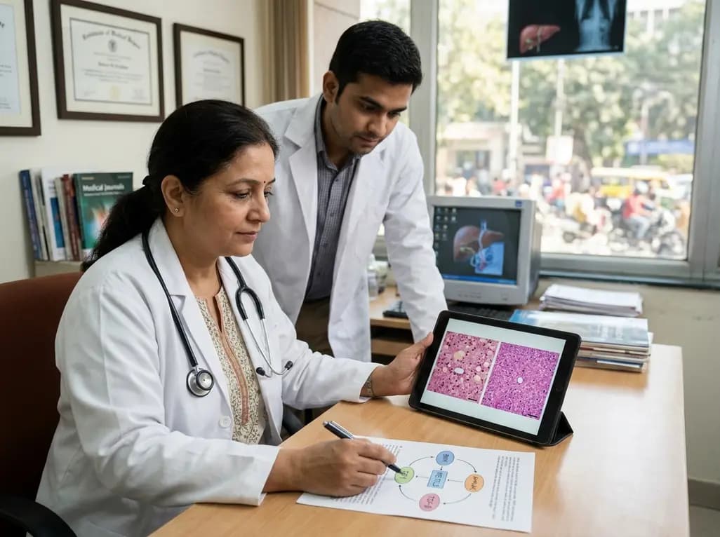
Preclinical research demonstrates that BMI1 overexpression alleviates metabolic syndrome-related liver injury. By triggering ubiquitin-mediated METTL3 degradation, BMI1 prevents AHNAK m6A methylation and suppresses fibrogenic TGF-β/SMAD signaling, significantly improving hepatic steatosis and insulin resistance.
Today
