.webp&w=384&q=75)

Loading, please wait...
Loading, please wait...
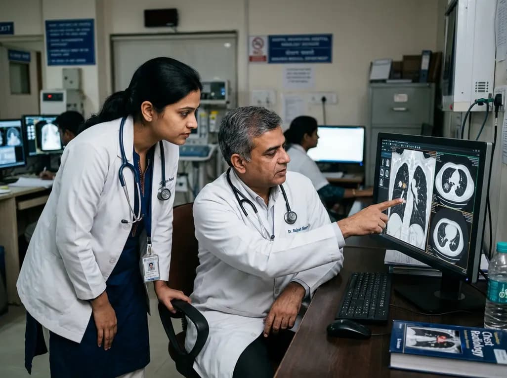
Lung cancer remains a leading cause of global cancer mortality. Fortunately, early detection can fundamentally transform clinical outcomes. A landmark evaluation by the International Agency for Research on Cancer confirms this reality. Specifically, low-dose CT screening reduces lung cancer mortality by 16% among high-risk individuals. Published in The New England Journal of Medicine, this assessment highlights the value of proactive screening. In addition, findings demonstrate a meaningful reduction in advanced-stage diagnoses. Consequently, these results reinforce the clinical necessity of targeted secondary prevention.
An international working group evaluated extensive clinical trials to assess cancer screening efficacy. Dr. Abhishek Shankar from the All India Institute of Medical Sciences contributed to this comprehensive global review. Primarily, researchers derived their conclusions from rigorous randomized controlled trials. In the United States National Lung Screening Trial, low-dose tomographic screening lowered relative mortality by 16% at seven years. Investigators compared screened participants directly against individuals who received standard chest radiographs. Similarly, the European NELSON randomized trial demonstrated substantial mortality reductions among screened men over a 10-year follow-up period. Moreover, trials consistently demonstrated significant downward stage migration at diagnosis. Consequently, patients presented with smaller, localized lesions rather than incurable metastatic neoplasms. Therefore, high-quality clinical trial data confirm that periodic tomographic screening directly alters the course of aggressive pulmonary tumors. However, clinicians must emphasize that these benefits apply strictly to selected high-risk cohorts. Overall, randomized evidence firmly establishes computed tomography as the standard of care for early detection. Furthermore, these findings align with international thoracic guidelines that endorse structured surveillance. Thus, robust data remove lingering doubts regarding radiographic mortality benefits.
Following a comprehensive systematic evaluation, the IARC working group assigned low-dose computed tomography to Group A. This classification signifies established scientific evidence that the modality reduces both lung cancer mortality and late-stage disease incidence. In contrast, researchers categorized standard chest radiography, performed with or without sputum cytology, as Group C. Therefore, evidence remains inadequate to demonstrate any meaningful mortality reduction from routine chest X-rays. Standard projection radiography lacks the spatial contrast resolution required to identify subtle, sub-centimeter parenchymal nodules. Consequently, radiographic films frequently miss early curable tumors until they progress to symptomatic, advanced stages. Furthermore, historical trials evaluating chest X-rays failed to demonstrate survival advantages, despite frequent screening intervals. Computed tomography overcomes these optical limitations by capturing high-resolution volumetric datasets of the pulmonary parenchyma. Thus, low-dose protocols provide exceptional diagnostic sensitivity while keeping ionizing radiation exposure remarkably low. Nevertheless, healthcare systems must avoid utilizing non-validated modalities like plain radiography for cancer detection. Instead, clinical directors should exclusively implement standardized helical computed tomography for eligible patients. Additionally, clear categorization helps clinicians guide patients away from ineffective diagnostic procedures.
Although low-dose imaging offers profound survival advantages, clinicians must carefully manage associated procedural harms. Notably, false-positive findings represented a prominent challenge across published trials, ranging from 1% to 42% depending on nodule management algorithms. In addition, between 5% and 32% of individuals with false-positive results subsequently underwent invasive diagnostic procedures. Unfortunately, complications occurred in 10% to 22% of these invasive interventions, including bleeding, pneumothorax, and infection. Moreover, the IARC report estimated that overdiagnosis accounts for 3% to 26% of screen-detected malignancies. Overdiagnosis represents the detection of indolent neoplasms that would never cause symptomatic illness or death during a patient's natural lifespan. Consequently, patients facing overdiagnosis risk experiencing unnecessary surgical resections, systemic therapies, and psychological distress. To minimize these harms, multidisciplinary teams must apply standardized reporting frameworks such as Lung-RADS. Furthermore, serial volumetric nodule assessment helps differentiate indolent lesions from aggressive malignancies before invasive biopsy. Hence, clinicians must conduct thorough shared decision-making discussions prior to imaging. In this manner, patients understand potential procedural risks alongside early-detection benefits. Similarly, rigorous quality control ensures that diagnostic workups proceed safely and judiciously. Ultimately, structured surveillance pathways prevent premature interventional biopsies in benign indeterminate nodules.
The IARC working group explicitly emphasized that effective screening requires comprehensive infrastructure beyond standalone CT scanners. Primarily, healthcare systems must establish robust patient identification registries and systematic risk-stratification pathways. Eligible candidates typically include individuals aged 50 to 80 years with at least a 20 to 30 pack-year smoking history. Furthermore, guidelines recommend annual screening for active smokers or former smokers who have quit within the preceding 15 years. However, technological access alone cannot produce optimal public health outcomes without integrated clinical workflows. Therefore, comprehensive programs must incorporate multidisciplinary nodule evaluation boards, rapid-access biopsy pathways, and coordinated oncology referral networks. Additionally, integrated smoking cessation assistance remains an indispensable cornerstone of any screening enterprise. Clinicians know that tobacco cessation substantially reduces cardiovascular and pulmonary morbidity independent of cancer detection. Moreover, organized initiatives require accredited medical imaging facilities, standardized low-dose acquisition protocols, and rigorous continuous quality assurance. Without equitable access and meticulous quality control, diagnostic disparities will widen. Consequently, institutions must invest in specialized personnel, including thoracic radiologists, nurse navigators, and pulmonary specialists. Thus, a multi-tiered infrastructure guarantees patient safety throughout the entire diagnostic continuum. Similarly, clear diagnostic reporting standardizes management decisions across diverse clinical centers.
Translating international screening frameworks into the Indian healthcare landscape introduces distinct epidemiological and diagnostic challenges. In India, Dr. Abhishek Shankar at AIIMS, New Delhi, is spearheading the Indian Lung Screening Trial. This pioneering initiative enrolls individuals aged 50 to 80 years with a 20 pack-year smoking history. Specifically, candidates must currently smoke or have quit within the preceding 15 years. Eligible individuals across the country can directly contact the trial team to access structured screening. Notably, Indian physicians encounter a unique diagnostic environment marked by a substantial burden of benign granulomatous diseases. In high-prevalence settings for tuberculosis and fungal infections, pulmonary nodules appear frequently on tomographic scans. Consequently, radiologists encounter higher baseline false-positive rates that demand nuanced radiological evaluation. Furthermore, India exhibits a high proportion of lung cancers among non-smokers. Biomass smoke and ambient air pollution drive this notable clinical trend. Therefore, ongoing research like the AIRCARE study at AIIMS actively explores predictive screening models that integrate particulate matter exposure. Ultimately, local trial data will provide critical evidence to formulate tailored national screening guidelines. Meanwhile, Indian clinicians should actively identify high-risk smokers and connect them with structured institutional screening protocols.
Q1: Who meets the eligibility criteria for low-dose CT screening?
Eligibility criteria target high-risk individuals aged 50 to 80 years with at least a 20 to 30 pack-year smoking history. Candidates must either be active smokers or former smokers who stopped smoking within the past 15 years. Conversely, screening is not recommended for asymptomatic individuals without substantial tobacco exposure. Clinicians must carefully evaluate patient age, comorbidities, functional status, and personal preferences before ordering screening. Furthermore, physicians should discuss follow-up testing protocols.
Q2: Why does the IARC classify chest X-rays as Group C for screening?
The International Agency for Research on Cancer classified chest radiography as Group C because clinical trials demonstrated no mortality reduction. Unlike low-dose computed tomography, plain projection radiographs lack sufficient spatial resolution to detect subtle, sub-centimeter pulmonary nodules. Consequently, tumors identified on routine radiographs are frequently advanced, invasive, and incurable. Therefore, professional guidelines firmly discourage using chest radiography for lung cancer screening purposes. Instead, physicians should recommend verified tomographic modalities.
Q3: How should clinicians address false-positive nodules detected on screening?
Clinicians must manage screen-detected pulmonary nodules using standardized radiologic protocols such as the Lung-RADS reporting classification. Structured frameworks guide appropriate follow-up intervals, typically recommending repeat scans to confirm nodule growth prior to biopsy. Furthermore, multidisciplinary tumor boards should review complex nodules to prevent unnecessary transthoracic biopsies and thoracic surgeries. Additionally, clinicians must educate patients regarding false-positive rates to reduce psychological distress during surveillance. Ultimately, structured monitoring safeguards patient health.
Disclaimer: This content is for informational and educational purposes only. It does not constitute medical advice or replace professional judgment. Refer to the latest local and national guidelines for clinical practice.
References

Read summarized clinical updates, watch expert medical content, and earn CME certifications right from your smartphone.
Open your smartphone camera to scan and install.





An International Agency for Research on Cancer working group has confirmed that low-dose CT screening cuts lung cancer mortality by 16% in high-risk populations. Learn how early detection reduces late-stage malignancy, alongside clinical insights for implementation, risk assessment, and screening protocols in India.
Today
India is advancing biomedical frontiers by integrating AI, genomics, and nanomedicine under supportive national policies like BioE3. Union Minister Jitendra Singh urges clinicians to balance cutting-edge innovations with bedside clinical acumen to preserve patient trust and deliver equitable precision healthcare.
Today

Novel preclinical research demonstrates that antisense oligonucleotides targeting essential genes such as DARS1, DYNC1I2, and EIF2S3 selectively induce apoptosis in ovarian and lung cancer cells. Conditional biomarker activation provides a viable strategy to spare normal tissue while overcoming therapy resistance.
Today

A pioneering Italian case series demonstrates that mobile heart teams can successfully implement TA-NRP heart procurement in peripheral noncardiac hospitals. Despite a mandatory 20-minute no-touch period and prolonged warm ischemia, 89.5% of hearts were transplanted with zero primary graft dysfunction.
Today

A 10-year Markov state-transition model shows minimally invasive tubular decompression dominates open laminectomy for pure lumbar spinal stenosis, yielding 5.95 vs 5.91 QALYs and saving $4,167 per patient through reduced complications and lower index costs.
Today

A systematic review evaluates the feasibility and safety of thyroidectomy after thermal ablation for thyroid nodules. Although procedures encounter significant fibrosis and surgical adhesions, permanent complication rates remain remarkably low in experienced centers.
Today
