.webp&w=384&q=75)

Loading, please wait...
Loading, please wait...

Clinicians frequently struggle to differentiate recurrent intracranial malignancy from treatment-induced cerebral injury following radiotherapy or stereotactic radiosurgery. Conventional neuroimaging modalities, including contrast-enhanced magnetic resonance imaging, frequently produce ambiguous findings that mimic neoplastic growth. Consequently, the adoption of liquid biopsy in brain tumors offers an essential, minimally invasive diagnostic pathway. By capturing circulating disease markers, clinicians can bypass repeat cranial surgery while personalizing subsequent therapeutic plans.
Malignant brain neoplasms, such as glioblastoma and secondary brain metastases, require intensive therapeutic regimens comprising surgical resection, localized irradiation, and cytotoxic chemotherapy. However, high doses of radiation often damage normal brain parenchyma, leading to localized inflammation, endothelial injury, and vessel thrombosis. These pathological processes manifest clinically as either pseudoprogression or radiation necrosis. Pseudoprogression typically presents within the first three months following chemoradiation, showing transient edema and contrast enhancement that gradually stabilize or resolve spontaneously. In contrast, radiation necrosis represents an irreversible, delayed necrotic reaction that emerges months or even years after stereotactic radiosurgery or fractionated therapy.
Standard follow-up neuroimaging protocols, such as conventional contrast-enhanced magnetic resonance imaging and metabolic positron emission tomography, often fail to reliably distinguish viable neoplastic tissue from radiation-induced inflammatory reactions. Consequently, radiologists and neuro-oncologists frequently face considerable diagnostic uncertainty. Performing a stereotactic brain biopsy provides histopathological confirmation, yet this procedure carries substantial morbidity, surgical risk, and economic cost. Therefore, clinicians urgently need reliable, noninvasive diagnostic surrogates to avoid premature discontinuation of active therapy or unnecessary surgical re-exploration.
To overcome tissue access limitations, researchers have focused on biofluid profiling to monitor tumor evolution. The integration of liquid biopsy in brain tumors provides real-time systemic access to tumor-derived biomolecules and immune constituents without cranial trephination. A recent comprehensive systematic review evaluated the diagnostic accuracy of fluid-based assays specifically applied to resolve ambiguous post-radiation neuroimaging findings. By examining records across PubMed, Embase, Scopus, Cochrane, and clinical registries, investigators systematically scrutinized 11 qualifying clinical studies assessing diverse biological markers.
Unlike single-point surgical biopsies, biofluid collection enables longitudinal disease monitoring across the entire post-treatment trajectory. Serial blood sampling allows clinicians to capture biological fluctuations as lesions evolve from early inflammatory states to active recurrent tumors. Furthermore, liquid sampling avoids spatial sampling bias, which frequently plagues focal stereotactic needle biopsies in heterogeneous intracranial lesions. Consequently, molecular surveillance via circulating biofluids offers an objective and dynamic framework that complements serial anatomical imaging.
The systematic review highlighted a remarkably broad spectrum of molecular and cellular analytes used to differentiate neoplastic progression from benign treatment effects. Investigators analyzed immune cell-based populations, focusing on circulating myeloid subsets. Specifically, research teams tracked HLA-DR-negative or low-expressing and VNN2-positive CD14-positive monocytic myeloid-derived suppressor cells. In addition, scientists quantified circulating inflammatory and tumor-associated proteins, most notably CXCL11 and MUC-16, to monitor systemic inflammatory cascades triggered by therapeutic radiation.
Beyond cellular and protein markers, genetic and epigenetic targets provide critical diagnostic insights. Researchers successfully isolated circulating tumor cells, cell-free nuclear DNA, mitochondrial DNA, repetitive B1-SINE genetic elements, microvesicles, and diverse microRNA species. Peripheral blood plasma served as the primary biofluid across the majority of identified studies due to ease of phlebotomy. However, several exploratory investigations analyzed cerebrospinal fluid and urine to assess whether proximal biofluids offer enhanced analytical sensitivity. These varied analytes reflect distinct facets of tumor recurrence versus radiation-mediated tissue destruction.
Across the analyzed cohorts, several isolated biomarkers demonstrated statistically significant differences between radiation necrosis, pseudoprogression, and verified tumor recurrence. For instance, elevated circulating tumor cell counts and elevated levels of cell-free tumor-specific DNA mutations strongly correlated with true disease progression. Conversely, circulating myeloid-derived suppressor cell fluctuations and specific cytokine releases reflected intense radiation-induced neuroinflammation.
Importantly, individual biomarkers frequently exhibited variable diagnostic performance when deployed in isolation. To enhance analytical precision, multiple investigator groups designed composite scoring indices. Prominent examples include the DR-VNN2 index and the Necrosis Prediction Index. These multifaceted algorithms synthesize cellular, immunological, and protein measurements into a single unified predictive score. As a result, these composite models showed superior discriminatory performance compared to individual biomarkers alone, minimizing ambiguous classifications and providing actionable diagnostic outputs for managing complex brain lesions.
Despite promising initial findings, several substantive methodological hurdles prevent immediate routine integration into daily clinical workflows. The systematic review identified significant cohort heterogeneity across the available medical literature. Most published studies evaluated relatively small, single-center patient cohorts with inconsistent reporting of critical diagnostic performance metrics, such as sensitivity, specificity, and area under the receiver operating characteristic curve. Moreover, the intact blood-brain barrier substantially restricts the passive egress of large macromolecular tumor fragments into peripheral venous circulation.
Additionally, isolation protocols, analytical assay platforms, and diagnostic reference thresholds vary widely between laboratory groups. High-throughput flow cytometry, digital droplet polymerase chain reaction, immunostaining fluorescence in situ hybridization, and multiplex protein assays each feature distinct detection limits. Therefore, the neuro-oncology community must conduct large, multi-center prospective validation trials to standardize biofluid collection procedures and validate composite predictive indices. Once standardized, these noninvasive assays will redefine the clinical management of post-radiation intracranial disease.
Pseudoprogression is an early, subacute inflammatory reaction that typically occurs within three months of chemoradiation, resolving without intervention. In contrast, radiation necrosis represents a delayed, chronic, and often progressive tissue injury occurring months or years after stereotactic radiotherapy, occasionally requiring surgical resection or targeted steroid therapy.
Both tumor recurrence and radiation damage disrupt the blood-brain barrier, causing contrast extravasation and vasogenic edema. Consequently, both entities present as growing contrast-enhancing lesions with surrounding hyperintensity on conventional T1- and T2-weighted magnetic resonance images, making accurate visual differentiation extremely difficult without advanced metabolic or molecular testing.
Peripheral plasma remains the most practical and widely studied biofluid due to simple, noninvasive collection. However, cerebrospinal fluid often provides higher concentrations of central nervous system tumor-derived DNA because it directly contacts the cerebral parenchyma, circumventing the restrictive filtration effects of the blood-brain barrier.
Disclaimer: This content is for informational and educational purposes only. It is not intended to provide medical advice or establish a standard of care. Healthcare professionals should exercise their independent clinical judgment. Refer to the latest local and national guidelines for clinical practice.
References
Izhar M et al. Clinical utility of liquid biopsy in distinguishing true progression from radiation necrosis and pseudoprogression in malignant brain tumor. J Neurooncol. 2026 May 25. doi: 10.1007/s11060-026-05618-z. PMID: 42185659.
Vellayappan B, Tan CL, Yong C, et al. Diagnosis and management of radiation necrosis in patients with brain metastases. Front Oncol. 2018;8:395.
Ellingson BM, Chung C, Pope WB, Boxerman JL, Kaufmann TJ. Pseudoprogression, radionecrosis, inflammation or true tumor progression? challenges associated with glioblastoma response assessment in an evolving therapeutic landscape. J Neurooncol. 2017;134(3):495-504.

Read summarized clinical updates, watch expert medical content, and earn CME certifications right from your smartphone.
Open your smartphone camera to scan and install.





Distinguishing true tumor progression from radiation necrosis and pseudoprogression remains a major hurdle in neuro-oncology. A systematic review reveals how emerging liquid biopsy biomarkers and composite indices provide noninvasive insights to guide critical treatment decisions after cranial irradiation.
last month

Recent epidemiological data reveals a dramatic surge in summer heat mortality driven by human-induced climate warming. This analysis explores the pathophysiological impacts on cardiac and renal systems, diagnostic dilemmas in death certification, and essential emergency cooling protocols for acute clinical care.
Today

Odisha has established India's first state-level clinical protocol specifically tailored for pregnant women with sickle cell disease. Developed with NHM and UNICEF, the framework provides structured antenatal screening, crisis mitigation, standardized referral pathways, and specialized interdisciplinary care.
Today

India is advancing preventive healthcare by integrating genomics, artificial intelligence, and quantum technologies. With the SANGAM programme sequencing one million Indian genomes, clinicians can unlock personalized therapies, detect cardiometabolic risks early, and curb the burden of diabetes and heart disease.
Today
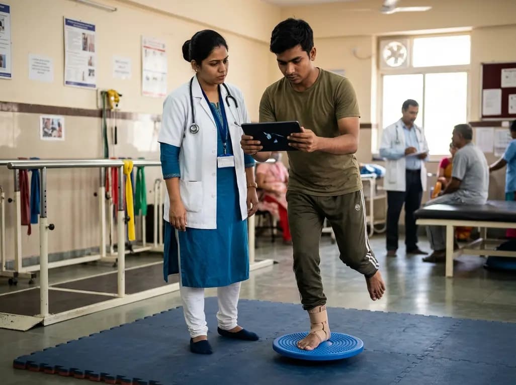
A randomized controlled trial demonstrates that an eight-week dual-task training intervention significantly improves static and dynamic postural control over standard balance exercises in individuals with chronic ankle instability, addressing critical neurocognitive deficits during athletic and daily activities.
Today
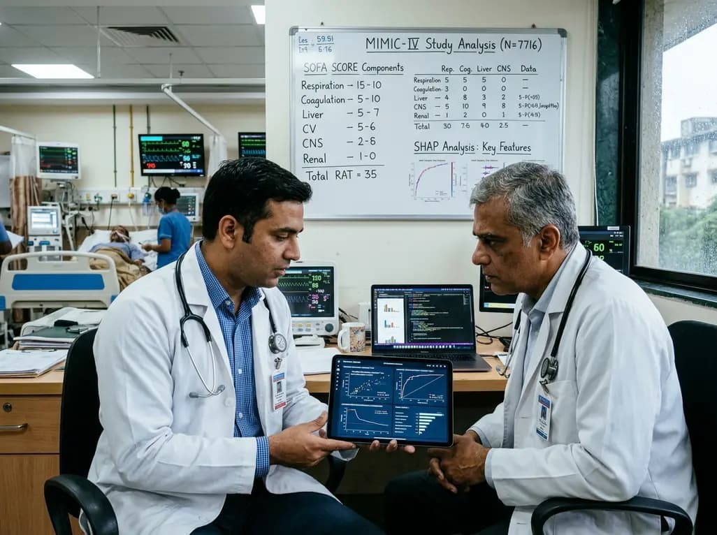
A retrospective cohort study of 7,716 ICU patients from the MIMIC-IV database demonstrates that Gradient Boosting Decision Trees significantly outperform traditional SOFA scores in predicting in-hospital mortality among patients with sepsis and diabetes, with SHAP analysis providing clinical interpretability.
Today
