.webp&w=384&q=75)

Loading, please wait...
Loading, please wait...
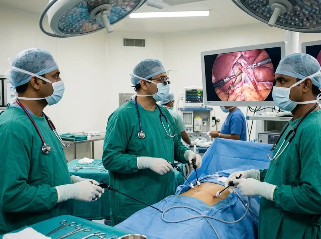
Performing a cholecystectomy in situs inversus presents an uncommon surgical scenario that demands spatial reorientation and customized technical modifications. Situs inversus abdominis with levocardia occurs when abdominal organs display mirror-image transposition while thoracic structures retain normal anatomical lie. Consequently, symptomatic gallstone disease in these patients requires careful preoperative planning. Surgeons must adapt standard operative steps to overcome unfamiliar ergonomics, altered dissection angles, and unexpected vascular branching patterns.
Situs inversus abdominis with levocardia represents a distinct developmental anomaly differing from complete situs inversus totalis. In complete situs inversus, both thoracic and abdominal viscera invert completely across the sagittal midline. In contrast, patients with levocardia retain a normal left-sided cardiac apex, whereas the liver, gallbladder, and spleen occupy mirrored abdominal positions. Therefore, symptomatic cholelithiasis manifests with atypical left upper quadrant or epigastric discomfort rather than traditional right hypochondriac pain. Because clinical signs present on the contralateral side, initial evaluation may inadvertently confuse treating clinicians. Meticulous preoperative imaging remains indispensable for clarifying spatial anatomy. Ultrasonography confirms gallstones and demonstrates left-sided hepatic lobar anatomy. Furthermore, contrast-enhanced computed tomography or magnetic resonance cholangiopancreatography defines anomalous vascular and biliary branching patterns before surgery. These cross-sectional imaging modalities prevent intraoperative disorientation. Ultimately, recognizing this anatomical variant prior to skin incision ensures rapid diagnosis and guides safe surgical planning.
Meticulous preparation is vital when organizing a cholecystectomy in situs inversus, because standard laparoscopic instincts cannot guide the procedure. Surgical teams must mentally rehearse reversed procedural steps before entering the operating suite. Furthermore, operating room geometry requires comprehensive realignment to maintain efficient communication and instrument handling. Clinicians reposition the primary laparoscopic monitor toward the patient's left shoulder, matching the direct visual line of sight toward the left upper quadrant. In addition, the surgical assistant and scrub nurse occupy adjusted locations to support fluid trocar handoffs. Preoperative multidisciplinary briefings ensure that all personnel recognize the mirror-image anatomy. Operating rooms must also prepare specialized instruments, including angled laparoscopes, atraumatic retractors, and advanced dissection devices. Moreover, anesthesiologists must monitor cardiopulmonary parameters attentively, because congenital visceral anomalies occasionally accompany latent cardiac conduction defects. By thoroughly coordinating equipment placement, monitor positioning, and team ergonomics, surgeons eliminate spatial disorientation and establish a controlled operative environment.
Conventional laparoscopic cholecystectomy relies on muscle memory that right-handed surgeons develop over years of practice. However, managing a left-sided gallbladder disrupts these ingrained motor patterns and creates substantial ergonomic difficulties. When surgeons attempt conventional port setups on mirrored anatomy, their instruments cross awkwardly, causing significant muscular fatigue and restricted dexterity. To resolve this dilemma, surgeons employ a modified four-port configuration with mirrored subcostal entry points. The primary surgeon stands on the patient's right side, directly facing the operative field in the left hypochondrium. Meanwhile, the assistant stands on the patient's left side to maneuver the camera and provide fundic traction. Placing mirrored subcostal ports along the left costal margin restores optimal triangulation toward Calot's triangle. Additionally, positioning the epigastric port slightly to the left of midline avoids falciform ligament interference. Therefore, customizing port geometry to individual patient habitus significantly improves operative comfort and visual clarity throughout difficult maneuvers.
Dissecting Calot's triangle in mirror-image anatomy challenges visual-motor coordination, particularly for right-handed surgeons. During standard operations, the right hand performs fine dissection while the left hand provides static countertraction. Conversely, operating on a left-sided gallbladder forces a reevaluation of manual roles. Many surgeons achieve superior stability by retracting Hartmann's pouch with their dominant right hand while dissecting Calot's triangle with their left hand through the epigastric port. Alternatively, ambidextrous instrument manipulation facilitates efficient tissue handling without crossed arms. Furthermore, surgeons must anticipate vascular anomalies in patients with situs variations. Congenital visceral rotation anomalies frequently alter arterial branching patterns. In the featured technical report, surgeons identified an anomalous cystic artery coursing to the right of the cystic duct rather than its expected medial position. Therefore, operators must dissect tissues meticulously, avoiding premature thermal coagulation before skeletonizing every vascular structure. Additionally, clinicians should consider intraoperative cholangiography whenever biliary anatomy remains uncertain.
Securing the critical view of safety represents an uncompromising imperative before clipping or dividing any tubular structure. Surgeons achieve this benchmark by clearing the hepatocystic triangle of all fibrofatty and fibrous tissue. In addition, they completely dissect the lower third of the gallbladder off the cystic plate to expose the liver bed directly. Consequently, only two structures—the cystic duct and cystic artery—should enter the gallbladder base. In mirror-image cases, verifying cystic plate clearance confirms orientation and rules out aberrant biliary branches. After achieving unambiguous visualization, the surgical team clips and divides the cystic artery and cystic duct individually. The surgeon then detaches the gallbladder from the liver bed and extracts the specimen in an endobag. Following thorough hemostasis, patients experience rapid recovery. Because minimally invasive access preserves abdominal wall integrity, patients mobilize early and resume oral intake quickly. Consequently, patients tolerate modified laparoscopic cholecystectomy exceptionally well, achieving uneventful discharge on postoperative day one.
Situs inversus totalis involves complete mirror-image transposition of all thoracic and abdominal viscera, including dextrocardia. In contrast, situs inversus abdominis with levocardia represents an isolated abdominal reversal where the abdominal organs sit in mirrored positions, but the heart remains in its standard left-sided thoracic location. Consequently, surgeons must navigate mirrored subdiaphragmatic anatomy while recognizing that cardiopulmonary mechanics, thoracic anatomy, and baseline vascular inflow patterns may retain traditional configurations.
Right-handed surgeons can position themselves on the patient's right side facing the left hypochondrium to improve instrument alignment. Furthermore, utilizing mirrored subcostal ports prevents instrument crossing and preserves triangulation. Clinicians often retract Hartmann's pouch with their dominant right hand while dissecting Calot's triangle with the left hand through the epigastric port. Alternatively, collaborating with a skilled assistant who provides infundibular traction enables the primary surgeon to dissect using their dominant hand.
Congenital situs abnormalities frequently correlate with aberrant biliary ductal branching and anomalous cystic arterial pathways, such as arteries coursing lateral to the cystic duct. Therefore, relying on standard anatomical assumptions invites severe bile duct or vascular injury. Achieving the critical view of safety ensures complete clearance of the hepatocystic triangle and partial liver bed exposure. Consequently, this rigorous landmark verification guarantees safe ligation of only the true cystic structures.
Disclaimer: This content is for informational and educational purposes only and should not be considered medical advice or substituted for professional clinical judgment. Diagnostic criteria, anatomical presentations, and procedural ergonomics may vary across individual surgical settings. Refer to the latest local and national guidelines for clinical practice.
References

Read summarized clinical updates, watch expert medical content, and earn CME certifications right from your smartphone.
Open your smartphone camera to scan and install.





Laparoscopic cholecystectomy in situs inversus with levocardia presents significant ergonomic challenges. This guide details modified port setups, surgeon positioning, and Calot's triangle dissection techniques to ensure the critical view of safety despite mirror-image anatomy and vascular anomalies.
Today
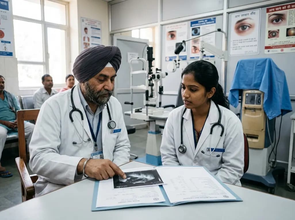
A multicenter study reveals that a significant proportion of HCV antibody-positive ophthalmic surgical patients remain untreated and eligible for direct-acting antivirals. Stratifying antibody titers and streamlining referral pathways can optimize hepatitis C micro-elimination efforts.
Today

The Delhi Cabinet has approved a key policy revision doubling the fixed monthly allowance for accredited social health activists. This financial enhancement bolsters grassroots maternal and child healthcare delivery, strengthens urban public health surveillance, and improves frontline community health worker retention.
Today
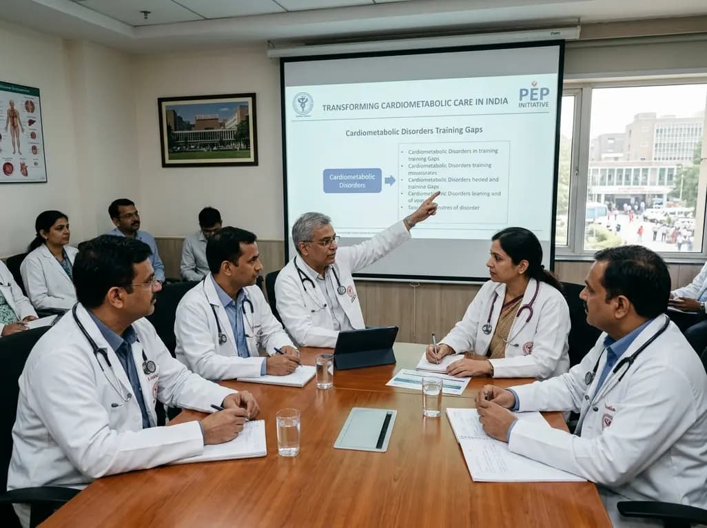
The All India Institute of Medical Sciences has convened national health experts, policymakers, and state nodal officers to transform medical training for cardiometabolic disorders. This collaborative effort under the PEP initiative addresses training gaps to elevate chronic disease care across Indian healthcare.
Today
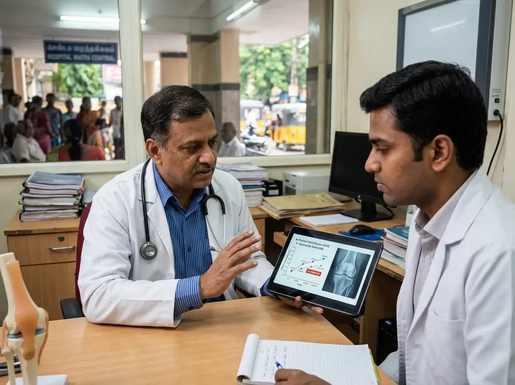
A systematic review and meta-analysis of 21 RCTs reveals that platelet-rich plasma provides small but statistically superior pain relief and functional improvements compared to hyaluronic acid at 12 months in knee osteoarthritis, influenced by platelet concentration and leukocyte enrichment.
Today

A multicenter real-world prospective study in Taiwan demonstrates that liraglutide effectively reduces body weight and BMI standard deviation score in adolescents with obesity. The therapy shows a favorable safety profile with mild gastrointestinal adverse events and zero treatment discontinuations due to intolerance.
Today
