

Loading, please wait...
Loading, please wait...

Modern restorative dentistry relies heavily on digital impression workflows to fabricate complex implant rehabilitations. However, determining true intraoral scanner accuracy for completely edentulous arches remains a formidable clinical dilemma. Clinicians frequently encounter unexpected framework misfits when restoring full-arch mandibular cases across multiple fixtures. While laboratory simulations often celebrate digital scans as reliable alternatives to conventional elastomeric impressions, extensive clinical experience reveals persistent discrepancies.
Completely edentulous arches lack stable anatomical landmarks like interdental papillae and distinct occlusal fissures. Furthermore, mobile mucosal tissues and salivary flow disrupt continuous optical tracking during image acquisition. Intraoral scanners capture long arches by sequentially stitching individual surface frames. Consequently, minor rotational or translational inaccuracies accumulate progressively from one quadrant to another. When clinicians position five implants across an edentulous mandible, cumulative stitching errors generate unpredictable spatial distortions. Therefore, dental researchers must critically examine how digital inspection software measures scanning precision. Establishing rigorous metrology protocols enables prosthodontists to understand true scanner limitations before undertaking complex full-arch restorations.
For decades, dental investigations evaluated optical scanning accuracy through best fit alignment and root mean square error. In this standard approach, inspection software superimposes test scans over a master reference scan by minimizing overall surface distances. However, this single mathematical metric creates an illusion of high precision. Best fit algorithms distribute dimensional discrepancies equally across all scanned contours. As a result, software inadvertently masks severe localized discrepancies located directly at critical implant scan bodies.
Moreover, root mean square equations calculate absolute distance vectors without indicating spatial direction. Positive and negative linear deviations mathematically offset one another during overall surface averaging. Consequently, an intraoral scan with substantial interimplant divergence can still yield an impressively low root mean square error value. Clinicians reviewing published data mistakenly assume that low surface deviation guarantees accurate screw-retained framework delivery. In reality, passive prosthetic fit depends on three-dimensional coordinate accuracy, interimplant distance, and exact angular orientation. Therefore, relying solely on surface root mean square calculations conceals profound translational and rotational shifts. Dental science requires sophisticated inspection techniques that decouple broad surface topography from precise fixture geometry.
To overcome the severe blind spots of global surface averaging, advanced industrial metrology utilizes geometric dimensioning and tolerancing protocols. Specialized inspection software applies reference point system alignment rather than arbitrary best fit algorithms. By locking coordinate datums onto stable scan bodies, this protocol isolates true linear and angular deviations without distributing errors. Furthermore, it measures spatial displacements along the x, y, and z axes independently.
A recent pivotal study applied this robust metrology protocol to evaluate three contemporary intraoral scanners on a five-implant mandibular model. The experimental model featured varied fixture depths, interimplant distances, and deliberate angulations to simulate authentic clinical challenges. Researchers captured repeated digital impressions using manufacturer-recommended scan paths for Primescan, TRIOS 5, and iTero Element 5D Plus. Subsequently, metrology inspection software quantified global linear distortion, absolute angular deviation, and three-dimensional interimplant distance shifts. Simultaneously, investigators computed conventional root mean square values via best fit alignment to compare methodological efficacy directly. This rigorous comparative design demonstrated that coordinate-based metrology exposes profound structural distortions that standard best fit alignment completely misses.
The experimental findings revealed striking differences among the investigated scanning systems and acquisition trajectories. Primescan achieved the lowest deviations whenever statistical significance occurred across global linear distortion, absolute angular distortion, and three-dimensional distance errors. In contrast, TRIOS 5 and iTero Element 5D Plus showed variable performance across distinct implant sites. Furthermore, scanning strategies exerted a major influence on optical accuracy. Specifically, scan pattern C generated significantly greater absolute angular distortion at posterior implant positions than alternative manufacturer strategies.
In addition to individual fixture discrepancies, the digital impressions exhibited macroscopic arch deformations. Scans produced by Primescan and TRIOS 5 showed a noticeable widening of total arch width across the posterior segments. Concurrently, Primescan and iTero scans exhibited significant reduction in arch length along the left quadrant. Most remarkably, TRIOS 5 generated a distinctive wave-like distortion pattern along the z-axis across the mandibular arch. Root mean square values ranged from 22.4 microns for Primescan to 53.6 microns for TRIOS 5. Although root mean square ranked device trueness neatly, it concealed these severe three-dimensional arch warpages. Consequently, clinicians must recognize that low root mean square values cannot guarantee undistorted full-arch digital representations.
Achieving passive prosthetic fit represents the cornerstone of long-term success in multi-unit implant therapy. When restorative frameworks exhibit structural misfit, tightening prosthetic retaining screws induces severe static stresses within the restorative complex. Over time, these mechanical strains cause screw loosening, component fracture, or framework fracture. Biologically, continuous mechanical strain around non-compliant implant fixtures can accelerate crestal bone resorption and endanger peri-implant tissue health.
Because digital scanners generate systemic arch widening and vertical wave-like warpage, fabricated frameworks often demonstrate unacceptable clinical misfit. An angular distortion of just one degree produces significant linear displacement at the opposite end of an edentulous arch. Therefore, dental practitioners must adopt validated clinical protocols to control digital impression inaccuracies. Operators should strictly execute manufacturer-recommended scanning patterns rather than random sweeping movements. Additionally, stabilizing movable oral mucosa with retractors and inserting rigid, high-contrast scan bodies substantially reduces optical stitching drift. Practitioners can also incorporate physical landmarks or splinted verification jigs to verify passive fit prior to final milling. Ultimately, understanding scanner limitations empowers clinicians to deliver durable, precisely fitting implant rehabilitations.
Root mean square error measures overall surface discrepancy by averaging deviations across an entire mesh surface. Consequently, positive and negative errors often cancel out during best fit alignment. This mathematical averaging conceals critical localized linear shifts and angular deviations of individual scan bodies. While an intraoral scanner might yield an impressively low root mean square value, substantial interimplant discrepancies can still persist and ruin prosthetic passive fit.
Angular distortions tilt the virtual orientation of implant fixtures relative to the baseline arch geometry. When technicians design full-arch frameworks over tilted scan bodies, screw channels and restorative interfaces become misaligned. Therefore, seated restorations generate non-axial loading, screw loosening, mechanical fracture, or biological crestal bone loss. Even sub-degree angular errors across multiple implants can generate massive misfit across full-arch prostheses, highlighting the necessity of angular tolerance testing.
Clinicians should utilize continuous, sweeping scanning paths recommended by scanner manufacturers to prevent tracking loss. Furthermore, stabilizing mobile soft tissues and maintaining steady scan speeds minimizes cumulative stitching errors. Scanning artificial landmarks, such as flowable composite dots on keratinized mucosa, provides distinct geometric topography for reliable registration. Additionally, selecting rigid scan bodies with high-contrast surfaces enables the algorithm to maintain tracking across featureless, smooth edentulous alveolar ridges.
Disclaimer: This content is for informational and educational purposes only and should not be considered medical advice. Always consult a qualified healthcare professional regarding any medical condition or treatment. While we strive to present accurate, up-to-date information based on reliable sources, medical knowledge is constantly evolving. Refer to the latest local and national guidelines for clinical practice.
References

Read summarized clinical updates, watch expert medical content, and earn CME certifications right from your smartphone.
Open your smartphone camera to scan and install.





Evaluating intraoral scanner accuracy with root mean square error conceals critical linear and angular distortions in full-arch implant scans. Enhanced metrology protocols reveal significant arch widening, length discrepancies, and wave-like deformations that directly threaten passive prosthetic fit.
Today
A prospective study introduces a clinical and biomarker-based score incorporating faecal calprotectin to predict microscopic colitis with high accuracy (AUC 0.90), outperforming older risk scores and preventing missed diagnoses during colonoscopy in chronic watery diarrhoea.
Today

Delhi is undergoing a major healthcare overhaul with enhanced Ayushman Bharat insurance up to ₹10 lakh, increased MBBS and PG medical training seats, expanded primary network mandirs, new tertiary neuro-ICUs, and public-private reproductive technology initiatives to build a resilient, preventative healthcare model.
Today
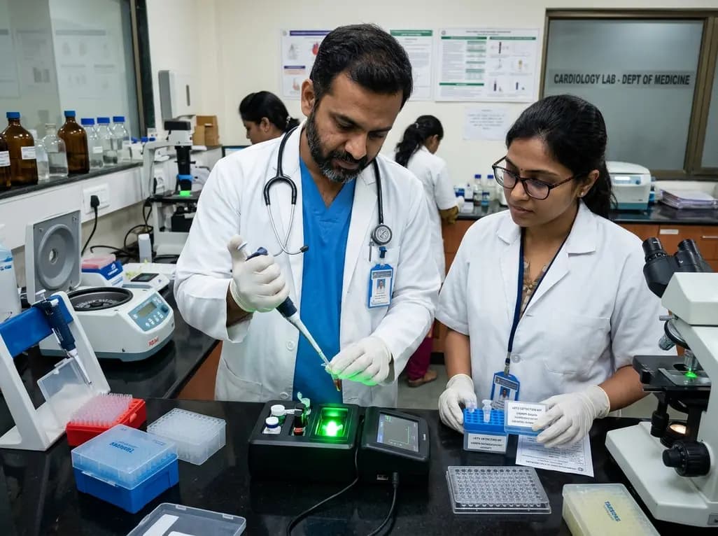
Researchers have engineered an innovative CRISPR-Cas12a fluorescent diagnostic kit for clinical sST2 detection. By combining nanotechnology with enzymatic trans-cleavage, this platform enables rapid, ultrasensitive heart failure diagnosis and risk stratification, outperforming conventional laboratory ELISA methods.
Today
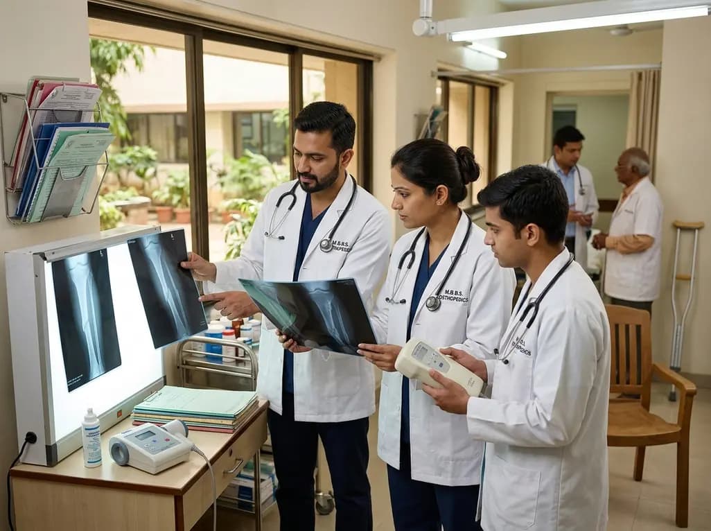
Non-union fractures occur in 2-5% of cases, causing significant disability and cost. Conservative modalities such as LIPUS, PEMF, and focused ESWT offer non-surgical healing options for defects up to 5-10 mm when adequate mechanical stability exists without infection.
Today
A Brazilian birth cohort reveals significant sex-specific associations between preterm birth and adolescent adiposity, underscoring the urgent need for targeted long-term cardiometabolic surveillance and tailored clinical monitoring.
Today
