

Loading, please wait...
Loading, please wait...
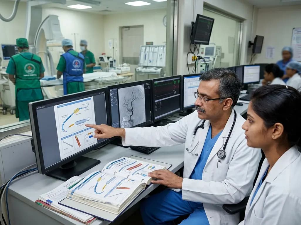
Modern endovascular practice requires rapid decision-making under fluoroscopic guidance. However, incoming trainees face a steep learning curve when navigating complex hardware inventories. Structured interventional radiology device education often lacks standard visual tools. Consequently, novice residents struggle to connect physical device appearance and cryptic packaging labels with functional mechanics and clinical indications. In busy catheterization suites, senior operators select microcatheters, wires, and coils with fluid intuition. In contrast, beginners must decipher French sizes, wire diameters in fractions of an inch, and needle gauges simultaneously. Moreover, inconsistent nomenclature across manufacturers worsens confusion during emergency interventions. When junior doctors cannot immediately identify compatible components, procedural delays occur and safety risks multiply. Traditional textbooks frequently emphasize procedural steps and pathophysiology while neglecting the tactile, physical properties of specific tools. Therefore, dedicated curricula focusing on foundational equipment literacy are essential for procedural safety. By addressing this instructional void, standardized training resources can empower trainees to develop procedural confidence, enhance catheterization fluency, and optimize patient outcomes in minimally invasive care.
To resolve these educational hurdles, educators designed The IR Table Book as a foundational visual atlas. The initial edition systematically organizes 171 interventional devices across 18 clinical sections within a compact 104-page reference. Specifically, the authors grouped medical hardware according to family, primary function, procedural application, and overall workflow role. Introductory modules provide clear orientation on package labeling, sizing conversions, and shelf organization. Trainees learn how to translate manufacturer specifications into actionable operative choices before scrub-in. Furthermore, the atlas outlines common catheter shapes, sheath designs, guidewire stiffness profiles, and embolic agents. Because catheterization laboratories stock extensive arrays of disposable items, this organized taxonomy demystifies the equipment storage room. Junior trainees can trace the step-by-step evolution of an intervention from vascular access to vessel closure. Additionally, the structured layout enables rapid bedside lookup when attending physicians request unfamiliar tools. As a result, this reference bridges the divide between theoretical radiology knowledge and tangible angio-suite mechanics, establishing a coherent baseline for novice operators.
The visual atlas adopts a standardized three-panel layout for every single entry, ensuring intuitive cognitive reinforcement. First, the Visual Recognition panel displays high-resolution imagery of the actual device alongside its sterile packaging and identifier tags. Learners quickly memorize external visual markers, hub colors, and tip profiles before touching sterile trays. Second, the Use and Procedural Context panel defines the explicit clinical role, mechanism of deployment, and anatomic target. This section clarifies when an operator selects a particular catheter shape, such as a Cobra or Simmons configuration, over alternative options. Third, the Key Distinctions panel highlights subtle variations among closely related devices, preventing common sizing errors and component mismatches. For example, it explains why a specific microwire pairs smoothly with one microcatheter lumen but jams within another. Furthermore, the guide emphasizes functional trade-offs regarding trackability, torque response, and radiopacity. Consequently, residents cultivate analytical judgment rather than relying on rote memorization during active operations, substantially shortening the early phase of technical training.
Educational accuracy in invasive medicine demands rigorous editorial and production scrutiny. Accordingly, the atlas underwent strict multi-tiered quality control during initial compilation. An interventional radiology resident coauthor independently scrutinized all 171 individual entries to ensure factual correctness, clinical utility, and clarity. Simultaneously, a senior physician coauthor evaluated the organizational framework, taxonomic logic, and overarching educational validity. Beyond medical verification, the development team implemented comprehensive technical quality checks. They audited alphanumeric tracking codes, standard device nomenclature, and section designations against physical mockups. In addition, quality checks confirmed the precise page coordinates across the entire 104-page print volume, eliminating rendering errors and digital display flaws. Although formal multi-institutional learner trials and external psychometric validations remain pending, this robust initial workflow established strong internal consistency. Consequently, educators produced an orderly, defect-free training manual ready for supervised departmental testing. Future planned studies will measure objective gains in resident diagnostic speed, equipment retrieval accuracy, and procedural confidence.
Mastering catheter and wire dynamics represents a critical safety imperative for interventional training programs worldwide. In developing healthcare environments, including teaching hospitals across India, access to hands-on endovascular simulation or standardized device libraries is often uneven. Therefore, a structured, portable visual atlas delivers immediate educational equity by democratizing foundational equipment knowledge. Trainees can study device characteristics independently before entering high-stress angiographic suites, reducing reliance on trial-and-error learning. Moreover, clear device literacy minimizes intraoperative miscommunication between fellows, primary operators, and catheterization laboratory technicians. When scrub teams communicate with precision, case turnover improves and fluoroscopy times decrease. Most importantly, selecting the proper wire stiffness or sheath size protects delicate endothelial walls, preventing arterial dissection, vessel thrombosis, and unnecessary puncture site trauma. Although virtual simulators and animal models offer interactive benefits, portable visual reference guides provide indispensable reinforcement during clinical rotations. Thus, incorporating systematic hardware atlases into medical curricula strengthens operative competence, protects patient safety, and cultivates the next generation of meticulous interventional radiologists.
Interventional radiology requires immediate familiarity with hundreds of specialized tools across diverse clinical scenarios. Trainees must navigate disparate measurement units, such as French sizes for sheaths, inches for guidewires, and gauges for needles. Furthermore, subtle nuances in wire stiffness, hydrophilic coatings, and catheter tip geometry determine procedural success. Without structured visual references, novice learners struggle to connect abstract packaging labels to physical hardware, creating cognitive overload during fast-paced invasive procedures.
The three-panel design delivers a comprehensive educational overview for every featured device. The first panel provides immediate visual recognition of physical appearance and packaging markings. Subsequently, the second panel explains clinical mechanisms, anatomic indications, and operational context. Finally, the third panel clarifies key distinctions among related instruments to prevent component mismatch. Consequently, this structured format strengthens visual memory, accelerates clinical recall, and reduces procedural errors during live catheterization cases.
Although the initial edition underwent thorough internal quality assurance and multi-tiered physician review, formal external validation remains vital. The authors plan multi-center evaluations to measure learner outcomes across varied training programs. Specifically, prospective studies will assess whether trainees achieve measurable gains in hardware recognition speed, component selection accuracy, and overall procedural efficiency. Additionally, learner feedback will guide subsequent revisions to incorporate emerging endovascular technologies and updated clinical protocols.
Disclaimer: This content is for informational and educational purposes only and does not substitute for formal clinical training or manufacturer instructions. Healthcare providers should verify device specifications, compatibility, and patient suitability independently. Refer to the latest local and national guidelines for clinical practice.
References

Read summarized clinical updates, watch expert medical content, and earn CME certifications right from your smartphone.
Open your smartphone camera to scan and install.





A structured visual atlas titled The IR Table Book organizes 171 interventional radiology devices into 18 sections using a three-panel framework, addressing critical knowledge gaps for early clinical trainees.
Today
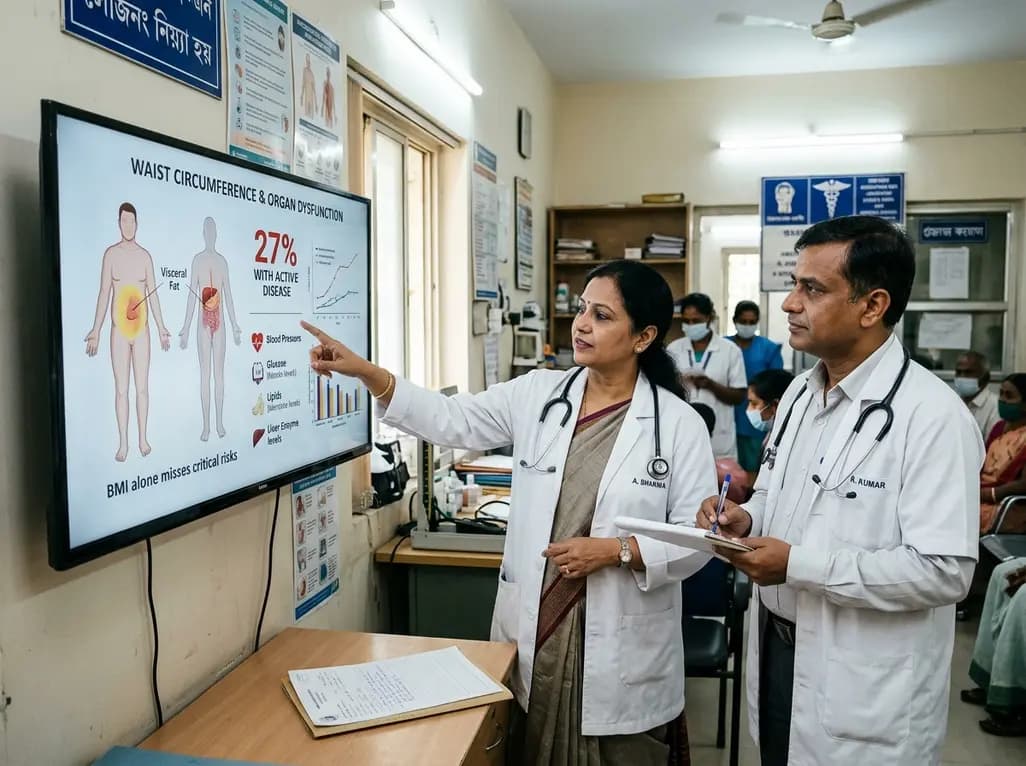
A landmark Nature study reveals that only 27% of individuals with obesity have active clinical disease. By incorporating waist circumference and organ dysfunction, this new diagnostic framework moves beyond BMI to prevent widespread misclassification and guide personalized metabolic intervention.
Today

A breakthrough blood test measuring the RNA biomarker Tu-Stroma detects microscopic serous tubal intraepithelial carcinoma (STIC) lesions with remarkable accuracy. This non-invasive diagnostic tool offers crucial biological insight to guide prophylactic surgery decisions for women carrying high-risk genetic mutations.
Today
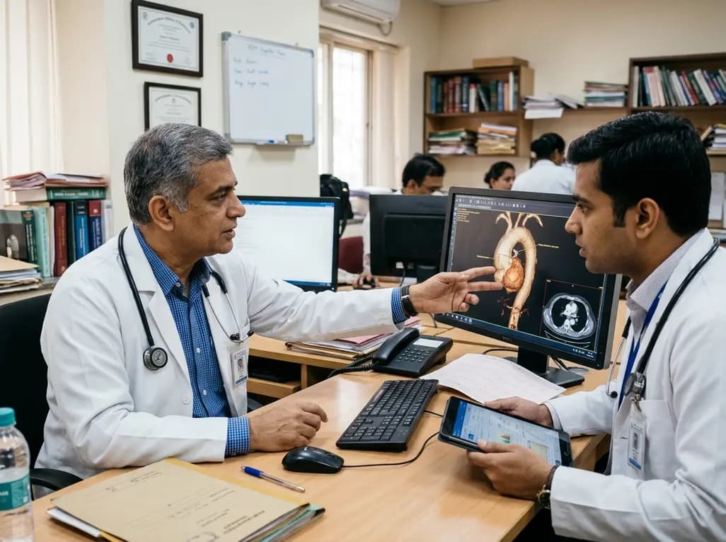
Thoracic aortic aneurysm lacks etiologic medical therapy. A new study reveals that targeting specific αV integrins with Cilengitide attenuates TGF-β and mechanotransduction pathways, effectively limiting pro-fibrotic remodeling and aortic bulb dilation in both syndromic and sporadic disease models.
Today

A nationwide observational study from Japan examines 10-year trends, regional variations, and mortality outcomes of cemented versus uncemented hemiarthroplasty in 296,841 femoral neck fractures, offering critical insights for orthopaedic practice and geriatric trauma care.
Today
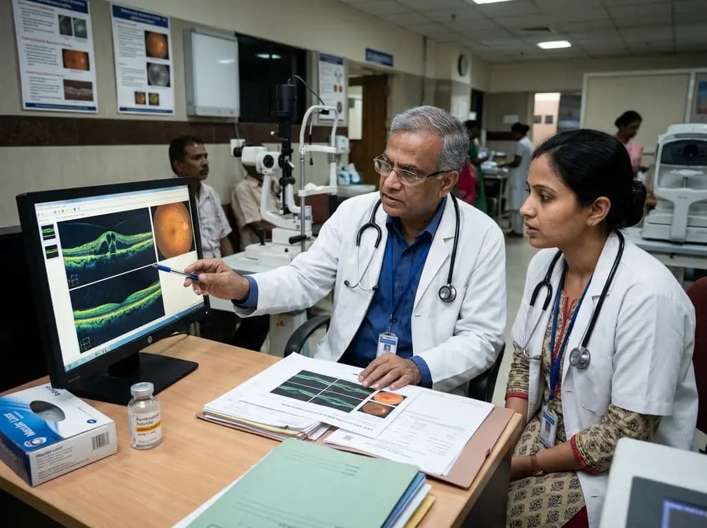
A real-world retrospective study demonstrates that combining intravitreal ranibizumab with focal/grid macular laser significantly improves visual acuity and central macular thickness compared to laser monotherapy in patients with center-involved diabetic macular edema.
Today
