

Loading, please wait...
Loading, please wait...

Ovarian malignancy remains one of the most lethal gynecological conditions due to late clinical detection. Historically, clinicians identified cases primarily at advanced stages when peritoneal seeding had occurred. Consequently, standard therapeutic interventions yielded poor long-term outcomes. Recently, investigators developed a groundbreaking ovarian cancer blood test capable of detecting premalignant alterations, establishing a vital diagnostic window. This breakthrough offers specialists an unprecedented opportunity to shift management from reactive treatment to proactive cancer interception.
For decades, diagnostic strategies focused on detecting established anatomical tumors through imaging and serum protein assays. However, this retrospective approach consistently failed to improve overall mortality across patient populations. Extensive research demonstrates that high-grade serous carcinomas rarely arise de novo within the ovary. Instead, these aggressive malignancies initiate within the distal fallopian tube as microscopic precursor lesions. Pathologists identify this earliest stage as serous tubal intraepithelial carcinoma. Because these focal cellular lesions measure mere microns, conventional pelvic ultrasound cannot visualize them. In addition, existing modalities lack the resolution needed to identify preinvasive cellular dysplasias. As a result, surgeons typically identified these lesions only during opportunistic prophylactic salpingectomy. Therefore, investigators shifted focus toward identifying early circulatory biochemical changes before invasive architectural growth begins. By tracking cell-to-cell competition within the tubal microenvironment, researchers discovered unique molecular signals entering circulation. Consequently, this breakthrough transforms early cancer screening from macroscopic tumor imaging to microscopic molecular surveillance. Furthermore, this paradigm allows proactive clinical management before invasive genetic subclones disseminate throughout the peritoneal cavity. Clinicians can therefore detect the earliest oncogenic trajectory when interventions offer the highest likelihood of cure. Early identification fundamentally halts the cascade toward widespread ovarian surface involvement. Ultimately, this conceptual evolution provides an actionable framework for preventing lethal gynecological malignancies.
The biological rationale behind this diagnostic advancement centers on cell competition within the tubal epithelium. Specifically, transformed epithelial cells actively weaken and eliminate neighboring normal cells to secure space for clonal proliferation. During this competitive process, nascent lesions release a distinct long noncoding RNA transcript designated as Tu-Stroma. Laboratory investigations confirmed that transformed cells package this molecule into extracellular vesicles that enter peripheral circulation. Moreover, elevated circulating concentrations occur exclusively when preinvasive or early invasive clones disrupt local cellular homeostasis. The novel ovarian cancer blood test utilizes targeted molecular amplification to detect and measure these specific vesicular transcripts. Unlike non-specific systemic markers that fluctuate during benign inflammation, this RNA biomarker reflects active oncogenic activity. Thus, the assay maintains exceptional diagnostic fidelity even when lesions comprise minimal cellular volumes. In fact, mechanistic studies show that Tu-Stroma directly alters surrounding stromal architecture and promotes host cell senescence. Consequently, detecting this transcript signals an active neoplastic process rather than an inert genetic predisposition. Clinicians therefore receive objective molecular insight into pathological events years before physical tumors appear. Furthermore, this non-invasive approach bypasses the anatomical barriers that traditionally concealed fallopian tube pathology from clinicians. Consequently, clinicians obtain a direct window into tissue-level competition before morphological disruption appears.
Rigorous validation studies demonstrate outstanding diagnostic accuracy across both high-risk and general patient cohorts. Specifically, investigators analyzed peripheral blood specimens from patients with histopathologically confirmed intraepithelial lesions alongside matched healthy controls. The liquid biopsy achieved an impressive sensitivity of 92.3% in identifying individuals harboring confirmed tubal precursor lesions. Furthermore, the assay demonstrated an exceptional specificity of 98.2%, accurately identifying healthy individuals without producing unwarranted positive signals. In oncological screening, false-positive findings trigger severe psychological distress and lead to invasive, unnecessary surgical procedures. Therefore, preserving high specificity remains vital for any diagnostic tool intended for widespread clinical adoption. Statistical modeling revealed that among women with known genetic mutations, the positive predictive value reached approximately 72%. Conversely, the negative predictive value approached nearly 100% across the evaluated risk populations. This exceptional negative accuracy reassures physicians when monitoring asymptomatic carriers who wish to delay preventive oophorectomy. In addition, receiver operating characteristic curve analysis confirmed superior discriminatory power compared to standard diagnostic parameters. Hence, these robust performance metrics provide compelling evidentiary support for integrating the assay into tertiary diagnostic workflows. Consequently, clinicians can utilize this objective molecular tool with substantial diagnostic confidence. Moreover, future multi-institutional validation will further establish its reproducibility across diverse clinical laboratories. Thus, these findings establish a transformative precedent for reliable non-invasive gynecologic cancer surveillance.
Management of patients carrying pathogenic BRCA1, BRCA2, or mismatch repair mutations remains a formidable clinical challenge. Currently, standard clinical guidelines recommend risk-reducing bilateral salpingo-oophorectomy between ages 35 and 45. However, this irreversible surgical intervention precipitates abrupt surgical menopause, causing vasomotor symptoms, cardiovascular risks, and bone loss. Many young women experience severe anxiety regarding the timing of castration, particularly while planning their families. At present, clinicians guide surgical timing based entirely on statistical age thresholds rather than demonstrable biological disease. This novel liquid biopsy introduces essential biological certainty into complex surgical counseling discussions. For instance, a confirmed negative test result allows clinicians to support delaying oophorectomy until childbearing is finished. In contrast, elevated Tu-Stroma levels indicate active tubal neoplasia, providing an unequivocal indication for immediate surgical intervention. Thus, physicians can transition from arbitrary population-based timelines to personalized, biology-driven clinical protocols. Additionally, objective molecular data alleviates patient apprehension by confirming that deferral remains biologically safe. Surgeons can also implement specialized pathology protocols during surgery when prior blood testing indicates high pre-test probability. Furthermore, this strategy empowers high-risk patients to make informed decisions without compromising their future overall health. Consequently, clinical discussions become collaborative evaluations rooted in real-time biology. Ultimately, this refined approach balances vital oncologic risk reduction with long-term hormonal and reproductive preservation.
For decades, clinicians relied primarily on serum CA-125 measurements and pelvic ultrasound to monitor ovarian cancer risk. Unfortunately, major trials demonstrated that these surveillance tools fail to decrease overall ovarian cancer mortality. Serum CA-125 exhibits poor diagnostic sensitivity for early disease, remaining unperturbed until massive tumor burden develops. Moreover, CA-125 frequently rises during benign conditions, including endometriosis and pelvic inflammatory infections. Consequently, non-neoplastic elevations frequently prompt unnecessary exploratory surgeries in healthy premenopausal individuals. In contrast, circulating Tu-Stroma reflects active cellular competition rather than gross macroscopic tissue volume. Extracellular vesicles actively encapsulate and shield these noncoding RNA transcripts against circulating enzymatic degradation. Therefore, vesicular Tu-Stroma exhibits robust analytical stability in routine peripheral blood specimens. Successfully translating this tool into everyday practice requires standardized laboratory protocols on automated platforms. Additionally, oncology societies must define evidence-based surveillance intervals for women enrolled in active monitoring protocols. Identifying lesions at the preinvasive stage also opens intriguing avenues for targeted chemoprevention before surgery becomes necessary. Furthermore, this technology eliminates longstanding diagnostic blind spots with remarkable precision. Consequently, integrating liquid biopsies into routine preventive care marks a decisive evolution in modern oncology. Therefore, precision screening will soon replace non-specific markers as the clinical gold standard. Ultimately, proactive precision diagnostics will redefine standard clinical management for high-risk women worldwide.
Q1: What biological precursor does the new test detect?
The test specifically identifies serous tubal intraepithelial carcinoma, commonly known as STIC. This microscopic lesion represents the earliest non-invasive precursor to high-grade serous ovarian carcinoma and originates in the fallopian tube epithelium. Because STIC lesions are microscopic and non-invasive, conventional radiological imaging cannot detect them. The test identifies elevated circulating levels of Tu-Stroma RNA released during early cellular competition, signaling tissue transformation long before an anatomical tumor forms.
Q2: How does Tu-Stroma compare to traditional CA-125 testing?
Traditional CA-125 is a high-molecular-weight glycoprotein that rises primarily after substantial tumor burden develops or in non-malignant inflammatory conditions like endometriosis. Consequently, CA-125 lacks both sensitivity for early precursor lesions and specificity in premenopausal women. In contrast, Tu-Stroma is an RNA transcript packaged in extracellular vesicles that reflects active cell-competition dynamics within the tubal microenvironment. This functional specificity allows Tu-Stroma to detect microscopic STIC with 92.3% sensitivity and 98.2% specificity.
Q3: How might this assay alter prophylactic surgical decisions for high-risk women?
Currently, carriers of BRCA1 or BRCA2 mutations undergo risk-reducing bilateral salpingo-oophorectomy based purely on age and statistical risk. This surgery triggers premature surgical menopause and adverse metabolic sequelae. Implementing this blood test provides individual biological data regarding the actual presence of preinvasive lesions. Consequently, patients with consistently negative results may safely delay surgery to complete childbearing, whereas a positive result provides clear clinical justification for immediate, targeted surgical intervention.
Disclaimer: This content is for informational and educational purposes only. It does not constitute medical advice or replace professional judgment. Refer to the latest local and national guidelines for clinical practice.
References

Read summarized clinical updates, watch expert medical content, and earn CME certifications right from your smartphone.
Open your smartphone camera to scan and install.





A breakthrough blood test measuring the RNA biomarker Tu-Stroma detects microscopic serous tubal intraepithelial carcinoma (STIC) lesions with remarkable accuracy. This non-invasive diagnostic tool offers crucial biological insight to guide prophylactic surgery decisions for women carrying high-risk genetic mutations.
Today

Ambient digital scribes promise effortless medical documentation, but a critical sociotechnical commentary reveals that AI alters rather than eliminates charting work. Discover how shifting cognitive burdens, oversight risks, and workflow redesign impact clinical safety and healthcare practice.
Today

Thoracic aortic aneurysm lacks etiologic medical therapy. A new study reveals that targeting specific αV integrins with Cilengitide attenuates TGF-β and mechanotransduction pathways, effectively limiting pro-fibrotic remodeling and aortic bulb dilation in both syndromic and sporadic disease models.
Today
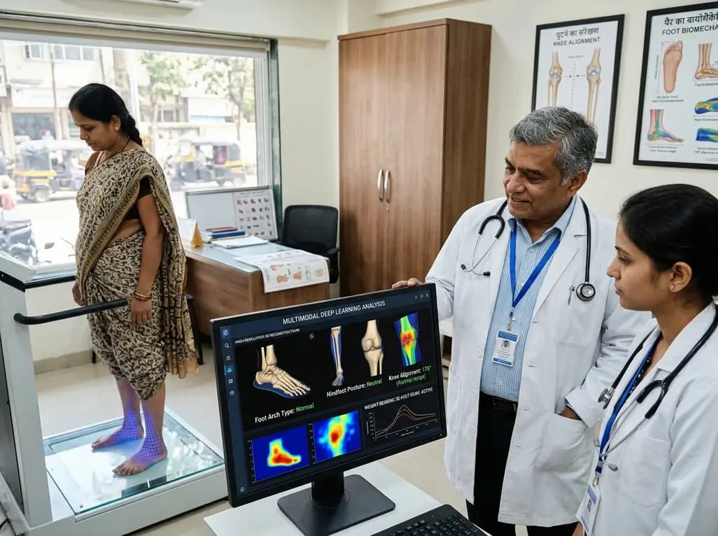
A multimodal deep learning framework utilizing weight-bearing 3D foot scans accurately screens lower-limb alignment, including arch type, hindfoot posture, and knee alignment, offering an accessible, radiation-free alternative to radiography for early biomechanical triage.
Today
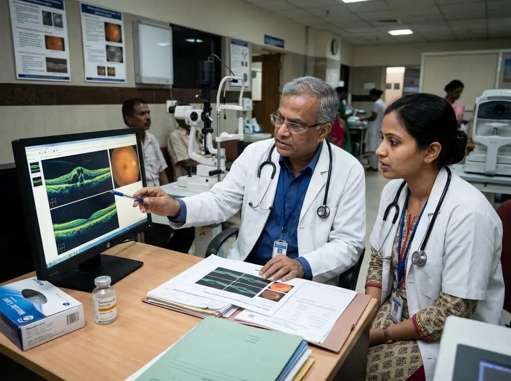
A real-world retrospective study demonstrates that combining intravitreal ranibizumab with focal/grid macular laser significantly improves visual acuity and central macular thickness compared to laser monotherapy in patients with center-involved diabetic macular edema.
Today
