

Loading, please wait...
Loading, please wait...
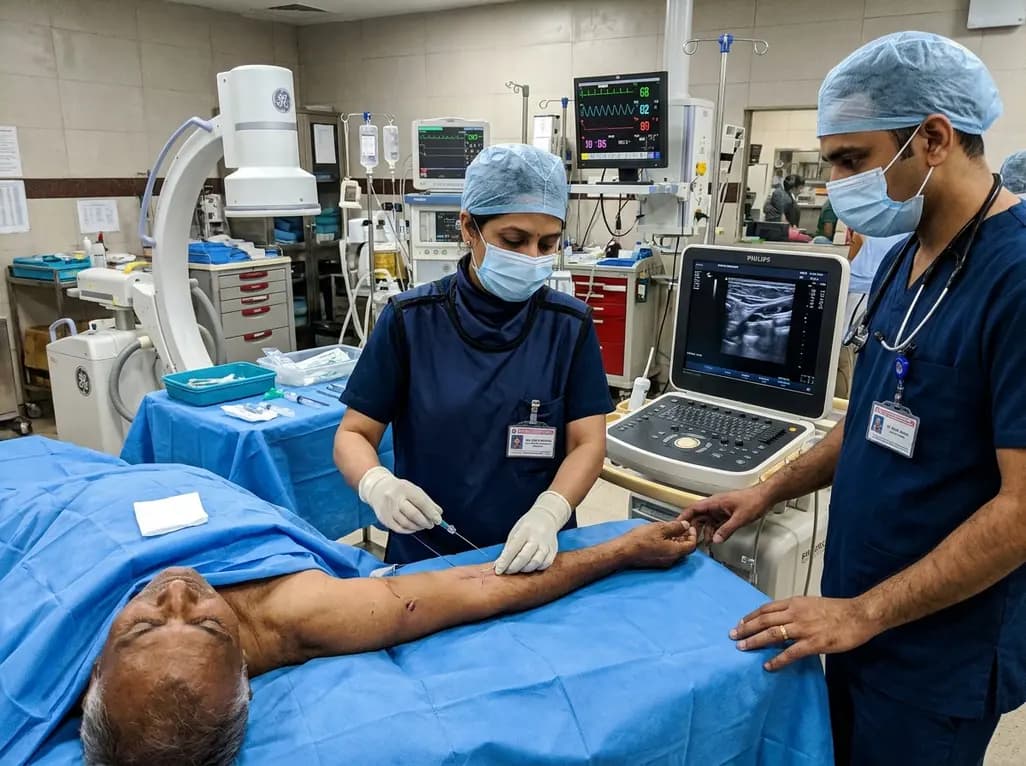
Modern inpatient care frequently requires prolonged intravenous access for targeted antimicrobial therapy and complex surgical interventions. Clinicians often encounter hospitalized patients with severe venous depletion, making standard peripheral cannulation impossible. In these scenarios, hybrid anterograde tunnelling offers an innovative procedural strategy to secure reliable central venous access. For instance, managing cervical spondylodiscitis requires weeks of parenteral antibiotics alongside major spinal stabilization surgery. However, repeated hospitalizations and prior venipunctures frequently leave peripheral veins sclerosed. Clinicians must balance multi-lumen access against the severe risks of central line-associated bloodstream infections. Therefore, teams often select peripherally inserted central catheters to decrease immediate insertion complications. Unfortunately, severe peripheral anatomical constraints can limit suitable puncture sites in the upper extremities. When optimal arm veins disappear, operators frequently face poor alternatives. They must either cannulate higher-risk central veins or accept sub-optimal peripheral vessels. Cannulating vessels near the axilla forces an exit site inside the moist axillary crease. Consequently, clinicians need dependable procedural strategies that safeguard both vessel health and the cutaneous exit site.
Vascular access teams rely heavily on Dawson's Zone Insertion Method to optimize catheter longevity and patient comfort. This systematic framework divides the upper arm into red, green, and yellow zones based on anatomical landmarks. Ideally, operators puncture veins and establish exit sites entirely within the middle third, known as the green zone. This middle region provides a flat, clean skin surface with low bacterial bioburden and minimal joint movement. In contrast, the proximal third, designated as the yellow zone, houses larger vessels like the proximal basilic and axillary veins. Cannulating yellow zone vessels provides sufficient luminal diameter, which prevents catheter-related thrombosis. However, placing the skin exit site in this proximal area causes major clinical challenges. The axillary fold generates continuous friction, heavy perspiration, and dense microbial colonisation. Additionally, surgical neck draping during cervical spine surgery directly compromises high axillary dressings. Dressings placed near the axilla peel rapidly, increasing accidental dislodgement rates. Therefore, clinicians must decouple the optimal deep vessel puncture from the superficial exit site. Tunnelling resolves this anatomical dilemma by bridging the two locations through healthy subcutaneous tissue.
Executing hybrid anterograde tunnelling allows operators to bridge distant anatomical zones without relying on specialized commercial tunnellers. The operator begins by performing comprehensive ultrasound evaluation of the deep brachial and axillary vessels. Once the team identifies a patent proximal basilic-axillary junction, they administer local anaesthesia along the planned subcutaneous path. First, the clinician utilizes a pseudo-tunnelling puncture technique. Under direct ultrasound guidance, the needle enters the skin distal to the target vein and traverses subcutaneous tissue before puncturing the vessel wall. Next, the practitioner advances a soft guidewire safely into the superior vena cava. To extend the exit site further distally into the ideal green zone, the team creates an anterograde subcutaneous tract. The clinician inserts a standard 14-gauge vascular cannula from the planned mid-arm exit site toward the initial venipuncture site. After withdrawing the needle stylet, the operator threads the 5-Fr catheter anterogradely through the plastic cannula sheath. Consequently, the catheter enters the green zone skin and reaches the venous entry tract. Finally, the operator introduces the catheter into the vein using a micro-introducer, securing an optimal mid-arm exit.
Commercial vascular access kits do not always contain specialized metallic blunt tunnellers, especially in resource-constrained hospital settings. Clinicians frequently encounter institutional supply shortages that demand safe procedural workarounds. Fortunately, standard vascular consumables can readily construct an effective anterograde subcutaneous conduit. Using an inexpensive 14-gauge intravenous cannula replicates the utility of commercial tunnellers without generating excess tissue trauma. The plastic sheath creates an atraumatic, smooth lumen that shields the catheter during advancement. Moreover, this hybrid anterograde technique eliminates forceful blunt dissection through fragile subcutaneous adipose tissue. Clinicians avoid creating large subcutaneous pockets that might collect haematomas. Additionally, operators must maintain continuous haemostatic control at the transition puncture point. Applying medical cyanoacrylate tissue adhesive at the puncture site effectively seals the tissue tract, preventing extraluminal bleeding. The adhesive also establishes a formidable barrier against external skin microbiota. Furthermore, using sutureless securement devices and transparent chlorhexidine dressings stabilizes the external line in the mid-arm. Thus, resourceful procedural adaptations ensure uncompromising patient safety, procedural efficiency, and reliable vascular preservation even when proprietary tools remain unavailable.
Relocating the exit site to the mid-upper arm provides profound mechanical and microbiological advantages for surgical patients. In clinical practice, catheters placed via this hybrid technique demonstrate outstanding stability throughout demanding inpatient stays. For example, cervical spine surgical procedures require intensive intraoperative patient positioning and postoperative rigid cervical orthoses. High axillary lines frequently catch on spinal braces or surgical drapes, resulting in premature accidental dislodgement. By contrast, a mid-arm exit site remains isolated from cervical surgical fields, preserving dressing integrity. Furthermore, clinical observation confirms that eliminating axillary moisture drastically reduces local skin maceration and microbial overgrowth. Daily catheter inspections reveal minimal erythema and no purulent discharge across the dwell period. Similarly, maintaining a favourable catheter-to-vein ratio in the larger basilic-axillary junction prevents venous stasis and secondary catheter-related thrombosis. Follow-up Doppler ultrasonography regularly confirms complete venous patency and normal phasic flow without mechanical catheter kinking. Consequently, patients receive full courses of intravenous antibiotic therapy without costly catheter exchanges. These reliable functional outcomes demonstrate that meticulous subcutaneous tunnelling preserves essential vascular access while minimizing secondary complications.
Conventional tunnelling creates a separate subcutaneous pathway using a specialized blunt metallic instrument before introducing the catheter into the vein. In contrast, pseudo-tunnelling uses a single long echogenic needle that enters the skin distally in the mid-arm and traverses deep subcutaneous tissue to puncture a proximal vein directly. This technique eliminates blunt dissection while effectively relocating the skin entrance away from moist flexural creases.
Clinicians should consider tunnelling whenever the most suitable vein resides exclusively in Dawson's yellow zone near the axilla. Puncturing proximal basilic or axillary veins ensures an adequate vein-to-catheter ratio, preventing thrombosis. However, leaving the exit site within the axilla significantly elevates infection and dislodgement risks. Tunnelling bridges the deep vessel puncture to a clean, flat exit site in the mid-arm green zone, maximizing stability.
Operators can safely construct an anterograde tunnel using a standard 14-gauge peripheral intravenous cannula sheath. After local anaesthesia, the clinician advances the cannula from the planned mid-arm exit site toward the initial venipuncture site. The operator removes the needle stylet, trims the plastic hub, and threads the catheter anterogradely through the sheath. This straightforward modification provides an atraumatic pathway without causing excessive tissue damage or haematoma formation.
Disclaimer: This content is for informational and educational purposes only. It is not intended to be a substitute for professional medical advice, diagnosis, or treatment. Always seek the advice of your physician or other qualified health provider with any questions you may have regarding a medical condition or procedural technique. Refer to the latest local and national guidelines for clinical practice.
References

Read summarized clinical updates, watch expert medical content, and earn CME certifications right from your smartphone.
Open your smartphone camera to scan and install.





This clinical review examines hybrid anterograde tunnelling for PICC insertion without dedicated blunt tunnellers. By combining pseudo-tunnelling with an anterograde subcutaneous tract, clinicians relocated the exit site to the mid-arm, ensuring optimal dwell time and zero complications.
Today

A landmark 12-year longitudinal study highlights significant reductions in opioid prescribing for nonspecific low back pain following clinical guidelines and prescription monitoring systems, emphasizing non-opioid multimodal pain care.
Today
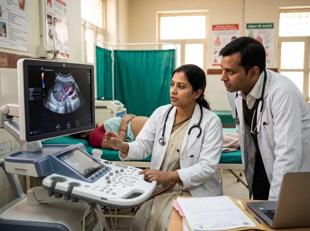
A study in Ultrasound in Obstetrics & Gynecology shows that quantitative vascularization of the anterior fornix/cervix with transvaginal ultrasound reliably identifies placenta accreta spectrum and predicts severe intraoperative hemorrhage, offering vital objective risk stratification.
Today

A landmark study details how an enterprise pediatric remote patient management model expanded from a single cardiology pilot to 16 diverse specialty programs by combining centralized digital operations with specialty clinical leadership.
Yesterday
A study evaluates vastus lateralis twitch kinetics, neural activation, and muscle architecture in 44 soccer players, showing dynamic neuromuscular measures predict jump and squat performance far better than ultrasound architecture alone.
Today

Protein O-GlcNAcylation couples cellular nutrient status to vital neuronal functions. This review explores how disrupted O-GlcNAc cycling drives both neurodevelopmental delays and neurodegenerative cascades, positioning metabolic post-translational modifications as therapeutic targets in clinical neurology.
Today
