

Loading, please wait...
Loading, please wait...

Modern sports medicine increasingly relies on advanced neuromuscular function testing to evaluate athletic capacity and guide return-to-play decisions. Although clinicians frequently examine gross structural attributes, muscular performance depends heavily on rapid neural activation and intrinsic contractile properties. Therefore, evaluating peripheral twitch dynamics provides crucial physiological insights that anatomical imaging cannot capture alone. Recently, investigators led by Artur Terbalyan resolved these complex relationships in competitive soccer players. Specifically, the researchers evaluated how muscle architecture, neural drive, and twitch kinetics interact to govern functional power and strength. Consequently, their groundbreaking trial establishes an objective framework for athletic profiling. By triangulating vastus lateralis function, clinicians can better understand dynamic muscular performance. In professional soccer, athletes constantly execute explosive ballistic actions such as sprinting, cutting, and jumping. As a result, assessing neuromuscular capability requires reliable modalities that isolate contractile velocity from voluntary effort. Furthermore, these diagnostic innovations bridge the gap between laboratory biomechanics and pitch-side rehabilitation. Ultimately, this integrated diagnostic strategy enables medical professionals to tailor precise conditioning programs.
To evaluate lower-extremity capacity comprehensively, the investigative protocol examined forty-four trained male soccer players. First, the researchers utilized diagnostic ultrasonography to assess the baseline architecture of the vastus lateralis muscle. This imaging measured muscle thickness, pennation angle, and estimated fascicle length. However, static ultrasound measurements reflect geometric structural orientation rather than dynamic cross-bridge recruitment during explosive activity. In addition, the researchers measured involuntary contractile properties using tensiomyography, an accurate non-invasive mechanical assessment tool. Tensiomyography quantifies radial muscle belly displacement, contraction velocity, and sustained twitch response following a standardized electrical stimulus. Consequently, it removes motivational confounders and assesses true peripheral contractile kinetics. Moreover, investigators recorded neural drive via surface electromyography normalized to maximal voluntary contraction. This electrical evaluation tracked voluntary central motor unit recruitment under maximal isometric conditions. Therefore, integrating these three distinct modalities provided a multi-dimensional biological portrait of the quadriceps. Sports clinicians can thus distinguish between structural hypertrophy, neural drive limitations, and peripheral mechanical deficiencies with remarkable fidelity.
Athletic performance testing in soccer commonly prioritizes dynamic power generation and maximal force output. To operationalize these functional capacities, the study evaluated countermovement jump height and half-squat one-repetition maximum strength. Countermovement jumps reflect fast stretch-shortening cycle mechanics, explosive acceleration, and elastic energy utilization. In contrast, the half-squat one-repetition maximum measures absolute lower-limb force production against heavy external resistance. Subsequently, the researchers performed stepwise multiple linear regression analyses to identify the physiological variables that most strongly predicted performance. Interestingly, the regression models accounted for 34% of the variance in countermovement jump height. Furthermore, the predictive models explained 42% of the variance observed in half-squat strength. Notably, static muscle thickness and pennation angle alone offered limited predictive power. Instead, incorporating twitch mechanics and normalized surface electromyography significantly improved model accuracy. Hence, functional jumping ability and squatting strength correlate far more strongly with contractile kinetics than with simple morphological dimensions. These statistical findings emphasize that muscle size does not equate directly to functional ballistic performance.
For decades, sports conditioning and clinical assessments focused heavily on cross-sectional muscle area and structural architecture. However, modern physiological evidence demonstrates that muscle architecture provides an incomplete depiction of real-world athleticism. Although fascicle length and pennation angle influence theoretical force-velocity curves, they remain static geometric parameters. In contrast, tensiomyography evaluates the rapid temporal dynamics of sarcoplasmic calcium release and actomyosin binding. Additionally, twitch kinetics capture intrinsic muscle fiber typing and muscle-tendon unit stiffness without requiring exhaustive voluntary testing. For example, faster contraction times and optimal radial displacement indicate higher proportions of fast-twitch glycolytic motor units. Furthermore, normalized surface electromyography reflects central descending command and motor unit firing rates. When athletes execute ballistic actions, high rate of force development depends on rapid neural recruitment rather than anatomical volume. Consequently, combining twitch mechanics with electromyography provides substantial incremental predictive value over ultrasonography alone. Because athletic actions occur in milliseconds, measuring kinetic twitch speed offers far superior functional insights for clinicians evaluating competitive athletes.
These comprehensive findings offer vital clinical applications for sports physicians, orthopaedic surgeons, and physical rehabilitation specialists. In India, competitive football and athletics are expanding rapidly, leading to increased demand for advanced musculoskeletal medicine. Frequently, clinicians evaluate athletes recovering from anterior cruciate ligament reconstruction, quadriceps strain, or patellar tendinopathy. Traditional return-to-play clearance relies predominantly on static imaging and subjective manual muscle testing. However, static healing does not guarantee the restoration of neuromuscular firing rates or fast-twitch mechanical kinetics. Therefore, incorporating tensiomyography and electromyography into clinical assessment batteries allows practitioners to identify subtle functional deficits. For instance, an athlete may demonstrate normal vastus lateralis thickness on ultrasound but retain persistent delays in twitch contraction time. As a result, premature return to competition could elevate re-injury risk during rapid decelerations. Moreover, sports medicine teams can utilize these objective metrics to design targeted strength, plyometric, or electrical stimulation interventions. Ultimately, objective neuromuscular profiling enhances rehabilitation precision, protects joint integrity, and accelerates safe return to high-level competition.
Tensiomyography evaluates muscle contractile kinetics non-invasively by applying a standardized electrical twitch stimulus directly to the muscle belly while the patient rests comfortably. A high-precision digital displacement sensor records muscle deformation, capturing parameters such as contraction time, delay time, and radial displacement. Consequently, this technique isolates intrinsic peripheral muscular properties from central voluntary effort. Furthermore, it provides objective diagnostic data regarding muscle stiffness and fiber-type characteristics without causing exercise-induced fatigue.
Muscle architecture measurements, including ultrasound-derived thickness and pennation angle, provide only static morphological information about tissue structure. Although structural cross-sectional area determines theoretical maximal force, dynamic athletic tasks depend heavily on temporal motor unit recruitment and cross-bridge cycling speed. In contrast, rapid ballistic actions like jumping and sprinting require instantaneous neural firing and rapid twitch kinetics. Therefore, structural dimensions alone cannot capture rate of force development, making dynamic physiological assessments indispensable for accurate functional evaluation.
Sports physicians can integrate twitch kinetics and electromyography to establish objective benchmarks during rehabilitation following knee injuries or surgeries. Specifically, these non-invasive tools identify persistent neuromuscular inhibition or prolonged contraction times that standard clinical examinations overlook. Consequently, rehabilitation specialists can prescribe targeted rate-of-force development exercises, eccentric overload training, or neuromuscular electrical stimulation. Ultimately, monitoring these mechanical properties ensures safe return to sport while significantly minimizing the risk of secondary reinjury.
Disclaimer: This content is for informational and educational purposes only... Refer to the latest local and national guidelines for clinical practice.
References

Read summarized clinical updates, watch expert medical content, and earn CME certifications right from your smartphone.
Open your smartphone camera to scan and install.





A study evaluates vastus lateralis twitch kinetics, neural activation, and muscle architecture in 44 soccer players, showing dynamic neuromuscular measures predict jump and squat performance far better than ultrasound architecture alone.
Today
A landmark 12-year longitudinal study highlights significant reductions in opioid prescribing for nonspecific low back pain following clinical guidelines and prescription monitoring systems, emphasizing non-opioid multimodal pain care.
Today
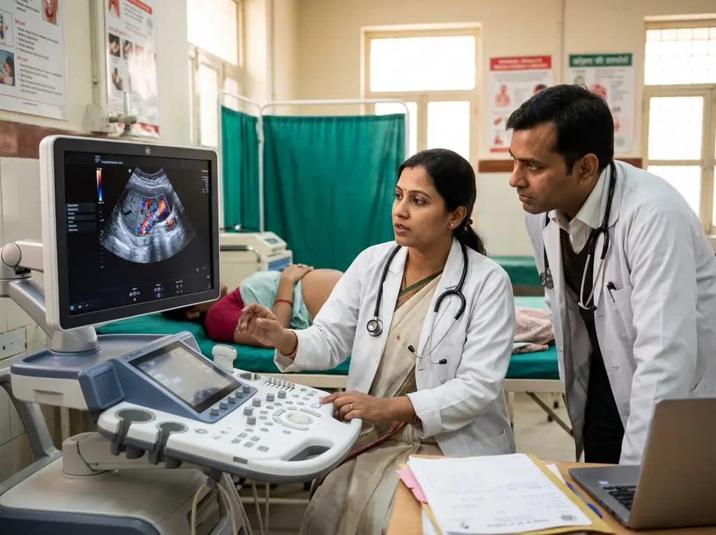
A study in Ultrasound in Obstetrics & Gynecology shows that quantitative vascularization of the anterior fornix/cervix with transvaginal ultrasound reliably identifies placenta accreta spectrum and predicts severe intraoperative hemorrhage, offering vital objective risk stratification.
Today

A landmark study details how an enterprise pediatric remote patient management model expanded from a single cardiology pilot to 16 diverse specialty programs by combining centralized digital operations with specialty clinical leadership.
Yesterday

Protein O-GlcNAcylation couples cellular nutrient status to vital neuronal functions. This review explores how disrupted O-GlcNAc cycling drives both neurodevelopmental delays and neurodegenerative cascades, positioning metabolic post-translational modifications as therapeutic targets in clinical neurology.
Today
