

Loading, please wait...
Loading, please wait...
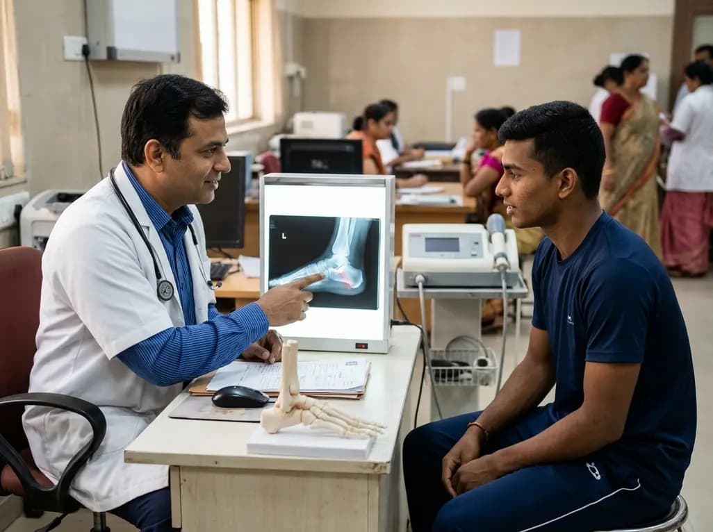
An anterior process calcaneus fracture represents a frequently overlooked musculoskeletal injury of the lateral hindfoot. Athletes typically sustain this injury during severe plantarflexion and inversion of the foot, which tensions the bifurcate ligament. Alternatively, forceful midfoot abduction may crush the anterior calcaneal process against the cuboid. Because clinical signs strongly mimic lateral ankle sprains, emergency clinicians frequently fail to identify the bony break on routine plain radiographs. Consequently, patients often receive treatment for routine ligamentous sprains while an intra-articular or extra-articular fracture goes unaddressed.
Delayed identification can result in chronic disabling morbidity, especially in active running athletes. In 1982, Degan and colleagues categorized these injuries into three distinct anatomical patterns. Type I lesions comprise non-articular avulsions of the tip. Type II injuries involve extra-articular fractures that do not disrupt the articular contour. In contrast, Type III fractures traverse the calcaneocuboid joint, creating substantial mechanical incongruity. Because conservative splinting frequently fails in neglected intra-articular injuries, symptomatic pseudoarthrosis and persistent lateral foot pain often follow. Therefore, early high-resolution computed tomography remains the gold standard to establish an accurate anatomical diagnosis and prevent prolonged disability.
A notable clinical scenario recently detailed the presentation of a 21-year-old collegiate hurdler who battled left lateral midfoot pain for two full years. The athlete initially suffered what appeared to be an ankle sprain during high-intensity track training. Despite undergoing extended conservative rehabilitation, his lateral foot discomfort persisted and prevented him from competing. When he presented for advanced orthopaedic evaluation, physical examination revealed point tenderness directly over the calcaneocuboid joint. His pain spiked significantly during terminal forefoot pushoff and running drills.
Subsequent computed tomography revealed an established, neglected Degan type III anterior process calcaneus nonunion with clear calcaneocuboid joint involvement. The persistent fibrous gap produced significant mechanical pain, reflected by a visual analog scale score of 75 out of 100. Historically, clinicians manage symptomatic type III nonunions through open reduction and internal fixation or excision of the ununited fragment. However, surgical interventions carry distinct risks, including nonunion recurrence, persistent joint instability, damage to adjacent tarsal ligaments, and prolonged athletic absence. Because the hurdler strongly wished to avoid operative reconstruction, the clinical team sought an innovative non-surgical pathway to stimulate bone healing.
Faced with surgical refusal, the clinical team turned to focused extracorporeal shock wave therapy as an innovative regenerative modality. Focused shockwave technology delivers acoustic energy pulses with high focal precision deep into musculoskeletal structures. Orthopaedic surgeons traditionally utilize this intervention for recalcitrant pseudoarthroses of the tibia, metatarsals, and scaphoid. At the cellular level, shockwaves trigger biological mechanotransduction. Consequently, this biomechanical stimulation upregulates vital vascular endothelial growth factor, accelerates regional microcirculation, and recruits osteogenic progenitor cells to recalcitrant bony margins.
The therapeutic regimen in this case consisted of nine focused shock wave sessions structured across three distinct treatment sets over nineteen weeks. The clinicians administered a peak energy flux density ranging between 0.07 and 0.25 mJ/mm². Clinicians precisely localized the shockwave focal zone to the anterior calcaneal process using anatomical landmarks and radiographic guidance. Furthermore, the low-to-moderate energy flux density minimized localized soft tissue irritation while adequately stimulating bone remodeling. By repeatedly delivering mechanical impulses across the sclerotic pseudoarthrosis interface, the protocol stimulated a dormant healing cascade without requiring open surgical exposure.
Biophysical stimulation alone rarely suffices without appropriate mechanical protection during athletic recovery. Therefore, clinicians coupled the shockwave sessions with a mandatory three-month athletic restriction from the onset of treatment. This load management protocol shielded the fragile anterior calcaneal process from cyclic shear forces generated by midtarsal rotation and aggressive forefoot push-off. During this rest period, the hurdler maintained cardiovascular conditioning through non-impact cross-training and targeted physical therapy that focused on ankle mobility and intrinsic foot muscle endurance.
The clinical results proved remarkably favorable as the therapeutic protocol advanced. The athlete reported steady pain relief, with his visual analog scale score dropping dramatically from 75 out of 100 before treatment to 0 out of 100 after therapy. Because his symptoms resolved completely, the athlete progressed through a structured running and hurdle drills program. At six months following the initiation of shock wave therapy, he successfully returned to full collegiate hurdling competition. Notably, he competed at his baseline pre-injury level without reporting residual pain, swelling, or secondary mechanical instability in the affected hindfoot.
Objective imaging confirmed the dramatic clinical turnaround observed during the collegiate athlete's recovery. Serial follow-up computed tomography performed at fourteen months after initiating shock wave therapy revealed robust bony bridging across the previous fracture gap. The scan confirmed consolidation across the Degan type III site and demonstrated smooth reconstitution of the calcaneocuboid joint line. Consequently, the radiographic findings established true structural osseous union rather than mere temporary symptomatic pain relief.
This remarkable outcome highlights valuable clinical lessons for sports medicine physicians and orthopaedic trauma specialists. While Degan type III fractures traditionally prompt surgical intervention due to articular involvement, invasive procedures carry prolonged postoperative downtime and risk arthritic degeneration. This case demonstrates that focused extracorporeal shock wave therapy can effectively induce osteogenesis in long-standing calcaneal nonunions. Although larger prospective clinical trials must validate these findings across broader populations, focused shockwave therapy represents an effective, organ-sparing alternative for competitive athletes who decline surgical intervention. Clinicians should maintain high clinical suspicion for anterior process nonunions in chronic sprains and consider biological acoustic therapy before scheduling invasive surgery.
Clinicians frequently miss these fractures because the injury mechanism closely mimics a routine lateral ankle inversion sprain. Standard anteroposterior and lateral ankle radiographs often obscure the anterior process due to overlapping midtarsal bones. Consequently, acute tenderness over the calcaneocuboid joint is mistakenly attributed to anterior talofibular ligament trauma. Therefore, persistent lateral midfoot swelling and inability to bear weight after conservative sprain management warrant dedicated oblique foot radiographs or computed tomography.
The Degan classification stratifies fractures based on fragment morphology and joint involvement. Specifically, type I involves a minor avulsion fracture of the anterior tip sparing the articular cartilage. Type II represents a larger extra-articular fragment that does not enter the calcaneocuboid joint. In contrast, type III fractures represent large, intra-articular fractures that breach the calcaneocuboid articular surface. Consequently, type III injuries create greater instability, carry substantial nonunion risk, and traditionally mandate open surgical reduction.
Focused shock wave therapy delivers high-energy acoustic pressure pulses directly to the sclerotic fracture gap. These acoustic waves induce mechanical microtrauma, triggering mechanotransduction within local osteoblasts and periosteal tissues. As a result, the biological cascade upregulates key osteogenic cytokines, including vascular endothelial growth factor and bone morphogenetic proteins. This biological stimulation accelerates microvascular ingrowth, clears interposed fibrous tissue, and restarts physiological osteogenesis, which successfully converts an established chronic nonunion into solid structural bony bridging.
Disclaimer: This content is for informational and educational purposes only... Refer to the latest local and national guidelines for clinical practice.
References

Read summarized clinical updates, watch expert medical content, and earn CME certifications right from your smartphone.
Open your smartphone camera to scan and install.





A collegiate hurdler with a 2-year neglected Degan type III anterior process calcaneus fracture achieved complete pain resolution and bony bridging using focused extracorporeal shock wave therapy, returning to competition without invasive surgery.
Today
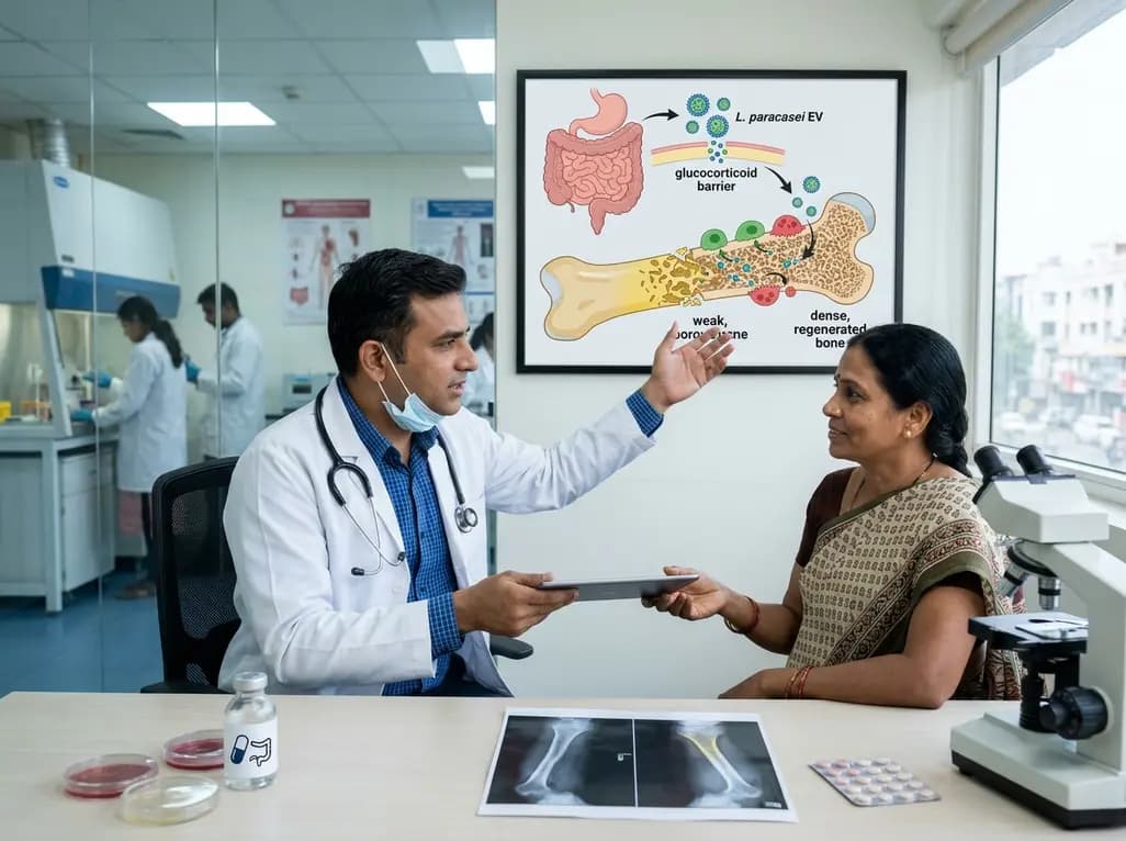
A novel oral nanoplatform using Lacticaseibacillus paracasei-derived extracellular vesicles delivers alendronate, reversing glucocorticoid-induced bone loss and restoring gut-immune-bone homeostasis.
Today

A rare presentation of cervical villoglandular adenocarcinoma metastasizing to the ovary mimicked a borderline ovarian tumor. Multimodal testing with immunohistochemistry and HPV16 DNA confirmed metastatic origin, guiding optimal surgical and chemoradiotherapy intervention.
Today

A landmark study details how an enterprise pediatric remote patient management model expanded from a single cardiology pilot to 16 diverse specialty programs by combining centralized digital operations with specialty clinical leadership.
Yesterday
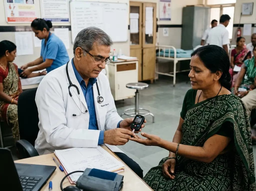
A real-world trial evaluating a near-infrared spectroscopy noninvasive glucose monitoring device in adults with type 2 diabetes demonstrated a combined MAD/MARD of 16.3 across 2,616 paired readings, highlighting its clinical feasibility.
Today
