

Loading, please wait...
Loading, please wait...

Clinical neuroscience increasingly recognizes the intricate interplay between supratentorial structures and the posterior fossa in neurodegenerative disorders. Investigating preclinical Alzheimer's disease has traditionally focused almost exclusively on the medial temporal lobe and cortical hubs. However, modern resting-state neuroimaging demonstrates that hippocampal-cerebellar functional connectivity serves as an essential functional axis during cognitive aging. Cerebrospinal fluid biomarkers directly reflect underlying neuropathology before overt dementia manifests. Similarly, the apolipoprotein E epsilon 4 allele represents the strongest genetic risk factor for sporadic Alzheimer's disease. Understanding how these biological determinants alter communication between the hippocampus and cerebellum provides vital insights into early compensatory mechanisms and progressive neural circuit failure.
Traditionally, clinicians regarded the cerebellum primarily as a coordinator of motor precision and gait stability. Nevertheless, contemporary neuroanatomical studies unequivocally confirm extensive reciprocal cerebro-cerebellar networks dedicated to higher-order cognition. The posterolateral cerebellar hemispheres, including Crus I and Crus II, communicate bidirectionally with the hippocampus and prefrontal cortex. Consequently, these pathways modulate spatial navigation, working memory, and episodic recall. In healthy aging adults, synchronized activity between these structures often strengthens over time. Clinicians interpret this heightened synchrony as an adaptive neurocognitive scaffolding mechanism. By recruiting additional cerebellar resources, the aging brain actively compensates for subtle structural or metabolic losses in the medial temporal lobe. Therefore, preserved functional coupling across this axis directly correlates with superior delayed recall and robust episodic memory performance. Conversely, when this coordinated interaction fails, subtle cognitive deficits emerge long before overt amnestic symptoms appear. Furthermore, longitudinal evaluations confirm that robust connectivity across this distributed network protects against executive and mnemonic decline. Specifically, functional coherence between contralateral cerebellar subregions and the anterior hippocampus reflects optimal network integrity during normal cognitive aging. Thus, monitoring this anatomical circuit provides essential information regarding baseline functional reserve in older patient populations.
The pathophysiological cascade of Alzheimer's disease begins decades before measurable clinical symptoms become evident. Evaluating the ratio of cerebrospinal fluid phosphorylated tau to amyloid beta provides an accurate index of early neuropathology. In individuals with normal biomarker levels, advancing chronological age correlates positively with synchronized network coherence between hippocampal subregions and cerebellar lobules. This positive trajectory reflects normal, non-pathological neural reorganization during healthy aging. However, an abnormal cerebrospinal fluid biomarker profile completely alters this physiological relationship. When elevated phosphorylated tau accompanies decreased amyloid beta, older adults display a marked disruption of this functional pathway. Instead of showing age-related compensatory gains, patients with abnormal biomarkers demonstrate a progressive decoupling of the circuit. Furthermore, this breakdown indicates that early soluble tau oligomers and diffuse amyloid pathology impair synaptically connected distant networks. Thus, resting-state fMRI captures subtle biomarker-driven disconnectivity syndromes long before structural magnetic resonance imaging detects gross parenchymal atrophy or hippocampal volume loss. In addition, these pathological alterations correlate with progressive declines in verbal recognition and delayed memory retrieval. As a result, assessing fluid biomarkers alongside functional imaging uncovers latent neurodegenerative vulnerability in asymptomatic patients.
The presence of the apolipoprotein E epsilon 4 allele significantly reshapes the landscape of cerebral functional connectivity. In healthy non-carriers, older chronological age corresponds with enhanced resting-state synchrony across cerebellar and hippocampal regions. This finding reinforces the concept that successful cognitive aging involves active functional reorganization. In sharp contrast, individuals carrying at least one epsilon 4 allele exhibit an inverse trajectory across their lifespan. For these genetically vulnerable individuals, older age correlates with declining functional coupling across the cerebellar axis. Moreover, epsilon 4 carriers demonstrate accelerated synaptic dysfunction and heightened neuroinflammatory susceptibility in both cortical and cerebellar microglial populations. As a result, the genetic predisposition prevents the brain from sustaining necessary compensatory scaffolding. Consequently, the convergence of advanced age and genetic vulnerability accelerates functional disconnection between the temporal lobe and cerebellum. Recognizing these distinct trajectory patterns allows clinicians to conceptualize genetic risk not merely as an isolated susceptibility factor, but as an active driver of early circuit-level vulnerability. Furthermore, asymptomatic carriers exhibit network degradation long before overt memory complaints surface. Therefore, genetic testing provides vital biological context when evaluating neuroimaging patterns in aging clinical cohorts.
Differentiating normal brain aging from insidious neurodegenerative disease remains a persistent challenge in outpatient clinical practice. Cognitive screening tools frequently fail to identify patients in the earliest preclinical phases of Alzheimer's disease. However, integrating advanced neuroimaging parameters with established fluid biomarkers offers a transformative diagnostic approach. When clinicians observe weakened functional interactions between the hippocampus and cerebellum, it signals pathological network breakdown rather than benign senescent change. Additionally, functional resting-state metrics correlate strongly with objective episodic memory scores across diverse patient cohorts. Identifying declining connectivity in cognitively intact adults carrying the epsilon 4 allele provides an objective rationale for closer longitudinal monitoring. Furthermore, identifying these circuit alterations enhances clinical trial stratification. Investigators can pinpoint individuals at greatest risk for rapid progression prior to irreversible neuronal loss. Consequently, evaluating distributed networks beyond traditional cortical regions helps physicians optimize risk assessments and tailor personalized management strategies well before clinical dementia manifests. In particular, this multimodal framework allows geriatricians to counsel high-risk families with greater precision. Therefore, neuroimaging protocols must expand beyond standard structural volumetry to incorporate functional network mapping.
Recognizing the active contribution of the cerebellum to episodic memory opens promising avenues for targeted neurorehabilitation. Because cerebellar networks demonstrate sustained neuroplastic potential, clinicians can design multimodal interventions to bolster functional scaffolding. For instance, structured cognitive training paired with aerobic physical exercise enhances functional synchrony between cerebellar lobules and the hippocampus. Additionally, non-invasive neuromodulation techniques, including repetitive transcranial magnetic stimulation and transcranial direct current stimulation, demonstrate growing efficacy in modulating cerebello-cerebral networks. Clinicians should also vigorously manage cardiovascular risk factors, as hypertension and microvascular disease exacerbate functional dysconnectivity in older adults. Furthermore, early therapeutic interventions using disease-modifying monoclonal antibodies targeting amyloid plaques may preserve network integrity more effectively when administered during this asymptomatic window. By proactively supporting these functional circuits through medical, lifestyle, and neuromodulatory interventions, clinicians can optimize brain resilience. Moreover, routine monitoring of motor and cognitive synchrony allows neurologists to measure real-world therapeutic responses with exceptional physiological sensitivity. Ultimately, preserving distributed network connectivity offers the greatest promise for delaying symptomatic cognitive impairment in high-risk patients. In conclusion, clinicians must adopt proactive multimodal strategies that harness cerebellar plasticity to safeguard cognitive longevity.
Coordinated activity between hippocampal regions and cerebellar lobules supports episodic memory consolidation and retrieval. Strong functional connectivity across this pathway provides neurocognitive scaffolding, which helps older individuals maintain superior memory performance. Conversely, dysconnectivity along this axis directly associates with measurable declines in delayed recall and memory retrieval accuracy.
The apolipoprotein E epsilon 4 allele accelerates synaptic dysfunction, impairs lipid metabolism, and promotes microglial neuroinflammation. Non-carriers exhibit adaptive increases in connectivity with age to compensate for neural changes. In contrast, carriers experience impaired synaptic resilience, preventing compensatory hyperconnectivity and causing functional network decoupling as they grow older.
Yes, resting-state functional MRI identifies subtle network alterations years before macroscopic parenchymal atrophy or hippocampal shrinkage becomes visible on structural scans. Disrupted functional synchrony between the hippocampus and cerebellum reflects early synaptic failure caused by amyloid and tau pathology, providing a sensitive physiological marker of preclinical neurodegeneration.
Disclaimer: This content is for informational and educational purposes only. It is not intended to be a substitute for professional medical advice, diagnosis, or treatment. Always seek the advice of your physician or other qualified health provider with any questions you may have regarding a medical condition. Refer to the latest local and national guidelines for clinical practice.
References

Read summarized clinical updates, watch expert medical content, and earn CME certifications right from your smartphone.
Open your smartphone camera to scan and install.





Recent neuroimaging findings demonstrate that hippocampal-cerebellar functional connectivity changes in preclinical Alzheimer's disease depending on APOE ε4 status and cerebrospinal fluid biomarker profiles.
Today

Ambient digital scribes promise effortless medical documentation, but a critical sociotechnical commentary reveals that AI alters rather than eliminates charting work. Discover how shifting cognitive burdens, oversight risks, and workflow redesign impact clinical safety and healthcare practice.
Today

A new comparative study demonstrates that intraoperative imprint cytology achieves 96.6% diagnostic accuracy in ovarian tumours, outperforming frozen section in sensitivity for borderline lesions and offering an accessible, rapid alternative for resource-constrained surgical centres.
Today

Thoracic aortic aneurysm lacks etiologic medical therapy. A new study reveals that targeting specific αV integrins with Cilengitide attenuates TGF-β and mechanotransduction pathways, effectively limiting pro-fibrotic remodeling and aortic bulb dilation in both syndromic and sporadic disease models.
Today
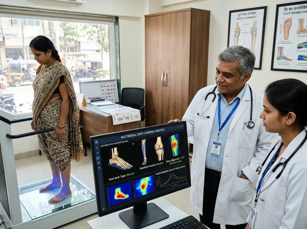
A multimodal deep learning framework utilizing weight-bearing 3D foot scans accurately screens lower-limb alignment, including arch type, hindfoot posture, and knee alignment, offering an accessible, radiation-free alternative to radiography for early biomechanical triage.
Today
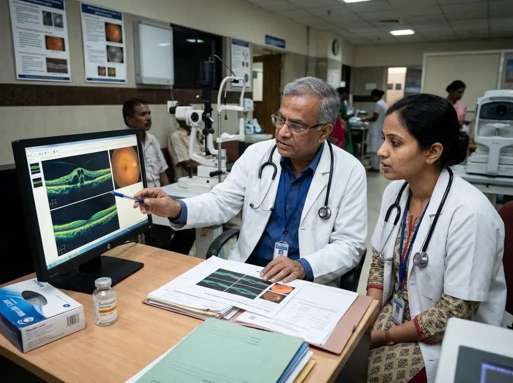
A real-world retrospective study demonstrates that combining intravitreal ranibizumab with focal/grid macular laser significantly improves visual acuity and central macular thickness compared to laser monotherapy in patients with center-involved diabetic macular edema.
Today
