

Loading, please wait...
Loading, please wait...

Diffusion MRI serves as an indispensable non-invasive tool for evaluating microstructural brain connectivity in clinical neurology and neurosurgery. However, cross-scanner variability significantly undermines multi-center clinical trials and diagnostic consistency across hospital networks. Modern quantitative neuroimaging critically relies on diffusion MRI harmonization to eliminate hardware-induced discrepancies between different imaging centers. A groundbreaking deep learning architecture named HARP now introduces a revolutionary phantom-only training methodology. Consequently, this technical milestone removes the historical requirement for traveling human cohorts across participating imaging facilities.
Multi-site neuroimaging studies frequently encounter substantial technical friction due to hardware disparities. Subtle differences in static magnetic field homogeneity, gradient coil specifications, radiofrequency receiver coils, and vendor reconstruction algorithms introduce systematic biases. As a result, identical biological tissues often generate divergent quantitative parameters across different magnetic resonance platforms. These site-dependent discrepancies confound longitudinal neurological investigations, multi-center stroke studies, neuro-oncology trials, and biomarker discovery in neurodegenerative diseases.
Historically, investigators attempted to overcome these distortions by scanning identical human volunteers across all participating medical institutions. However, organizing traveling human subject cohorts creates significant logistical complications, escalates institutional expenditures, and imposes heavy ethical burdens on participants. Furthermore, normal physiological variations and subtle aging effects within human subjects over time can inadvertently obscure genuine scanner-related differences. Traditional statistical post-processing approaches like ComBat attempt to remove batch effects retrospectively, yet they often risk scrubbing true biological heterogeneity or failing to address complex non-linear distortions. Consequently, clinicians require an automated, objective framework that stabilizes measurements without demanding traveling cohorts.
To address the logistical barriers of traveling human subjects, researchers developed HARP as a phantom-based deep learning pipeline. The framework trains a specialized neural network entirely on an easily transportable physical diffusion phantom rather than human brains. Because physical phantoms maintain absolute microstructural consistency over time, they provide an unvarying empirical baseline across disparate scanner environments.
Furthermore, this physical phantom travels effortlessly between diagnostic facilities without biological decay, physiological motion artifacts, or scheduling conflicts. Participating hospital centers scan the phantom using their standard single-shell diffusion protocols. Therefore, the network accurately learns the underlying mathematical transformations required to reconcile inter-scanner discrepancies. By decoupling algorithm training from human neuroanatomy, HARP completely eliminates the financial expenses and ethical challenges linked to traveling volunteer cohorts. Clinicians can readily integrate newly installed or updated imaging centers into active research consortia simply by scanning the transportable phantom. Consequently, multi-centric clinical studies can scale globally with unprecedented operational efficiency and dependable protocol consistency.
The core computational innovation of HARP lies in its voxel-wise one-dimensional neural network architecture. Instead of processing entire three-dimensional volumetric images, the model analyzes diffusion signals at an isolated voxel level. Specifically, the network maps mathematical relationships between spherical harmonics coefficients across distinct imaging facilities.
By focusing exclusively on spherical harmonics representations, the network learns directional diffusion transformations without memorizing anatomical structures or patient-specific brain geometry. Therefore, the algorithm cannot overfit to specific anatomical topographies or introduce hallucinated structural patterns into pathological scans. Additionally, spherical harmonics efficiently capture complex angular profiles of water diffusion within highly organized white matter tracts. The one-dimensional neural framework maintains exceptionally low computational overhead, facilitating rapid processing on standard hospital workstations. Furthermore, this compact network architecture allows rapid re-calibration whenever clinical scanners receive hardware upgrades or gradient maintenance. Thus, the system preserves raw diffusion physics while robustly standardizing signal properties across disparate vendor platforms.
Rigorous quantitative evaluation confirms that HARP achieves remarkable measurement stability across varied imaging platforms. When evaluated against scan-rescan standard error baselines, HARP significantly curtails inter-scanner variability across foundational diffusion tensor metrics. In quantitative analyses, the framework reduced inter-scanner variability by 12% in fractional anisotropy and by 10% in mean diffusivity.
Most notably, generalized fractional anisotropy demonstrated an impressive 30% reduction in inter-scanner standard error across sites. These improvements prove that the model successfully strips away scanner-induced noise without compressing physiological diffusion differences. Crucially, the harmonization process preserves underlying white matter fiber orientations with exquisite anatomical fidelity. Detailed tractography evaluations demonstrate that critical white matter tracts retain their directional integrity, spatial trajectory, and connectivity matrices post-harmonization. Consequently, neurosurgeons and radiologists can confidently utilize post-processed diffusion tractography for delicate pre-operative planning and stereotactic navigation. Clinicians gain reproducible quantitative biomarkers that accurately reflect underlying tissue microarchitecture rather than scanner discrepancies.
The deployment of phantom-only training offers transformative advantages for healthcare ecosystems across India. Indian tertiary hospitals, regional cancer centers, and government medical colleges typically operate a heterogeneous fleet of MRI scanners from multiple manufacturers. Consequently, pooling quantitative neuroimaging data across different diagnostic centers has historically proven exceedingly difficult.
By utilizing transportable diffusion phantoms, Indian clinical trial networks can now harmonize quantitative imaging datasets across major hubs like Mumbai, Delhi, Bengaluru, and Hyderabad. For instance, multi-center studies investigating traumatic brain injury, stroke recovery, and tuberculous meningitis will gain massive statistical power through standardized diffusion metrics. Furthermore, neurosurgical units performing tractography-guided resection of eloquent brain tumors can achieve consistent white matter tract delineation regardless of scanner brand. In addition, the method substantially reduces operational costs for academic research consortia by eliminating travel stipends and volunteer compensation. Ultimately, this scalable harmonization approach empowers Indian clinicians to participate seamlessly in global neuroimaging initiatives and precision neuroscience research.
Traditional methods require scanning the same human volunteers across multiple imaging sites to identify machine differences. Conversely, HARP utilizes a transportable diffusion phantom with known physical properties across all participating scanners. The neural network learns scanner-to-scanner transformation functions from phantom signals alone. Consequently, this innovative approach removes human recruitment burdens, eliminates biological variability over time, and radically lowers institutional travel costs.
HARP achieves significant error reductions compared to scan-rescan baselines across primary diffusion indices. Specifically, the framework decreases inter-scanner variability by 12% in fractional anisotropy and by 10% in mean diffusivity. Furthermore, generalized fractional anisotropy displays a 30% reduction in inter-scanner standard error. Crucially, these quantitative gains occur while preserving fine white matter fiber orientations and complex tractography pathways.
Yes, clinical teams can readily incorporate HARP without disrupting routine diagnostic schedules. Technicians scan a standardized, transportable diffusion phantom during routine quality assurance intervals. The voxel-wise one-dimensional neural network requires minimal computing resources and processes datasets rapidly on existing picture archiving and communication systems. Therefore, hospitals can easily standardize multi-vendor scanner fleets without purchasing specialized proprietary hardware.
Disclaimer: This content is for informational and educational purposes only and should not be considered as medical advice. Always consult a qualified healthcare professional regarding any medical condition or treatment. Refer to the latest local and national guidelines for clinical practice.
References

Read summarized clinical updates, watch expert medical content, and earn CME certifications right from your smartphone.
Open your smartphone camera to scan and install.





HARP is a novel deep learning framework that achieves multi-site diffusion MRI harmonization using phantom-only training. By replacing traveling human cohorts with a transportable physical phantom, HARP reduces inter-scanner variance in FA, MD, and GFA while preserving tractography for clinical studies.
Today
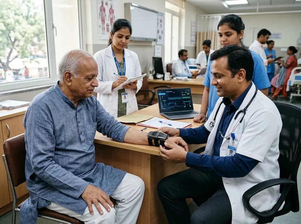
The TREM-AB open-label pilot study evaluated a wearable passive nonlinear vibration absorber for essential tremor. The Nonlinear Energy Sink bracelet reduced clinical Fahn-Tolosa-Marin tremor scores by 32.1% and electrophysiological tremor power by 49.2% with no adverse effects reported.
Today

A population-based study of 20,928 children reveals that small vulnerable newborns face sex-divergent mental capital outcomes at ages 6-7: term SGA girls show higher behavioral difficulties and lower resilience, while preterm non-SGA boys experience increased behavioral problems.
Today

A landmark study details how an enterprise pediatric remote patient management model expanded from a single cardiology pilot to 16 diverse specialty programs by combining centralized digital operations with specialty clinical leadership.
Today

A comparative review of tension band wiring and intramedullary screw fixation in thumb metacarpophalangeal arthrodesis, evaluating union rates, hardware complications, and clinical outcomes.
Today
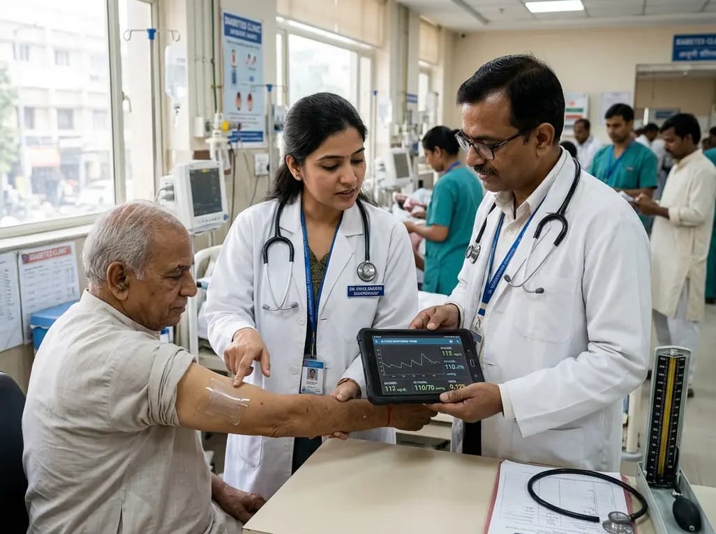
A breakthrough ultrathin multimodal sensor decouples physical hemodynamic monitoring from disposable electrochemical calibration, achieving long-term wearable glucose prediction with 81.36% accuracy and a 9.12% MARD. This offers a robust, drift-free solution for outpatient metabolic surveillance.
Today
