

Loading, please wait...
Loading, please wait...

Clinicians frequently encounter atypical degenerative joint disorders that defy standard osteoarthritis patterns. Historically, physicians linked severe joint destruction to classic hereditary hemochromatosis driven by homozygous C282Y mutations. However, emerging multicentre data demonstrates that haemochromatosis arthropathy can develop even in patients who lack classic systemic iron overload. Consequently, medical teams must rethink the pathogenesis linking HFE gene mutations to articular cartilage degeneration. The HFE gene encodes an atypical major histocompatibility complex class I molecule that regulates iron homeostasis by modulating hepcidin. When pathogenic variants occur, cellular iron transport shifts substantially. Historically, researchers believed that systemic iron accumulation directly caused synovial and chondrocyte toxicity. In contrast, recent clinical investigations reveal that abnormal HFE genotypes trigger distinctive arthropathic phenotypes independently of marked hyperferritinemia. Therefore, clinicians must recognize that musculoskeletal manifestations may precede or completely bypass overt systemic iron storage disease. Furthermore, heterozygous states and alternative variant combinations frequently manifest with disabling joint pain. Because early recognition prevents inappropriate interventions, rheumatologists must reassess their diagnostic algorithms to identify high-risk individuals before irreversible structural deterioration occurs.
To clarify these complex relationships, investigators recently evaluated 373 participants from the European Alliance of Associations for Rheumatology inception cohorts. Specifically, the study analyzed four distinct clinical groups to assess criteria fulfillment across diverse genotypes and iron statuses. Group 1 comprised 154 C282Y homozygous patients displaying significant systemic iron overload. Meanwhile, group 2A contained 27 C282Y homozygotes who lacked significant iron overload. In addition, group 2B encompassed 72 individuals carrying other recognized HFE genotypes regardless of systemic iron levels. Finally, group 3 included 120 control patients diagnosed with standard osteoarthritis or calcium pyrophosphate crystal arthropathy without iron overload. The researchers applied the validated EULAR classification criteria to each cohort systematically. The primary results revealed striking distinctions among the groups. Remarkably, 71.4% of patients in group 1 and 22.2% in group 2A fulfilled the classification criteria. Moreover, 43.1% of participants in group 2B met these criteria, compared to only 6.7% among control subjects. Thus, the data confirms that alternative HFE genotypes frequently drive typical arthropathy patterns without requiring traditional systemic iron overload thresholds.
Perhaps the most transformative finding of this multicentre study centres on circulating iron parameters. Within group 2, detailed multivariable regression analyses assessed whether serum ferritin concentrations or transferrin saturation levels predicted criteria fulfillment. Interestingly, neither elevated ferritin nor increased transferrin saturation demonstrated any statistically significant association with disease classification. Consequently, these findings challenge the long-held dogma that articular pathology directly reflects circulating iron burden. Instead, localized mechanisms within the synovial joint likely drive cartilage breakdown and subchondral bony remodeling. Clinicians have long recognized that therapeutic phlebotomy successfully clears hepatic and cardiac hemosiderin but fails to alleviate arthritic pain. This therapeutic disconnect strongly supports the biological independence of joint pathology from circulating iron pools. Furthermore, mutated HFE proteins may directly impair chondrocyte metabolism or disrupt matrix metalloproteinase regulation within articular tissues. Alternatively, microenvironmental iron deposition inside articular chondrocytes might occur without provoking abnormal serum indices. Therefore, physicians cannot rely solely on normal blood tests to exclude genetic articular involvement. Recognizing this decoupling prevents clinicians from falsely reassuring patients whose symptoms originate from genetic predisposition.
Because clinical presentations often overlap with common rheumatologic conditions, diagnosing HFE-related joint disease remains challenging. Typical structural changes frequently mimic primary osteoarthritis, rheumatoid arthritis, or calcium pyrophosphate deposition disease. However, specific anatomical distributions provide crucial diagnostic clues during physical examination and imaging reviews. For example, bilateral involvement of the second and third metacarpophalangeal joints strongly suggests metabolic arthropathy rather than nodal osteoarthritis. In addition, radiographic imaging often demonstrates distinctive hook-like osteophytes, uniform joint space narrowing, and pronounced subchondral cyst formation. Radiologists may also observe chondrocalcinosis within the triangular fibrocartilage complex or knee menisci. Nonetheless, clinicians frequently misclassify these individuals as having idiopathic chondrocalcinosis or premature degenerative osteoarthritis. Furthermore, inflammatory signs can fluctuate, occasionally generating synovial effusions that resemble seronegative rheumatoid arthritis. Because misdiagnosis delays appropriate supportive management, practitioners must actively seek multi-joint involvement patterns. Combining targeted radiographic screening of the hands with detailed family histories substantially improves diagnostic precision. Therefore, when middle-aged patients present with persistent wrist, ankle, or knuckle pain, clinicians must maintain a high index of clinical suspicion.
These breakthrough insights hold vital practical implications for general physicians, internists, and rheumatologists worldwide. First, practitioners should consider HFE genetic testing for patients with unexplained symmetrical polyarthropathy, even if baseline serum ferritin remains completely normal. Previously, normal iron parameters routinely dissuaded physicians from pursuing genetic evaluation. However, the evidence now indicates that non-homozygous genotypes and normoferritinemic individuals experience substantial articular morbidity. Consequently, clinicians must expand their diagnostic framework beyond classical genetic hemochromatosis. Second, managing this distinct entity requires targeted symptomatic and structural strategies rather than relying on iron depletion alone. While phlebotomy protects vital organs, it does not halt preexisting cartilage deterioration. Accordingly, therapeutic regimens should emphasize joint protection, physical therapy, judicious use of nonsteroidal anti-inflammatory drugs, and intra-articular therapies. Furthermore, emerging chondroprotective strategies warrant prospective clinical trials in genotype-stratified cohorts. By identifying HFE-associated joint disease early, doctors can prevent inappropriate immunosuppressive therapies often prescribed for presumed inflammatory arthritis. Ultimately, integrating clinical criteria, genetic testing, and meticulous radiographic evaluation ensures timely, accurate patient care across primary and specialty medical settings.
Yes, robust clinical evidence confirms that haemochromatosis arthropathy can develop in patients with completely normal serum ferritin and transferrin saturation. Genetic mutations in HFE may alter chondrocyte cellular mechanics and trigger localized joint damage independently of systemic iron storage. Therefore, clinicians must never rely exclusively on standard iron panels. When patients exhibit persistent, unexplained polyarthropathy affecting typical joints, medical providers should pursue genetic testing and comprehensive radiographic evaluations regardless of baseline iron concentrations.
HFE-related joint pathology classically targets the second and third metacarpophalangeal joints, wrists, hips, and ankles in a symmetrical pattern. Unlike conventional nodal osteoarthritis, which primarily affects distal interphalangeal joints, this condition exhibits distinct radiographic features. Specifically, radiographs reveal hook-like osteophytes, uniform joint space loss, subchondral cysts, and chondrocalcinosis. Consequently, bilateral wrist or hand pain in middle-aged individuals warrants thorough diagnostic investigation to distinguish this genetic disorder from standard degenerative or inflammatory rheumatic conditions.
No, therapeutic venesection does not reverse or significantly relieve established articular destruction, despite effectively reducing visceral iron stores. While phlebotomy prevents life-threatening hepatic, endocrine, and cardiac complications, joint degeneration often progresses independently once structural changes occur. Therefore, medical management must prioritize rheumatologic interventions. Physicians should combine tailored physical therapy, nonsteroidal anti-inflammatory agents, joint protection measures, and occasional intra-articular steroid injections. Orthopedic surgical consultation becomes necessary when advanced structural deterioration severely limits mobility and patient quality of life.
Disclaimer: This content is for informational and educational purposes only and does not constitute medical advice, diagnosis, or treatment. Always consult qualified healthcare professionals for clinical decisions. The views expressed are those of the authors and do not necessarily reflect the official policy or position of any affiliated organizations. Refer to the latest local and national guidelines for clinical practice.
References

Read summarized clinical updates, watch expert medical content, and earn CME certifications right from your smartphone.
Open your smartphone camera to scan and install.





A multicentre EULAR study reveals that haemochromatosis arthropathy can develop in patients carrying HFE genotypes without overt systemic iron overload, challenging conventional clinical reliance on serum ferritin.
Today

A major ABCD study analysis reveals that prenatal co-exposure to cannabis and tobacco significantly impairs fluid cognitive abilities in middle childhood, whereas crystallized intelligence remains unaffected. The findings highlight urgent needs for targeted antenatal screening and intervention.
Today
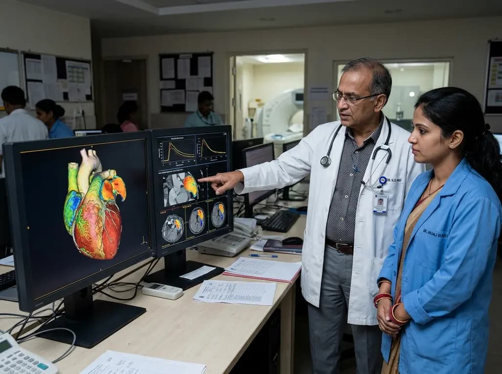
Dynamic contrast 4DCT enables precise mapping of left atrial appendage blood transport in atrial fibrillation. By quantifying regional stasis, wash-in kinetics, and residence time, this imaging advance offers clinician-tailored thromboembolic risk stratification beyond standard clinical scores.
Today
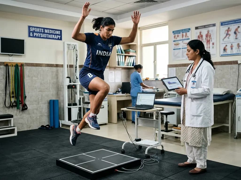
A systematic review and meta-analysis highlights the substantial impact of plyometric jump training on jump performance, reactive strength, and landing kinematics in female team-sport athletes, providing evidence-based programming guidelines for sports medicine and orthopedics.
Today
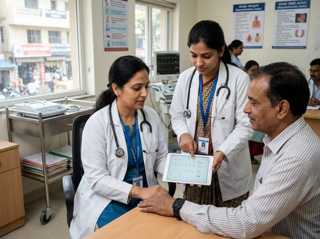
India is poised to transform chronic disease management through wearable health technology. Discover how continuous remote monitoring and predictive algorithms empower clinicians to detect early cardiovascular signals, personalize metabolic interventions, and deliver equitable preventive care across populations.
Today
