

Loading, please wait...
Loading, please wait...

Congenital head and neck swellings present unique diagnostic and therapeutic challenges for surgeons worldwide. Among these conditions, submandibular dermoid cysts represent exceptionally uncommon developmental malformations that arise from sequestered ectodermal elements. Although clinicians frequently observe dermoid and epidermoid lesions in pediatric populations, large manifestations can occasionally persist unrecognized into adult life. A recent clinical report highlighted a 39-year-old male who presented with an extraordinary thirty-year history of bilateral neck masses, illustrating the insidious natural progression of these slow-growing floor-of-mouth lesions.
Congenital floor-of-mouth cysts originate from abnormal embryonic development during early fetal life. During the third to fifth gestational weeks, the first and second branchial arches fuse along the midline. Consequently, trapped ectodermal cells can survive beneath the closing surface tissue. These cellular rests gradually expand as epithelial desquamation and glandular secretions accumulate. Therefore, lesions grow at an indolent pace across decades without triggering significant inflammation. Furthermore, acquired forms can also develop following accidental trauma or surgical puncture, which implants epithelial cells into deeper cervical tissues. Nevertheless, the congenital variety remains far more common in routine clinical practice. Histologists classify these developmental lesions into three distinct categories: epidermoid cysts, true dermoid cysts, and teratoid teratomas. While epidermoid cysts contain only stratified squamous epithelium, dermoid lesions possess adnexal structures like sebaceous glands. Meanwhile, complex teratoid varieties encompass tissues derived from all three germ layers. Understanding these precise distinctions helps clinicians determine the exact embryonic timing of cellular entrapment.
Because these lesions enlarge at an exceptionally gradual rate, patients frequently adapt to their physical presence over many years. In this notable clinical case, the patient managed his neck swelling for thirty years before seeking formal treatment. Consequently, the mass eventually reached impressive dimensions and crossed anatomical compartments bilaterally. Typically, lesions situated superior to the mylohyoid muscle push the tongue upward and backward, thereby causing dysarthria, dysphagia, or snoring. Conversely, cysts located inferior to the muscular floor bulge predominantly into the submental and submandibular triangles. Therefore, patients usually complain of an unsightly submandibular fullness rather than immediate functional distress. However, sudden volumetric expansion can occur if secondary bacterial infection develops. Furthermore, hormonal surges during puberty occasionally stimulate sebaceous secretions within the cyst lumen, accelerating enlargement. Fortunately, physical examination typically reveals a soft, non-tender, doughy mass that remains unattached to the overlying skin. Systemic symptoms remain absent unless infectious cellulitis supervenes, which makes careful physical palpation essential for diagnostic differentiation.
Comprehensive radiographic imaging plays an indispensable role in defining lesion boundaries and planning operative intervention. Clinicians usually perform bedside ultrasonography during initial consultations to confirm the lesion's fluid-filled architecture. However, magnetic resonance imaging provides the gold standard for mapping complex submandibular dermoid cysts. Specifically, MRI clarifies the critical spatial relationship between the lesion and adjacent musculature, particularly the genioglossus, geniohyoid, and mylohyoid muscles. T1-weighted sequences characteristically demonstrate low to intermediate signal intensity, whereas T2-weighted scans display prominent hyperintensity that reflects thick proteinaceous content. Additionally, post-contrast images reveal a thin, smoothly enhancing fibrous capsule without nodular invasion into surrounding neurovascular structures. Computed tomography serves as an acceptable alternative when clinicians need to evaluate cortical bone remodeling along the lingual mandibular plate. Furthermore, fine-needle aspiration cytology can extract thick, cheesy keratinaceous debris, which effectively rules out vascular malformations and salivary carcinomas before definitive operative exploration.
Pathologists provide the definitive diagnosis through systematic microscopic evaluation of the excised surgical specimen. Macroscopically, these unilocular masses exhibit a fibrous outer wall and contain pasty, pale yellowish keratinaceous material. Microscopically, the specimen from this reported case demonstrated an epidermoid cyst lined completely by well-differentiated, orthokeratinized stratified squamous epithelium. Notably, pathologists observed a prominent granular layer beneath lamellar flakes of shed keratin. True dermoid variants display distinctive skin appendages, including mature hair follicles, sebaceous glands, and occasional eccrine ducts within the surrounding fibrovascular wall. Conversely, pathologists identify respiratory epithelium, smooth muscle, and bone elements only when evaluating complex teratoid lesions. Malignant transformation into squamous cell carcinoma represents a rare occurrence, appearing in less than two percent of recorded literature. Therefore, thorough microscopic sampling across the entire capsule remains mandatory. By identifying clear surgical margins, pathologists confirm complete enucleation and reassure clinicians regarding the lack of residual malignant potential.
Complete surgical enucleation serves as the definitive treatment of choice for extensive floor-of-mouth cysts. Surgeons must tailor their operative approach according to the relationship of the mass with the mylohyoid muscle. For smaller cysts situated entirely in the sublingual space, a transoral incision minimizes visible scarring. However, giant lesions extending into bilateral submandibular spaces require a trans-cervical extraoral approach. In this patient, surgeons performed an external submandibular incision under general anesthesia to preserve adjacent marginal mandibular nerves, lingual vessels, and hypoglossal fibers. Careful blunt dissection along the fibrous capsule prevents accidental cyst rupture, which could otherwise induce intense chemical cellulitis in deep cervical fascial spaces. Following total capsule excision, surgeons place active suction drains to prevent dead-space hematoma formation. Consequently, postoperative recovery proceeds smoothly, and recurrence rates remain exceptionally low after complete capsular removal. Patients achieve complete functional recovery with excellent cosmetic outcomes and full preservation of tongue motility.
Clinicians differentiate these lesions by evaluating tissue consistency and anatomical origin. Ranulas typically exhibit a bluish, translucent hue within the oral mucosa and fluctuate rapidly in volume due to saliva extravasation from the sublingual gland. Conversely, dermoid cysts present as uniform, doughy masses containing keratin debris. Magnetic resonance imaging reliably distinguishes their borders, whereas histopathology provides final confirmation.
Dermoid cysts represent congenital developmental anomalies rather than true neoplasms. Because these lesions enlarge predominantly through the mechanical accumulation of shed keratin, sebum, and cellular debris rather than uncontrolled cellular proliferation, they exhibit strictly benign biological behavior. Malignant transformation remains exceedingly rare, thereby allowing long periods of clinical quiescence without aggressive tissue invasion.
Surgeons choose the operative corridor based on the relationship between the cyst and the mylohyoid muscle. Small intraoral lesions permit transoral resection without cutaneous scars. However, giant cysts that plunge into bilateral submandibular and submental triangles require an extraoral submandibular approach to ensure complete capsular enucleation while protecting vital cranial nerves and lingual vessels.
Disclaimer: This content is for informational and educational purposes only and should not be considered medical advice. It does not replace professional clinical judgment, diagnosis, or treatment. Refer to the latest local and national guidelines for clinical practice.
References
Zhai Y et al. Giant Bilateral Submandibular Dermoid Cysts. J Craniofac Surg. 2026 Oct 26. doi: 10.1097/SCS.0000000000013550. PMID: 42930056.
Pirgousis P, Fernandes R. Giant submental dermoid cysts with near total obstruction of the oral cavity: report of 2 cases. J Oral Maxillofac Surg. 2011;69(2):532-535.
Jain H, Singh S, Singh A. Giant Sublingual Dermoid Cyst in Floor of the Mouth. J Maxillofac Oral Surg. 2012;11(2):235-237.

Read summarized clinical updates, watch expert medical content, and earn CME certifications right from your smartphone.
Open your smartphone camera to scan and install.





Dermoid cysts in the submandibular space represent rare developmental entities. This case highlights a 39-year-old male presenting with a 30-year history of bilateral submandibular swelling, detailing the diagnostic MRI protocols, trans-cervical surgical resection, and definitive histopathological differentiation.
Today

A prospective study using wireless sensors reveals that smaller physician workrooms significantly increase contact density and respiratory virus transmission risks despite lower average occupancy, underscoring the urgent need for better architectural design and infection control in healthcare duty rooms.
Today

A KGMU study reveals over half of women with breast abnormalities fail to undergo diagnostic confirmation, leading to advanced-stage detection. Discover the socio-cultural barriers, clinical risks of incomplete resection, and community-level solutions needed to bridge the critical screening-to-diagnosis gap in India.
Today

A comparative non-contact study reveals that high-power radiofrequency delivery with the QDOT MICRO catheter induces an artifactual contact force rise. Electrophysiologists must understand this power-dependent thermal and electrical phenomenon to avoid inadvertent catheter withdrawal and ineffective ablation.
Today
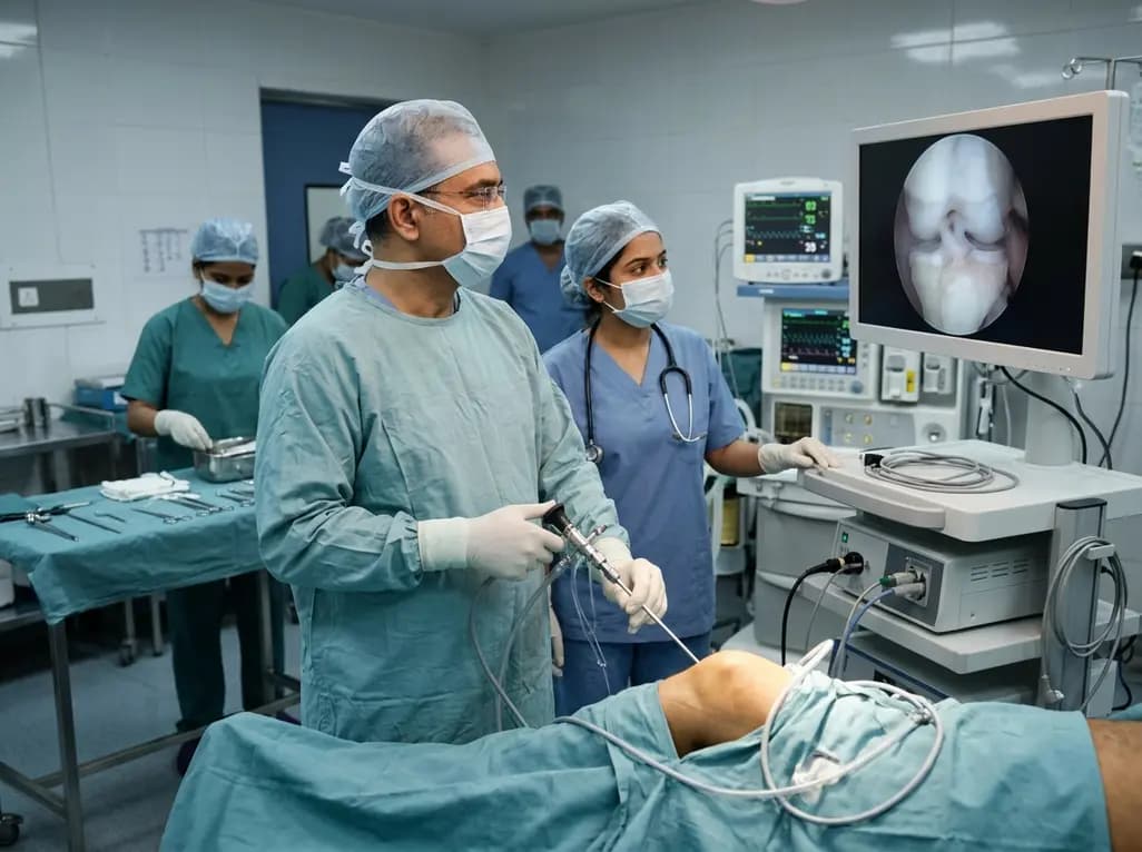
Healthium Medtech has transitioned its arthroscopy vertical into SIRONIX Orthopedics, aiming to capture key segments of the global joint care market. The strategic shift introduces dedicated portfolios in sports medicine, joint regeneration, surgical reconstruction, and digital operating technologies.
Today

Preclinical research demonstrates that BMI1 overexpression alleviates metabolic syndrome-related liver injury. By triggering ubiquitin-mediated METTL3 degradation, BMI1 prevents AHNAK m6A methylation and suppresses fibrogenic TGF-β/SMAD signaling, significantly improving hepatic steatosis and insulin resistance.
Today
