.webp&w=384&q=75)

Loading, please wait...
Loading, please wait...
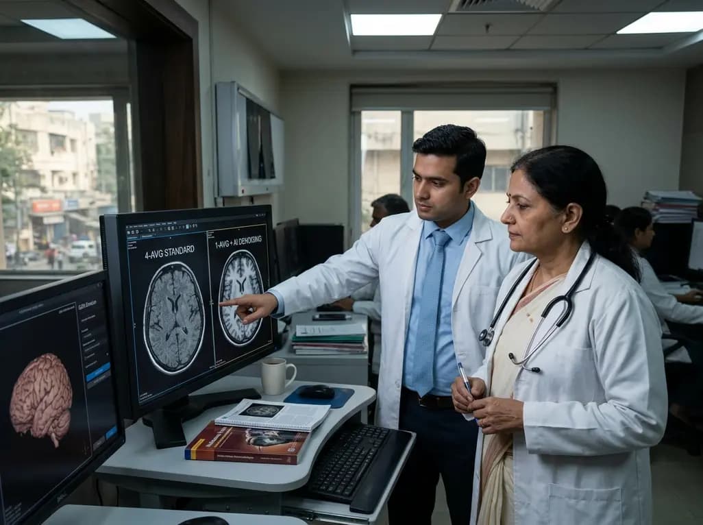
Recent innovations in ultra-high-field neuroimaging offer unprecedented anatomical visualization, yet extended scan durations limit clinical throughput and patient compliance. To solve this dilemma, clinicians increasingly explore generative AI MRI denoising to accelerate acquisition pipelines without sacrificing diagnostic clarity. Researchers have introduced a specialized conditional diffusion framework that transforms noisy, rapid scans into high-definition clinical images. Consequently, this computational breakthrough provides radiologists and neurologists with exceptional structural clarity while significantly reducing the physical strain experienced by vulnerable patients during lengthy diagnostic protocols.
Ultra-high-field 7 Tesla magnetic resonance imaging delivers superior spatial resolution and heightened susceptibility contrast. Therefore, it provides incomparable sensitivity for detecting cortical microinfarcts, subtle hippocampal alterations, and microscopic iron deposits. However, achieving this extraordinary image fidelity traditionally demands multi-repetition acquisitions. These extended acquisition times often cause severe patient fatigue and involuntary head motion. Consequently, motion artifacts frequently degrade diagnostic accuracy, particularly in elderly populations suffering from cognitive deficits.
In clinical radiology environments, technologists must constantly balance signal-to-noise ratios against overall examination length. When clinics shorten gradient-echo sequences to mitigate motion, the resulting images suffer from heavy thermal noise and severe contrast loss. Therefore, standard acceleration methods often fail to deliver diagnostic quality. Deep learning methods offer a compelling alternative, but older algorithms frequently introduce unnatural smoothing or erase crucial microvascular markers. To overcome these constraints, investigators have developed generative models that actively preserve sub-millimeter anatomical architecture while suppressing background thermal noise. Thus, machine learning creates practical opportunities for ultra-high-field clinical neuroimaging.
The newly developed 7T Conditional Diffusion Model, designated 7TCDM, represents a substantial engineering leap forward. Rather than relying on synthetic simulations, developers trained the model on native single-acquisition two-dimensional gradient-echo brain scans. Furthermore, the training pipeline incorporated multi-repetition examinations as an empirical reference standard. Through this rigorous design, the diffusion architecture learns the underlying probability distributions of high-frequency anatomical signals alongside scanner noise patterns.
Consequently, the conditional framework iteratively reverses noise corruption while strictly maintaining spatial fidelity. By conditioning the generative reverse steps directly on the acquired raw data, the algorithm prevents uncontrolled image hallucinations. Moreover, this mathematical framework outperforms traditional deep learning models by preserving subtle edge gradients and tissue boundaries. Standard algorithms frequently blur critical gray-white matter interfaces, but 7TCDM sharpens microstructural features. As a result, the model delivers reliable signal restoration across diverse tissue contrasts. Clinicians can therefore trust the visual integrity of these reconstructed scans for complex neurodegenerative evaluations.
To validate performance, researchers directly benchmarked the generative AI MRI denoising platform against standard reconstruction techniques. Specifically, they compared 7TCDM with unprocessed scans, convolutional neural networks, vision transformers, and generative adversarial networks. Objective evaluations utilized Mean Squared Error, Peak Signal-to-Noise Ratio, and the Structural Similarity Index Measure. Furthermore, board-certified neuroradiologists performed rigorous blind perceptual evaluations to confirm authentic diagnostic value.
The quantitative results demonstrated decisive clinical superiority across all parameters. Referencing the multi-repetition reference standard, the diffusion model improved the single-acquisition baseline image by 31.7 percent in Mean Squared Error. Additionally, the platform increased Peak Signal-to-Noise Ratio by 4.9 percent and boosted Structural Similarity by 7.5 percent. Most importantly, 7TCDM outperformed all alternative machine learning architectures with statistical significance. Neuroradiologists consistently awarded the highest qualitative ratings to the diffusion reconstructions, citing superior tissue contrast and lack of artificial blurring. Consequently, these metrics confirm that conditional diffusion constitutes the most reliable computational method for accelerating high-field neuroimaging.
Translational medical innovations require robust validation across symptomatic clinical cohorts. Therefore, investigators tested 7TCDM on nineteen human subjects, comprising healthy controls, patients with mild cognitive impairment, and individuals diagnosed with Alzheimer’s disease. In neurodegenerative disorders, accurate visualization of subcortical nuclei, microbleeds, and cortical laminar architecture dictates therapeutic planning. However, patients with cognitive impairment frequently struggle with confinement inside narrow bore scanners, triggering severe motion artifacts.
By deploying the generative framework, clinicians successfully transformed rapid, single-acquisition gradient-echo sequences into scans matching full-length multi-repetition quality. Furthermore, the model preserved vital diagnostic markers of neurodegeneration, such as microvascular iron accumulation and hippocampal laminar boundaries. The algorithm avoided erasing tiny hemorrhagic foci, which represents a persistent risk in conventional denoising filters. Moreover, the enhanced signal clarity enabled clinicians to distinguish early degenerative changes from benign senescent atrophy with heightened confidence. Consequently, this acceleration approach substantially enhances diagnostic feasibility for vulnerable geriatric patients who cannot endure conventional, prolonged ultra-high-field scanning sessions.
Although generative artificial intelligence offers immense clinical potential, physicians rightfully demand rigorous safeguards against computational hallucinations. When synthetic models invent fictitious anatomical structures, patient safety suffers catastrophic risks. Fortunately, 7TCDM mitigates these concerns through deterministic conditional guidance tied directly to raw k-space representations. Neuroradiologic evaluations confirmed that the platform did not generate spurious lesions or obscure existing pathological markers. Thus, the model achieves unprecedented fidelity without compromising patient safety.
Looking ahead, integrating diffusion-based denoising into commercial imaging consoles will revolutionize neuroimaging workflows. Hospitals can significantly increase patient throughput while expanding access to ultra-high-field diagnostic investigations. Furthermore, adopting this computational framework may reduce the necessity for repeat examinations and sedation in uncooperative patients. Continued multicenter trials across diverse scanner hardware will solidify regulatory approval and widespread adoption. Ultimately, generative artificial intelligence bridges the long-standing gap between cutting-edge physics and routine bedside clinical care, ushering in a safer, faster diagnostic era.
Generative AI MRI denoising substantially shortens acquisition times by reconstructing crystal-clear images from rapid, single-acquisition scans. Consequently, patients spend far less time immobilized within the confined scanner bore. This operational speed drastically minimizes physical discomfort, claustrophobia, and involuntary motion artifacts. Therefore, the technology proves particularly advantageous for elderly individuals, young children, and patients diagnosed with painful neurological conditions or progressive cognitive impairment.
The 7T conditional diffusion model utilizes strict conditional guidance tied directly to the acquired raw imaging data. Unlike unguided generative algorithms, this architecture repeatedly anchors each mathematical reverse step to physical sensor measurements. Furthermore, extensive blinded neuroradiologic evaluations confirmed that the model preserves authentic pathological features without introducing spurious tissue abnormalities. Consequently, clinicians obtain verified signal restoration that respects patient anatomy while eliminating disruptive thermal noise.
Although researchers optimized this specific conditional model for ultra-high-field 7T systems, the underlying mathematical architecture easily adapts to routine 1.5T and 3T platforms. In addition, lower-field scanners frequently experience acute signal-to-noise limitations during rapid imaging protocols. Consequently, applying generative diffusion algorithms can boost structural contrast and accelerate clinical examinations across diverse field strengths. Hospitals can therefore enhance overall diagnostic productivity without purchasing costly new imaging hardware.
Disclaimer: This content is for informational and educational purposes only and should not be considered professional medical advice. Always seek the advice of a qualified healthcare provider with any questions you may have regarding a medical condition. Refer to the latest local and national guidelines for clinical practice.
References

Read summarized clinical updates, watch expert medical content, and earn CME certifications right from your smartphone.
Open your smartphone camera to scan and install.





A novel 7T conditional diffusion model leverages generative AI to denoise short 2D gradient-echo brain MRI acquisitions. It matches multi-repetition quality in Alzheimer's and cognitive impairment cohorts, drastically reducing scan times.
Today

New evidence from the J-ECOH study demonstrates that exclusive and dual use of heated tobacco products significantly increases sleep latency and restless sleep among workers. These findings refute harm reduction claims, highlighting critical sleep health risks relevant to clinical counseling in India.
Today

A multiparametric MRI approach combining quantitative T2 shading, apparent diffusion coefficient (ADC) measurements, and morphological signs provides superior diagnostic accuracy over subjective evaluation to differentiate ovarian endometriomas from benign hemorrhagic cysts.
Today

Discover a novel laparoscopic approach for iatrogenic diaphragmatic hernia repair following pedicled omentoplasty. This modified Sugarbaker technique integrates dorsal pedicle lateralization and round ligament reinforcement to secure the diaphragmatic defect while preserving vital omental vascularity.
Yesterday

A new randomized crossover trial reveals that a hinged ulnar gutter orthosis significantly improves hand function and weight-bearing tolerance compared to standard volar splints in patients with degenerative triangular fibrocartilage complex lesions, supporting functionally permissive orthotic designs.
Today

New research reveals that M1 macrophage-derived exosomes aggravate diabetic nephropathy by transferring WTAP to stabilize S1PR2 mRNA. Silencing WTAP in these vesicles attenuates endothelial injury and renal fibrosis, pointing toward innovative nanomedicine therapies.
Today
