

Loading, please wait...
Loading, please wait...

Cognitive vitality in late life represents a major clinical priority worldwide. While typical aging trajectories involve gradual memory and executive decline, select older adults maintain peak cognitive performance. Understanding the neural mechanisms underlying successful cognitive aging helps clinicians distinguish physiological senescence from early neurodegeneration. Functional neuroimaging modalities, particularly fMRI and EEG, provide critical insights into these divergent cognitive pathways. By monitoring cerebral metabolism and functional network organization, researchers identify key features of resilient brains. These clinical biomarkers guide early risk stratification and targeted geriatric interventions.
Older individuals present strikingly divergent cognitive trajectories across their lifespans. Many older adults experience progressive reductions in episodic memory and processing speed. In contrast, cognitively resilient individuals sustain baseline cognitive performance matching younger cohorts. Clinicians historically attributed this sustained performance to dynamic neural compensation or high cognitive reserve. However, contemporary neuroimaging indicates that successful longevity primarily reflects resistance to structural brain pathology. When individuals avoid microvascular ischemia, amyloid accumulation, and severe cortical thinning, primary functional networks remain intact. Furthermore, maintaining educational attainment, active lifestyle habits, and metabolic health directly buffers central nervous system integrity. Thus, cognitive decline is not an inevitable consequence of human aging. Differentiating healthy senescence from insidious neurodegeneration allows clinicians to offer timely prognostic evaluations in outpatient geriatric clinics.
Recent scientific consensus highlights brain maintenance as the primary driver of preserved late-life cognition. This theoretical paradigm posits that successful cognitive aging does not require novel compensatory wiring. Instead, it reflects the sustained preservation of youthful brain structure, functional connectivity, and cellular metabolism. Neuroimaging studies confirm that high-performing older adults display metabolic profiles resembling young cohorts. Specifically, functional connectivity within the default mode network and executive control network remains robustly segregated in resilient individuals. When brain regions maintain functional specialization, they prevent the neural dedifferentiation that degrades memory retrieval. Furthermore, structural integrity in the medial temporal lobe preserves hippocampal volume. Consequently, older adults who avoid synaptic degradation recruit canonical circuits without invoking atypical pathways. Clinical evidence therefore confirms that preventing neurobiological decay remains the ultimate foundation for enduring cognitive health.
Functional magnetic resonance imaging offers profound insights into large-scale network architecture during cognitive tasks. Blood-oxygen-level-dependent signaling reveals how healthy older brains coordinate distributed cerebral networks during memory retrieval. Importantly, cognitively resilient seniors exhibit preserved modularity across cortical systems. In typical senescence, the brain experiences dedifferentiation, characterized by diffuse functional connectivity and blurred system boundaries. In contrast, successful cognitive aging correlates with distinct boundaries between task-positive regions and intrinsic baseline networks. Furthermore, high performers effectively suppress default mode network activity during demanding cognitive tasks. When older individuals preserve dynamic modulation, their task-relevant circuits execute complex operations without interference. Consequently, longitudinal fMRI analyses indicate that youth-like resting-state architecture serves as a dependable functional biomarker. These neuroimaging metrics help clinicians evaluate subclinical changes before overt deficits appear.
Electroencephalography provides millisecond-level temporal resolution to assess dynamic oscillatory activity across the adult lifespan. Oscillatory synchronization across specific frequency bands governs information routing, attention allocation, and working memory operations. Typically, chronological aging shifts dominant posterior alpha rhythms toward slower frequencies and blunts event-related desynchronization. However, individuals demonstrating successful cognitive aging preserve robust posterior alpha peak frequencies alongside sharp frontal theta dynamics. In addition, resilient adults maintain healthy phase-amplitude coupling between low-frequency theta oscillations and high-frequency gamma bursts. This intact coupling coordinates local cortical processing with distant associative networks during encoding. Furthermore, electrophysiological studies reveal that preserved neural signal variability reflects adaptive flexibility in dynamic environments. Therefore, electroencephalographic profiling delivers cost-effective neurophysiological indicators of brain health that clinicians can integrate into routine screening protocols.
For decades, researchers debated whether older brains maintain performance by recruiting supplementary brain regions. Classic cognitive aging models suggested that bilateral frontal recruitment represented functional compensation for compromised circuits. However, contemporary neuroimaging investigations challenge this compensatory recruitment hypothesis. Many scientists now argue that additional bilateral activation often represents inefficient neural dedifferentiation or failed inhibitory control. In many cases, increased regional activation fails to correlate directly with improved behavioral performance. Moreover, establishing true neural compensation requires strict criteria, including proving that alternate recruitment directly enhances cognitive success. When researchers control for task difficulty and baseline atrophy, compensatory effects frequently diminish. Consequently, functional preservation of canonical youthful networks consistently emerges as the genuine correlate of cognitive longevity. Clinicians must view widespread overactivation cautiously, recognizing that hyperactivation may signify neural vulnerability.
Translating neuroimaging discoveries into routine clinical practice remains essential for aging societies facing rising dementia burdens. Because cognitive resilience relies heavily on brain maintenance, preventive clinical strategies must prioritize neuroprotection across the lifespan. Clinicians should proactively manage vascular risk factors, including hypertension, diabetes, dyslipidemia, and sleep apnea, which accelerate cerebral microvascular pathology. Furthermore, regular physical exercise, cognitively demanding activities, and balanced diets directly promote neuroplasticity and preserve functional brain network integrity. Integrating neuroimaging biomarkers like resting-state functional connectivity and electroencephalographic dynamics into preventive screening protocols helps clinicians identify vulnerable patients early. In addition, these functional markers will enhance patient stratification in clinical trials evaluating novel neuroprotective therapeutics. Ultimately, promoting brain maintenance empowers physicians to help patients preserve executive function throughout late life.
Brain maintenance refers to the relative preservation of youth-like brain structures, neurochemistry, and functional networks free from neuropathology. In contrast, cognitive reserve represents the brain's capacity to optimize cognitive performance through alternative strategies or flexible neural networks despite accumulating structural damage or pathology. Consequently, while maintenance emphasizes resisting physical brain changes, reserve reflects functional resilience when underlying neurological damage has already occurred.
Yes, electroencephalography provides sensitive physiological markers capable of distinguishing normal aging from prodromal dementia. While healthy cognitive agers preserve posterior alpha peak frequencies and robust phase-amplitude coupling, early neurodegenerative conditions typically exhibit excessive diffuse theta-delta slowing alongside disrupted functional connectivity. Furthermore, quantitative electroencephalography detects subtle synchronization breakdowns during complex memory challenges well before standard structural neuroimaging reveals evident cortical atrophy, thereby enabling timely clinical diagnosis and personalized intervention.
Clinical evidence demonstrates that aggressive vascular risk management preserves neural dynamics most effectively. Specifically, controlling chronic hypertension, insulin resistance, and dyslipidemia directly limits microvascular ischemic injury that degrades white matter networks. Furthermore, regular aerobic physical exercise stimulates neurotrophic factor release, whereas structured intellectual engagement and active social connection support large-scale network integrity. Together, these multimodal lifestyle interventions delay neurobiological decay, effectively sustaining youth-like cerebral dynamics.
Disclaimer: This content is for informational and educational purposes only. It is not intended to be a substitute for professional medical advice, diagnosis, or treatment. Always seek the advice of your physician or other qualified health provider with any questions you may have regarding a medical condition. Refer to the latest local and national guidelines for clinical practice.
References

Read summarized clinical updates, watch expert medical content, and earn CME certifications right from your smartphone.
Open your smartphone camera to scan and install.





This clinical review evaluates functional brain signatures underlying successful cognitive aging. By examining fMRI and EEG evidence, it contrasts youth-like neural maintenance against functional compensation, highlighting neurobiological insights for geriatric and neurological practice.
Today
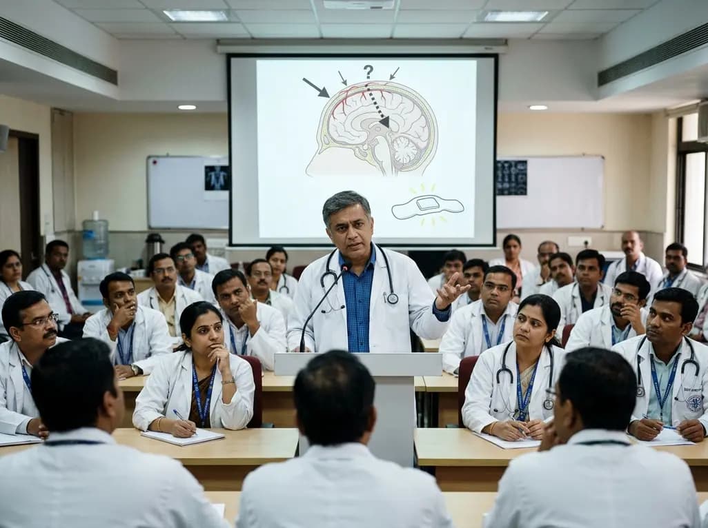
The $900 Temple wearable launched by Deepinder Goyal promises real-time tracking of cerebral blood flow and metabolic entropy. However, prominent clinicians warn that optical scalp sensors merely reflect superficial arterial circulation rather than intracranial hemodynamics, raising major regulatory concerns.
Today

A new comparative study demonstrates that intraoperative imprint cytology achieves 96.6% diagnostic accuracy in ovarian tumours, outperforming frozen section in sensitivity for borderline lesions and offering an accessible, rapid alternative for resource-constrained surgical centres.
Today

A study reveals that extracellular vesicle-derived miR-144-5p is a highly predictive diagnostic biomarker for carotid artery plaques (AUC 0.907). Correlating with IL-1β and TNF-α, it opens new avenues for liquid biopsy-based stroke risk assessment and vascular monitoring.
Today
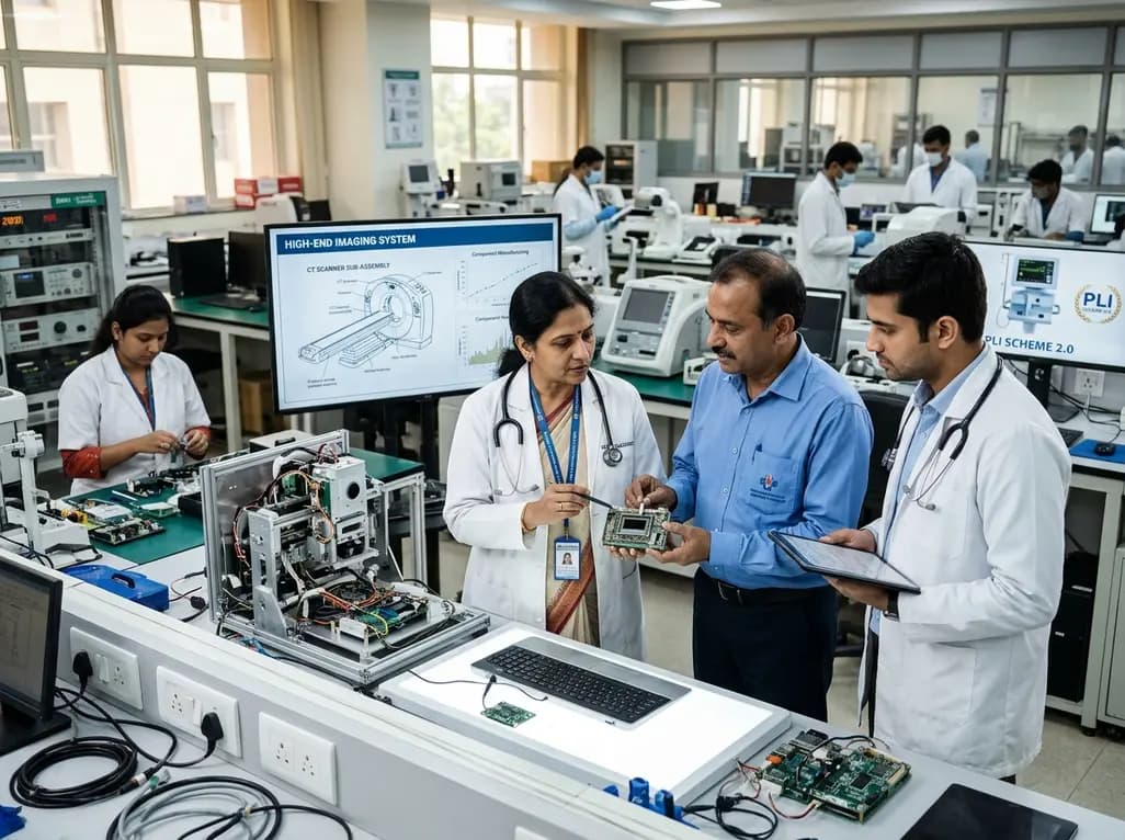
The Department of Pharmaceuticals has extended the medical devices PLI scheme to 2035-36 with a Rs 2,700 crore outlay. Featuring relaxed investment barriers and eliminated sales thresholds, the revised framework targets deep manufacturing, high-end diagnostics, critical MedTech components, and supply resilience.
Today

A systematic review evaluates psychological interventions for smoking cessation in diabetes, showing short-term success but highlighting challenges in long-term abstinence.
Yesterday
