

Loading, please wait...
Loading, please wait...
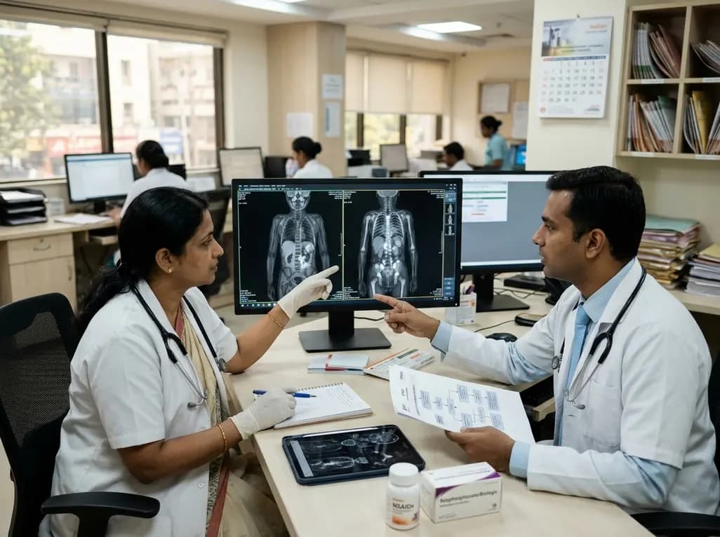
Chronic nonbacterial osteomyelitis is an autoinflammatory bone disorder that predominantly affects children and adolescents. Clinicians frequently encounter significant diagnostic dilemmas when evaluating pediatric patients who present with unexplained bone pain and localized swelling. Because early clinical signs closely resemble bacterial osteomyelitis or skeletal malignancies, affected children often endure unnecessary courses of antibiotics and diagnostic delays. Consequently, standardized national and international protocols provide essential clarity for early diagnosis, non-invasive imaging, and structured medical therapy. By adopting these evidence-based recommendations, healthcare practitioners can promptly identify sterile bone inflammation, prevent permanent skeletal damage, and enhance overall quality of life.
Chronic nonbacterial osteomyelitis characteristically displays a relapsing-remitting course characterized by inflammatory bone pain. The median age at onset centers around ten years, with registries documenting a consistent female preponderance. Patients typically experience dull, aching bone discomfort that frequently awakens them at night. Furthermore, localized swelling, erythema, and mild warmth often overlay the active bony lesion. Additionally, physical examinations may reveal restricted joint movement when lesions arise adjacent to major synovial articulations. The metaphyses of the femur, tibia, and fibula constitute the most common anatomical sites. However, lesions also frequently involve the clavicle, mandible, pelvis, and feet.
In addition, clinicians must recognize that axial skeleton lesions can develop silently. Spinal involvement frequently progresses without obvious local tenderness until vertebral compression or deformity emerges. Patients may also exhibit associated extra-skeletal inflammatory disorders, linking this condition to SAPHO syndrome. Cutaneous features such as palmoplantar pustulosis, severe acne, and psoriasis occur in a notable subset of children. Similarly, peripheral arthritis and chronic inflammatory bowel disease occasionally coexist with bone inflammation. Therefore, general practitioners must maintain heightened diagnostic suspicion whenever a child reports recurrent or multifocal bone aches without infectious causes.
Contemporary diagnostic pathways establish whole-body magnetic resonance imaging as the gold standard modality for pediatric evaluations. Conventional plain radiographs frequently fail to detect early active lesions, displaying only subtle lucency or sclerosis months into the illness. Conversely, magnetic resonance imaging provides exceptional sensitivity for bone marrow edema and surrounding soft tissue changes without delivering ionizing radiation. Short tau inversion recovery sequences highlight active sterile osteitis with outstanding clarity. Consequently, pediatric radiologists strongly endorse magnetic resonance imaging as an indispensable whole-body diagnostic and staging tool.
Crucially, whole-body imaging routinely discovers subclinical, asymptomatic skeletal lesions that extend well beyond the primary painful complaint. Even when a child presents with isolated limb pain, whole-body scanning frequently reveals multiple symmetric lesions across contralateral bones. Demonstrating multifocal bone involvement strongly supports the diagnosis of sterile osteitis and effectively excludes solitary bone malignancies. Additionally, routine laboratory testing usually reveals only mild elevations in inflammatory markers, including erythrocyte sedimentation rate and C-reactive protein. Because blood cultures, synovial fluids, and autoimmune serologies remain persistently negative, laboratory investigations primarily serve to rule out active bacterial infection and systemic sepsis. Thus, comprehensive radiologic assessment provides the definitive foundation for prompt diagnosis.
Historically, orthopedic teams routinely performed surgical bone biopsies on nearly every suspected patient to rule out acute infection or malignant neoplasms. Today, updated consensus protocols strictly reserve bone biopsy for cases with genuine diagnostic uncertainty. Clinicians should primarily mandate a bone biopsy when imaging reveals strictly unifocal disease, aggressive cortical destruction, or soft tissue masses that suggest osteosarcoma or Ewing sarcoma. Similarly, systemic red flags, including unexplained nocturnal fevers, night sweats, marked lymphadenopathy, or significant weight loss, require prompt histological exclusion of leukemia or lymphoma.
Conversely, when a child presents with classic multifocal clavicular, mandibular, or symmetric lower limb lesions without systemic alarm symptoms, clinicians can safely avoid invasive procedures. Thus, multidisciplinary review panels increasingly replace routine diagnostic surgery with comprehensive imaging surveillance in straightforward pediatric presentations. When surgeons do perform a biopsy, histological examination reveals nonspecific sterile inflammatory changes. Acute lesions show neutrophilic infiltration and bone resorption, whereas chronic lesions exhibit osteoclasts, lymphocytes, plasma cells, and reactive osteosclerosis. Most importantly, microbiological cultures from sterile bone tissue yield negative results. Therefore, avoiding unnecessary bone biopsies reduces surgical morbidity, spares young children unnecessary hospital trauma, and conserves healthcare resources.
The primary objectives of medical management are controlling inflammatory pain, restoring physical function, and preserving skeletal integrity. Non-steroidal anti-inflammatory drugs represent the universal first-line pharmacological treatment for all diagnosed children. These medications inhibit cyclooxygenase enzymes, thereby decreasing inflammatory prostaglandin production and curbing abnormal osteoclastic activity. Physicians typically prescribe scheduled, full-dose non-steroidal anti-inflammatory agents for an initial period of at least one to three months before judging therapeutic efficacy. Moreover, treating physicians should evaluate both clinical symptom resolution and inflammatory marker normalization during regular scheduled reviews.
Fortunately, approximately half of all pediatric patients achieve rapid symptom relief and radiologic stabilization using non-steroidal anti-inflammatory drugs alone. During this initial phase, clinicians should carefully monitor children for potential gastrointestinal upset and ensure appropriate hydration. Furthermore, physical therapy serves as an essential adjunct to prevent joint stiffness, maintain muscle conditioning, and support healthy mobility. However, if a patient demonstrates persistent pain, frequent inflammatory flares, or objective radiologic progression despite continuous anti-inflammatory therapy, clinicians must immediately initiate second-line options. Consequently, proactive clinical re-evaluations ensure that refractory patients receive timely therapeutic escalation without experiencing prolonged disability.
When patients fail first-line medications or exhibit vertebral involvement, clinicians must step up therapy immediately to prevent permanent skeletal sequelae. Vertebral lesions present an exceptionally high risk for vertebral collapse, kyphosis, and neurological compromise. Therefore, pediatric rheumatologists favor intravenous bisphosphonates, such as pamidronate or zoledronic acid, as preferred second-line agents. Bisphosphonates inhibit osteoclast activity, effectively alleviate inflammatory bone pain, and promote vertebral remineralization. Alternatively, tumor necrosis factor inhibitors, such as adalimumab or infliximab, provide powerful anti-inflammatory control, particularly for patients with coexisting arthritis or inflammatory bowel disease.
Although long-term overall prognosis remains generally favorable, with many patients reaching spontaneous remission after four years, up to fifty percent of children still encounter flares after five years. Persistent bone inflammation may lead to limb length discrepancies, joint contractures, and spinal deformities. Consequently, long-term multidisciplinary surveillance involving rheumatologists, orthopedic surgeons, radiologists, and general practitioners remains paramount. Regular follow-up with whole-body magnetic resonance imaging ensures early detection of asymptomatic relapses. Moreover, supporting the child's psychological health and academic engagement remains vital for holistic recovery. Ultimately, coordinated protocol-driven care prevents irreversible orthopaedic disability and ensures optimal long-term developmental outcomes.
Whole-body magnetic resonance imaging serves as the premier diagnostic modality by identifying both symptomatic and clinically silent skeletal lesions. The imaging technique accurately depicts bone marrow edema and surrounding tissue inflammation without exposing pediatric patients to harmful ionizing radiation. Furthermore, revealing multifocal, symmetric lesions often eliminates the necessity for painful, invasive bone biopsies. Consequently, whole-body scanning accelerates accurate diagnostic confirmation and guides comprehensive baseline staging.
Clinicians must actively evaluate the spine even when children report no localized back pain, because vertebral lesions frequently remain clinically silent. Physicians should routinely suspect spinal involvement during baseline whole-body magnetic resonance imaging or whenever patients develop subtle posture abnormalities, localized tenderness, or reduced spinal mobility. Identifying these lesions early prevents catastrophic structural collapse, persistent kyphosis, and permanent neurological impairment, prompting immediate therapeutic escalation to intravenous bisphosphonates or biologics.
Specialists justify stepping up therapy beyond non-steroidal anti-inflammatory drugs when patients experience ongoing bone pain, recurrent inflammatory flares, or objective radiologic progression after three months of compliant first-line therapy. Additionally, the initial discovery of vertebral osteitis mandates immediate second-line treatment, regardless of initial symptom severity. Under these critical circumstances, pediatric rheumatologists introduce intravenous bisphosphonates or tumor necrosis factor inhibitors to halt bone resorption, resolve inflammation, and avert long-term skeletal morbidity.
Disclaimer: This content is for informational and educational purposes only... Refer to the latest local and national guidelines for clinical practice.
References

Read summarized clinical updates, watch expert medical content, and earn CME certifications right from your smartphone.
Open your smartphone camera to scan and install.





Chronic nonbacterial osteomyelitis (CNO) is a pediatric autoinflammatory bone disorder. This review covers the French protocol, emphasizing whole-body MRI as the diagnostic gold standard, prudent biopsy avoidance, first-line NSAID treatment, and second-line biologic or bisphosphonate therapies for spinal lesions.
Today
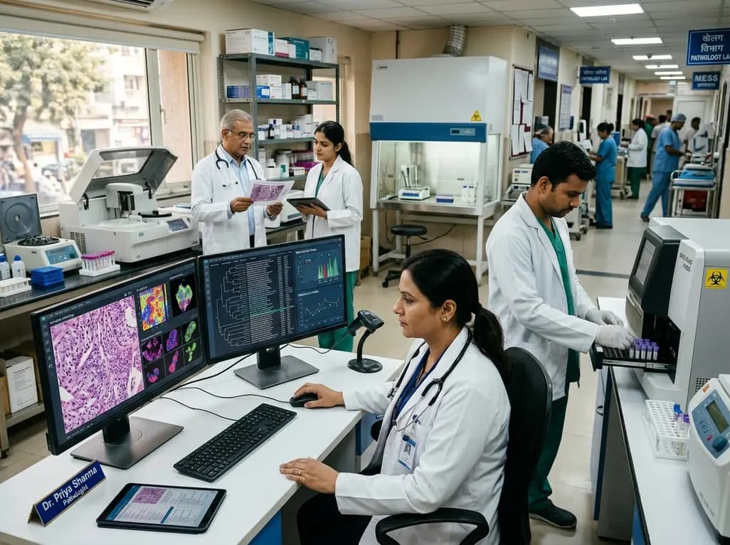
Diagnostic medicine in India is experiencing a paradigm shift towards high-complexity pathology, preventive health, and automated workflows. We review emerging molecular panels, AI-assisted cytogenetics, and the regulatory clarifications from the National Medical Commission regarding digital report authentication.
Today

A breakthrough blood test measuring the RNA biomarker Tu-Stroma detects microscopic serous tubal intraepithelial carcinoma (STIC) lesions with remarkable accuracy. This non-invasive diagnostic tool offers crucial biological insight to guide prophylactic surgery decisions for women carrying high-risk genetic mutations.
Today
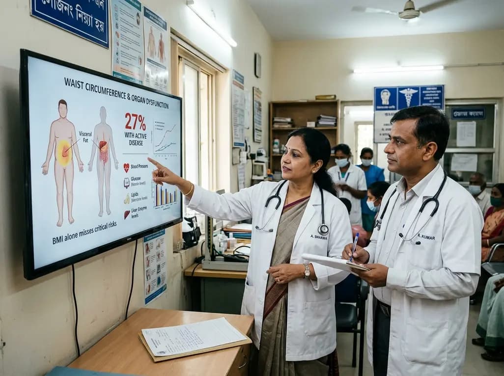
A landmark Nature study reveals that only 27% of individuals with obesity have active clinical disease. By incorporating waist circumference and organ dysfunction, this new diagnostic framework moves beyond BMI to prevent widespread misclassification and guide personalized metabolic intervention.
Today
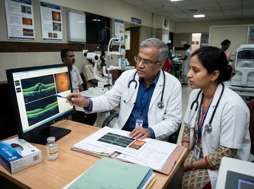
A real-world retrospective study demonstrates that combining intravitreal ranibizumab with focal/grid macular laser significantly improves visual acuity and central macular thickness compared to laser monotherapy in patients with center-involved diabetic macular edema.
Today
