

Loading, please wait...
Loading, please wait...

Managing intraventricular extension of hemorrhagic stroke remains a major challenge in neurocritical care. Clinicians frequently encounter severe complications when intraventricular blood obstructs cerebrospinal fluid pathways. Fortunately, newer therapeutic advancements allow continuous active irrigation, thereby accelerating clot clearance. However, rapid fourth ventricular recanalization can introduce unexpected secondary complications that obscure clinical recovery. A recent report highlights how restoring ventricular patency may displace inflammatory breakdown products into distant anatomical spaces. Consequently, patients can present with debilitating symptoms despite pristine intracranial imaging.
Secondary fourth ventricular hemorrhage occurs when aneurysmal subarachnoid hemorrhage or deep parenchymal bleeding breaches the ependymal lining. As extravasated blood enters the ventricular system, it forms dense casts that rapidly impede cerebrospinal fluid transit. Consequently, obstructive hydrocephalus develops and triggers a sudden rise in intracranial pressure. Beyond acute physical obstruction, erythrocytes in the subarachnoid space release toxic breakdown products over subsequent days. Specifically, hemoglobin degradation generates extracellular free heme, iron, and methemoglobin. These breakdown molecules stimulate an intense inflammatory reaction across the leptomeninges and ventricular lining. Moreover, inflammatory cytokines peak sharply during the first week following the initial hemorrhage. This inflammatory cascade frequently induces cerebral vasospasm, blood-brain barrier disruption, and persistent meningeal irritation. In typical neurocritical scenarios, severe headache correlates directly with intracranial hypertension or progressive ventriculomegaly. Therefore, clinicians instinctively look for intracranial causes when headache worsens. Nevertheless, localized ventricular events represent only one component of global cerebrospinal fluid dynamics. Neurointensivists must recognize that blood clearance initiates complex physical and chemical shifts throughout the entire craniospinal axis.
Traditional external ventricular drains clear intraventricular hematomas slowly because they rely purely on passive gravitational outflow. As a result, blood clots frequently occlude drainage catheters and delay anatomical restoration. Newer dual-lumen irrigation and drainage systems address this limitation through active, automated fluid exchange. In this published case, neurosurgeons utilized an active irrigation system in a 61-year-old woman presenting with severe secondary fourth ventricular hemorrhage. Consequently, the automated irrigation cleared cisternal blood rapidly and achieved complete fourth ventricular recanalization by day 3. Furthermore, basal cisternal hemorrhage resolved almost entirely by day 5. This rapid restoration of flow re-established the natural communication between the ventricular cavities and the spinal subarachnoid space. Active irrigation significantly reduces the duration of catheter dependence and mitigates ventricular infection risks. Furthermore, early patency protects the periventricular brain parenchyma from prolonged mechanical distension. Neurointensivists celebrated the rapid radiological clearance as a therapeutic triumph. However, expedited recanalization fundamentally alters the trajectory of remaining degradation products. Rather than evacuating entirely through the catheter, residual debris travels downward through the newly cleared foramina of Luschka and Magendie.
On post-bleed day 7, the patient suddenly developed an excruciating, intractable headache. Naturally, the medical team suspected acute complications such as recurrent hemorrhage, catheter failure, or delayed cerebral vasospasm. Surprisingly, repeated non-contrast head computed tomography demonstrated completely open ventricles without residual cisternal or intraventricular blood. In addition, continuous intracranial pressure monitoring revealed stable, completely normal values throughout the day. Serum inflammatory markers and vital signs also failed to demonstrate any infectious or systemic derangement. Despite aggressive intravenous multimodal analgesia, the patient experienced unremitting, intense head pain. Clinicians often consider headache following aneurysmal hemorrhage to reflect intracranial hypertension or vasospasm. Therefore, when neuroimaging and intracranial monitors show pristine parameters, clinicians encounter a diagnostic conundrum. This clinical scenario represents a true oy-ster in neurocritical care. Physicians may mistakenly attribute such intractable pain to psychogenic distress or drug-seeking behavior. However, severe intracranial symptoms can arise from inflammatory processes located entirely outside the cranial vault. Clinicians must realize that normal cranial parameters do not exclude significant downstream pathology.
The underlying etiology of this perplexing headache was severe caudal sac inflammation. When active irrigation reopened the fourth ventricular outflow, gravity transported residual toxic blood products into the dependent spinal subarachnoid space. Consequently, hemoglobin breakdown products accumulated within the lumbosacral thecal sac. Extracellular heme and hemate products trigger potent chemical inflammation along the lower leptomeninges and cauda equina roots. Because the spinal subarachnoid space possesses extensive sensory afferent networks, local chemical arachnoiditis produces intense discomfort. In addition, inflammation within the caudal thecal sac alters craniospinal compliance and stretches dural sleeves. As a result, afferent pain signals travel rostrally through spinal sensory pathways to generate severe referred headache. Furthermore, dependent settling occurs silently without causing classic focal neurologic deficits in the lower extremities. Head computed tomography cannot capture this spinal compartment pathology. Thus, clinicians overlook the caudal sac unless they deliberately investigate the spinal canal. The phenomenon proves that rapid fourth ventricular recanalization creates a physical conduit for downstream inflammatory displacement. Patency resolves ventricular obstruction but deposits inflammatory triggers directly into the caudal thecal reservoir.
When a patient develops unremitting headache after intraventricular hemorrhage clearance, physicians must apply a disciplined diagnostic algorithm. First, clinicians must thoroughly exclude life-threatening cranial pathologies such as rebleeding, hydrocephalus, or vasospasm. If cranial neuroimaging, intracranial pressure readings, and laboratory profiles remain normal, attention must immediately shift to the spinal axis. Clinicians should evaluate for subtle radicular irritation and obtain contrast-enhanced magnetic resonance imaging of the lumbar spine. This imaging typically reveals smooth leptomeningeal enhancement along the cauda equina and caudal sac. Furthermore, diagnostic lumbar puncture can confirm persistent xanthochromia or inflammatory pleocytosis. Effectively managing caudal sac inflammation requires targeted therapies directed at both leptomeningeal inflammation and neuropathic irritation. Conventional opioids often fail to relieve chemical arachnoiditis. Therefore, neurointensivists should initiate anti-inflammatory therapies, including short courses of systemic corticosteroids, combined with neuropathic agents such as gabapentin. In refractory cases, temporary lumbar drainage physically evacuates irritating degradation products from the caudal reservoir. Ultimately, recognizing caudal sac irritation transforms an agonizing, unexplained headache into a manageable, treatable clinical entity.
Caudal sac inflammation represents a sterile chemical arachnoiditis affecting the lumbosacral thecal sac. It occurs when blood breakdown products, such as free heme and methemoglobin, clear from the cerebral ventricles and pool dependently in the lower spinal space. This accumulation triggers severe leptomeningeal irritation and debilitating referred head pain.
Intracranial pressure remains normal because the active irrigation system successfully clears ventricular obstruction, restoring open cerebrospinal fluid circulation. The headache does not arise from elevated cranial tension or mass effect. Instead, it stems from chemical inflammation and radicular irritation in the spinal thecal sac transmitting sensory pain upward.
Clinicians should initiate targeted anti-inflammatory therapy, such as systemic corticosteroids, alongside neuropathic pain medications like gabapentin or pregabalin. Furthermore, when conservative pharmacotherapy proves insufficient, clinicians may consider temporary lumbar cerebrospinal fluid drainage. This intervention physically removes residual irritative blood products from the dependent spinal canal to provide definitive symptom relief.
Disclaimer: This content is for informational and educational purposes only. It is not intended to be a substitute for professional medical advice, diagnosis, or treatment. Always seek the advice of your physician or other qualified health provider with any questions you may have regarding a medical condition. Refer to the latest local and national guidelines for clinical practice.
References

Read summarized clinical updates, watch expert medical content, and earn CME certifications right from your smartphone.
Open your smartphone camera to scan and install.





A clinical review of post-stroke headache after fourth ventricular recanalization. Discover how active irrigation can lead to caudal sac inflammation and how to treat it.
Today

A retrospective study of 6,113 adults shows that combining GLP-1 receptor agonists with digital health apps nearly doubles the odds of achieving significant weight loss. Frequent progress logging serves as a primary behavioral driver of long-term metabolic success.
Today

Sexual dysfunction affects a high proportion of reproductive-age females with multiple sclerosis. This comprehensive review highlights multi-tier etiologies, proactive clinical screening tools, preconception counseling considerations, and integrated therapeutic options across multidisciplinary care pathways.
Today
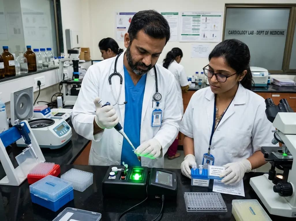
Researchers have engineered an innovative CRISPR-Cas12a fluorescent diagnostic kit for clinical sST2 detection. By combining nanotechnology with enzymatic trans-cleavage, this platform enables rapid, ultrasensitive heart failure diagnosis and risk stratification, outperforming conventional laboratory ELISA methods.
Today
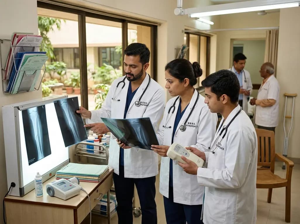
Non-union fractures occur in 2-5% of cases, causing significant disability and cost. Conservative modalities such as LIPUS, PEMF, and focused ESWT offer non-surgical healing options for defects up to 5-10 mm when adequate mechanical stability exists without infection.
Today

India is poised to transform chronic disease management through wearable health technology. Discover how continuous remote monitoring and predictive algorithms empower clinicians to detect early cardiovascular signals, personalize metabolic interventions, and deliver equitable preventive care across populations.
Yesterday
