

Loading, please wait...
Loading, please wait...
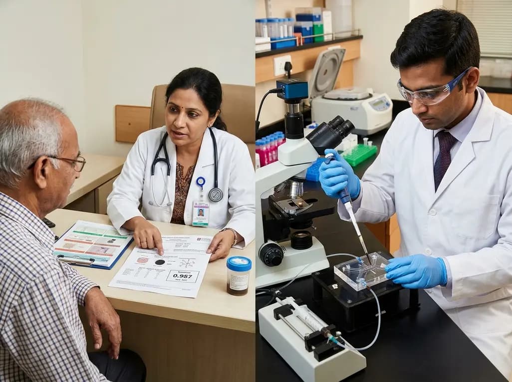
Colorectal cancer remains a leading cause of oncological morbidity and mortality worldwide. Although early detection improves long-term survival, patient adherence to invasive colonoscopy screening remains low. Non-invasive screening approaches utilizing faecal extracellular vesicles provide an exciting diagnostic alternative. These nanoscale membrane vesicles carry informative molecular cargo directly from tumor tissue into the bowel lumen. Consequently, profiling their surface proteins offers a promising path for accurate, non-invasive colorectal cancer detection.
Extracellular vesicles serve as vital mediators of cellular communication, carrying specific proteins, lipids, and nucleic acids. In colorectal neoplasms, malignant cells release vesicles directly into the digestive tract. Therefore, faecal extracellular vesicles preserve molecular signatures that accurately mirror the underlying tumor tissue. Because these vesicles enter the colonic lumen directly, they provide early neoplasia indicators before circulating tumor cells enter the blood.
However, isolating vesicles from human stool poses formidable technical challenges. Faecal samples contain complex matrices loaded with bacteria, digestive enzymes, and food debris. Furthermore, neoplastic vesicles exist in very low concentrations relative to background biological noise. Traditional laboratory methods like ultracentrifugation require extensive processing times and large sample volumes. Moreover, conventional assays often lack the analytical sensitivity needed to detect low-abundance surface markers. To overcome these limitations, microfluidic platforms isolate target vesicles directly from crude samples. As a result, microfluidic platforms effectively preserve critical diagnostic proteins. Thus, profiling faecal vesicle proteins provides a reliable avenue for early cancer detection.
To achieve high analytical sensitivity, bioengineers designed an integrated microfluidic biosensor that combines capture, amplification, and optical readout. First, the device captures nanovesicles using Y-shaped micropillar arrays functionalized with CD63 aptamers. Because CD63 is a common tetraspanin, these microstructures efficiently isolate vesicles from complex faecal suspensions. Additionally, the unique micropillar geometry promotes fluid mixing, which increases physical contact between passing vesicles and aptamer surfaces.
Following vesicle capture, the biosensor initiates an innovative dual-stage cascade amplification process. Target-specific aptamer conjugates bind to designated surface proteins, converting target recognition into nucleic-acid output strands. Subsequently, these output strands drive entropy-driven catalysis and DNAzyme reactions on chip. This catalytic cascade amplifies faint biological signals without needing external thermal cyclers or costly polymerase enzymes.
Finally, the device removes unreacted probes through magnetic separation to reduce background noise. The remaining output strands then enter a detection chamber to trigger secondary DNAzyme amplification. Consequently, this secondary reaction generates amplified fluorescence signals for precise quantification. Thus, the integrated workflow achieves exceptional signal resolution.
Colorectal tumors exhibit substantial molecular heterogeneity, which often limits the clinical utility of single-biomarker assays. To address this challenge, researchers developed a modular target-trigger platform for multiplexed biomarker profiling. Clinicians can readily adapt the aptamer probes to detect diverse surface signatures without changing the microfluidic architecture.
In this study, the biosensor successfully evaluated three essential tumor-associated proteins: EpCAM, EGFR, and HSP70. Specifically, EpCAM promotes malignant cell proliferation during tumor development. Similarly, EGFR overexpression frequently correlates with disease progression and altered cell survival pathways. Moreover, HSP70 acts as a molecular chaperone that neoplastic cells overexpress during cellular stress.
Remarkably, the platform achieved ultra-low limits of detection across all three targets. The biosensor detected EpCAM at 98 particles per millilitre, EGFR at 115 particles per millilitre, and HSP70 at 103 particles per millilitre. Therefore, the cascade amplification mechanism resolves minute vesicle concentrations in complex samples. Furthermore, this multiplexed panel captures a comprehensive molecular profile of colorectal tumors.
Translating microfluidic biosensors into clinical applications requires validation using primary patient specimens. Accordingly, investigators tested faecal samples obtained from 13 patients with colorectal cancer and 12 healthy control donors. Stool specimens present difficult bioanalytical conditions, including variable enzymatic activity and high matrix background.
Despite these biological challenges, the microfluidic platform demonstrated outstanding diagnostic accuracy. While individual markers showed meaningful signal differences, combining EpCAM, EGFR, and HSP70 provided superior analytical discrimination. Specifically, the three-marker combined panel yielded an area under the receiver operating characteristic curve of 0.987 within this cohort. This high metric highlights clear signal separation between cancer patients and healthy individuals.
Furthermore, the entire analytical process operates rapidly within sealed microfluidic chambers. By automating capture and amplification, the device minimizes handling errors and contamination risks. Thus, this integrated biosensor offers a reliable molecular approach for non-invasive clinical evaluation. In the future, such platforms could significantly strengthen standard colorectal screening protocols.
Effective cancer management depends heavily on widespread patient participation in routine screening programs. Although colonoscopy remains the definitive clinical standard, high costs and bowel preparation requirements often reduce patient compliance. Meanwhile, conventional fecal tests frequently produce false-positive results due to dietary factors or non-malignant bleeding. Therefore, advanced biosensors targeting specific tumor-derived vesicles represent a vital leap forward in non-invasive diagnostics.
Additionally, the modular design of this microfluidic system creates exciting opportunities for broader clinical utility. Because engineers can reconfigure aptamer sequences, the platform can adapt to detect gastric and pancreatic cancers. Moreover, serial monitoring of vesicle surface proteins could help clinicians evaluate therapeutic efficacy and detect disease recurrence early.
However, moving this technology into clinical laboratories requires further validation. Researchers must validate these findings in larger, multicenter cohorts that include patients with precancerous polyps. Furthermore, manufacturers must scale up device production to maintain low costs and high reproducibility. Ultimately, accessible microfluidic liquid biopsy tools could enhance early cancer detection worldwide.
Conventional tests like the fecal immunochemical test detect trace hemoglobin caused by mucosal bleeding. However, benign conditions such as hemorrhoids or ulcers can also produce bleeding, leading to false-positive results. In contrast, faecal extracellular vesicles carry specific surface proteins shed directly by neoplastic cells. Analyzing these membrane proteins provides molecular insight into tumor biology, offering higher specificity for early colorectal cancer detection than standard occult blood tests.
Clinical stool specimens contain low concentrations of tumor vesicles surrounded by dense biological debris. To overcome this limitation, the microfluidic biosensor integrates entropy-driven catalysis with dual-stage DNAzyme amplification. Target recognition initiates a molecular cascade that multiplies fluorescence signals without requiring expensive thermal cycling. Furthermore, magnetic separation removes unbound probes, eliminating non-specific background noise. Consequently, the biosensor achieves single-vesicle detection sensitivity, resolving low-abundance proteins even in crude faecal matrices.
Yes, the microfluidic biosensor features an adaptable target-trigger architecture that supports modular reconfiguration. Clinicians can replace the existing aptamers with alternative recognition sequences that bind different cancer-associated surface antigens. Therefore, researchers can apply this diagnostic design to study biomarkers from esophageal, gastric, or pancreatic cancers. This modular capability establishes the microfluidic platform as a versatile liquid biopsy tool for various gastrointestinal malignancies beyond colorectal cancer alone.
Disclaimer: This content is for informational and educational purposes only. It does not constitute medical advice, clinical guidance, or diagnostic recommendation. Refer to the latest local and national guidelines for clinical practice.
References

Read summarized clinical updates, watch expert medical content, and earn CME certifications right from your smartphone.
Open your smartphone camera to scan and install.





Researchers developed a cascade-amplified microfluidic biosensor that isolates faecal extracellular vesicles and profiles surface EpCAM, EGFR, and HSP70. In clinical stool tests, this non-invasive liquid biopsy achieved an AUC of 0.987, opening new possibilities for early colorectal cancer screening.
Today

Following regulatory scrutiny from FSSAI regarding promotional representations and lean muscle preservation claims, Dr. Reddy's Laboratories and Nestlé Health Science are currently reviewing the branding and marketing strategy for their joint nutritional supplement Celevida GLP+.
Today

Demanding exhaustive hormonal profiling can inadvertently discourage clinical studies involving females. Methodological standards must align with the specific scientific question to expand, not restrict, equitable evidence for women.
Today

The Directorate General of Health Services directs Indian states to halt the hazardous reuse and reprocessing of single-use medical devices. Discover regulatory implications under the Drugs and Cosmetics Act, critical infection risks, legal liabilities, and operational changes required across clinical specialties.
Today

A novel calcium polyphosphate coacervate enables injectable bone implants with in situ hierarchical porosity via phase separation and gas foaming, significantly enhancing cell proliferation, energy metabolism, and osteogenic differentiation for minimally invasive bone defect repair.
Today

India is advancing preventive healthcare by integrating genomics, artificial intelligence, and quantum technologies. With the SANGAM programme sequencing one million Indian genomes, clinicians can unlock personalized therapies, detect cardiometabolic risks early, and curb the burden of diabetes and heart disease.
Today
