

Loading, please wait...
Loading, please wait...

Digital prosthodontics and reconstructive maxillofacial therapies increasingly rely on comprehensive digital workflows to optimize patient outcomes. Consequently, capturing precise 3-dimensional virtual patient representations has become an essential pillar for predictable aesthetic and functional planning. Clinicians routinely superimpose extraoral optical data onto intraoral scans to evaluate dental proportions alongside facial dynamics. However, optical artifacts frequently arise when scanning irregular cutaneous structures, which introduces questions regarding overall facial scanner accuracy. Modern demographic trends display a substantial rise in male patients maintaining diverse facial hair styles, including dense stubble and extensive beards. Because hair filaments absorb, scatter, and reflect light unpredictably, surface stitching algorithms often encounter technical registration errors. Therefore, dental practitioners must understand how distinct beard patterns interact with various hardware configurations to maintain high diagnostic precision.
Integrating 3-dimensional facial digitization into daily restorative workflows has reshaped modern smile design and orthodontic diagnostics. In routine clinical settings, practitioners merge stereophotogrammetry or structured-light facial scans with precise intraoral datasets. Consequently, dental teams obtain dynamic virtual twins that assist in determining incisal edge positioning, midline alignment, and occlusal planes. Nevertheless, achieving optimal facial scanner accuracy demands an undisturbed optical surface during image capture. While smooth dermal contours facilitate seamless point-cloud alignment, dense follicular coverage frequently introduces geometric noise. Furthermore, optical scanners record surface textures through distinct illumination patterns, which dark or coarse hair shafts can easily distort. When optical reflections encounter micro-shadows created by facial hair, stitching software may miscalculate spatial coordinates. Therefore, understanding the technological limitations imposed by patient facial hair helps prosthodontists choose the most dependable capture protocols for restorative execution.
To quantify these technical discrepancies, researchers executed a rigorous observational clinical trial involving ten adult male participants. The investigative protocol compared four distinct scanning devices, incorporating three dedicated professional facial scanners and one mobile smartphone-based scanning system. Specifically, the clinical team utilized the MetiSmile structured light scanner, the Rayface multi-camera photogrammetry unit, the Morpheus structured system, and the Qlone mobile software. In addition, examiners evaluated participants across three distinct follicular presentations: clean-shaven, short stubble, and full beard growth. Before initiating image acquisition, the operators strategically bonded four identifiable fiducial landmarks to stable extraoral anatomical sites. The clinicians recorded scans under two controlled functional states, capturing both dynamic full smiles and lip-retracted expressions. Subsequently, digital technicians aligned the resulting facial point clouds with matched intraoral digital impressions. Investigators determined actual control values by manually measuring 20 linear distances with precision digital calipers, comparing them directly against metrology software evaluations.
The clinical trial revealed critical findings regarding the relationship between facial expression and follicular interference. Interestingly, statistical analysis using 2-way ANOVA indicated that facial hair style significantly altered scan trueness only during smiling acquisitions. During dynamic smiling scans, cutaneous soft tissues stretch while exposing varying underlying hair density and altering reflective angles. Consequently, optical sensors struggle to distinguish fluctuating lip borders from adjacent hair follicles. In contrast, facial hair styles did not significantly compromise diagnostic accuracy when operators captured virtual representations under retracted lip conditions. When clinicians utilized cheek retractors, the tension isolated the anterior dentition cleanly from adjacent cutaneous hair. Therefore, the superimposition algorithms successfully matched the exposed teeth and stable landmarks without experiencing optical confusion from surrounding beard structures. Clinicians can capitalize on this observation by always obtaining retracted baseline registrations before initiating dynamic aesthetic assessments.
The investigation demonstrated that hardware architecture substantially influences virtual patient trueness across different facial hair environments. Notably, the MetiSmile structured-light device achieved superior trueness when recording individuals presenting with stubble. Conversely, the Morpheus facial scanner generated its highest trueness measurements exclusively on clean-shaven surfaces. Meanwhile, the smartphone-based Qlone application yielded acceptable generalized meshes, but professional units routinely provided tighter dimensional tolerances. Furthermore, the specialized software algorithm embedded within the MetiSmile hardware revealed intriguing nuances. Activating the dedicated beard scan mode significantly enhanced overall trueness for individuals displaying thick, full beards. However, enabling this identical mode on patients with short stubble inadvertently worsened digital trueness. This performance divergence occurs because algorithms optimized for deep volumetric hair clusters misinterpret the subtle micro-contours of light stubble as computational noise. Thus, clinicians must select software modes tailored specifically to each patient's exact beard density.
Given the increasing popularity of facial hair, dental teams must establish standardized optical capture protocols in routine digital practices. First, clinicians should carefully examine the patient's beard architecture before selecting digital scanning modalities. If a patient displays a voluminous beard, the dental team should engage dedicated beard filtering modes in supported hardware systems. Conversely, operators must deactivate these advanced filtering algorithms when recording clean-shaven individuals or individuals with light stubble to prevent software overcompensation. Moreover, dental professionals should always prioritize lip-retracted facial acquisitions as their primary structural registration source. Because retracted acquisitions eliminate beard-related interference across the dentofacial border, they guarantee reliable alignment with intraoral scan bodies. Finally, practitioners should utilize high-resolution professional scanners whenever comprehensive maxillofacial surgeries or complex full-mouth rehabilitations demand millimeter-level precision. Adopting these methodical steps guarantees predictable dentofacial workflows while accommodating modern patient grooming preferences without technical compromise.
Yes, facial hair can significantly compromise smile design workflows if clinicians rely exclusively on smiling facial scans. Uncontrolled light refraction from beards disrupts surface stitching algorithms around dynamic perioral tissues. However, practitioners can easily mitigate these dimensional discrepancies by acquiring an initial lip-retracted scan, which isolates the dental landmarks from surrounding follicular interference and preserves restorative precision.
Dedicated beard scanning modes modify the sensor threshold and computational meshing algorithms to accommodate non-reflective, porous hair volumes. These adjustments significantly improve scanning trueness in individuals with full, dense beards. Nevertheless, activating these filtering protocols on light stubble often degrades accuracy, because the software erroneously smooths out fine anatomical features as background image artifacts.
Smartphone scanners provide accessible, cost-effective digital records for preliminary patient education and simple aesthetic visualisations. However, professional structured-light and photogrammetry units consistently offer superior geometric trueness and precision. For complex prosthetic rehabilitations or orthognathic planning involving bearded patients, clinicians should utilize dedicated professional scanning systems to avoid cumulative registration inaccuracies during digital dataset superimposition.
Disclaimer: This content is for informational and educational purposes only. Refer to the latest local and national guidelines for clinical practice.
References

Read summarized clinical updates, watch expert medical content, and earn CME certifications right from your smartphone.
Open your smartphone camera to scan and install.





A clinical study evaluated how clean-shaven, stubble, and full beards affect facial scanner accuracy across professional and smartphone devices. Facial hair significantly influenced trueness during smiling scans but not retracted smiles, highlighting key scanning protocols for digital dentistry.
Today
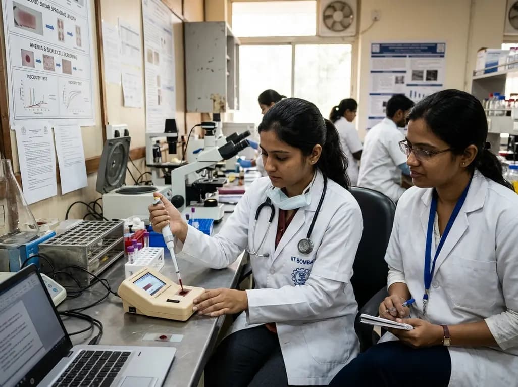
Researchers at IIT Bombay have engineered an automated, cost-effective point-of-care device that diagnoses anaemia and sickle cell disease from a single drop of blood. By measuring blood smear spreading length, this portable tool assesses blood viscosity to offer rapid screening in resource-limited healthcare centers.
Today
A population-based study of 20,928 children reveals that small vulnerable newborns face sex-divergent mental capital outcomes at ages 6-7: term SGA girls show higher behavioral difficulties and lower resilience, while preterm non-SGA boys experience increased behavioral problems.
Today
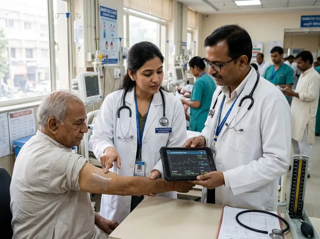
A breakthrough ultrathin multimodal sensor decouples physical hemodynamic monitoring from disposable electrochemical calibration, achieving long-term wearable glucose prediction with 81.36% accuracy and a 9.12% MARD. This offers a robust, drift-free solution for outpatient metabolic surveillance.
Today

A comparative review of tension band wiring and intramedullary screw fixation in thumb metacarpophalangeal arthrodesis, evaluating union rates, hardware complications, and clinical outcomes.
Today
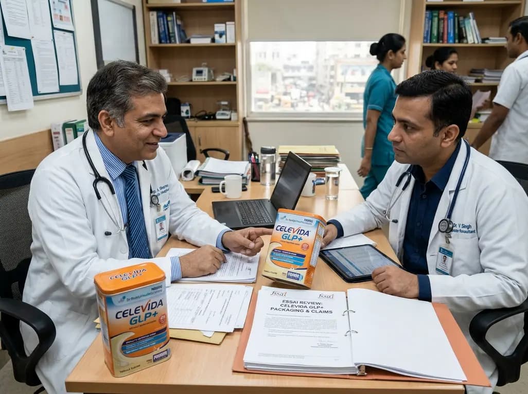
Following regulatory scrutiny from FSSAI regarding promotional representations and lean muscle preservation claims, Dr. Reddy's Laboratories and Nestlé Health Science are currently reviewing the branding and marketing strategy for their joint nutritional supplement Celevida GLP+.
Today
