

Loading, please wait...
Loading, please wait...

Modern digital dentistry relies heavily on creating an accurate virtual patient representation to execute comprehensive dentofacial diagnostics and aesthetic restorative planning. Clinicians routinely integrate three-dimensional facial scans with detailed intraoral digital impressions. However, achieving precise spatial superimposition between soft facial tissue and rigid dental arches remains technically demanding. Dental teams frequently adopt an extraoral scan body or leverage natural tooth morphology to merge these disparate digital datasets.
Digital prosthodontics has transformed conventional treatment planning by unifying extraoral aesthetics with functional occlusion. Clinicians construct a comprehensive digital twin to preview restorative outcomes, plan orthognathic surgeries, and align smile parameters with facial dynamics. Consequently, dental practitioners require reliable workflows that consistently align intraoral dental arches with dynamic facial topographies. Natural dentition often serves as the anatomical reference for this integration process. Nevertheless, limited labial exposure, patient movement, or restricted optical access can compromise direct tooth registration. To overcome these hurdles, engineers developed the extraoral scan body as a geometric transfer medium. This device protrudes beyond the lips to offer clear fiducial markers for optical surface tracking. Furthermore, clinicians can connect these devices to maxillary or mandibular splints to stabilize registration. Therefore, understanding whether geometric variation in scan body architecture improves alignment precision represents a vital clinical question for restorative specialists seeking dependable aesthetic predictability.
A recent clinical study led by Ntovas and colleagues investigated how geometric configurations affect superimposition quality. The researchers evaluated ten human participants using one professional facial scanner alongside three smartphone-based scanning applications. Specifically, they tested the MetiSmile professional scanner against mobile applications comprising EM3D, Heges, and Qlone. For every scanning system, the investigators assessed six experimental subgroups to evaluate alignment consistency. These subgroups included one direct tooth-based integration group and five distinct extraoral scan body configurations. The evaluated hardware designs featured a single sphere, a single cube, a three-sphere array, a three-cube array, and a rectangular block. Furthermore, the operators repeated every scanner and alignment protocol twelve times to ensure rigorous data reproducibility. Before scanning, researchers placed four stable fiducial landmarks across each participant's face. The investigators subsequently superimposed every facial scan onto its corresponding intraoral surface model to construct the final composite files.
To determine spatial accuracy, investigators compared virtual landmark measurements directly against physical control dimensions. Specifically, operators used precision digital calipers to acquire twenty linear distances between facial landmarks and intraoral tooth coordinates. The team then mirrored these identical linear measurements digitally using industrial metrology inspection software. Two-way analysis of variance evaluated trueness and precision metrics across all testing conditions. The resulting clinical data demonstrated striking outcomes regarding registration strategy. Direct tooth-based digital integration achieved superior trueness compared with all extraoral scan body workflows when using both the professional scanner and the EM3D mobile tool. In addition, the geometric design of the extraoral scan body significantly influenced virtual patient trueness across all four optical scanning devices. Geometrically complex arrays did not consistently outperform simpler anatomical alignments. Consequently, relying on visible tooth structure provides higher registration trueness whenever dental visibility permits direct optical capture.
The research highlighted crucial performance differences across hardware modalities. Professional facial scanners utilize dedicated structured-light stereophotogrammetry, which captures crisp surface topography rapidly. In contrast, smartphone applications harness mobile TrueDepth optical sensors or photogrammetry algorithms. Although smartphone technology democratizes facial digitization by reducing clinical equipment costs, software algorithms handle geometric scan bodies with varying reliability. For example, the EM3D application matched professional scanning trends by exhibiting higher trueness during tooth-based alignment. However, applications like Qlone and Heges demonstrated variable registration discrepancies across different scan body shapes. Spherical and cubical landmarks often created optical artifacts or registration drift during mobile sensor sweeps. Furthermore, ambient clinical lighting and minor patient head micro-movements can degrade smartphone tracking. Therefore, while mobile facial scanning offers practical preliminary chairside visualizations, clinicians should remain cautious when using smartphone workflows for complex full-arch reconstructions requiring tight spatial tolerances.
Restorative dentists must balance clinical convenience against digital precision when assembling multi-source 3D models. When anterior teeth provide sufficient optical visibility during smiling or lip retraction, clinicians should prioritize tooth-based superimposition. Direct tooth alignment minimizes intermediate errors caused by mechanical flexure, seating discrepancies, or scan body segmentation errors. Conversely, an extraoral scan body remains valuable in completely edentulous patients where stable anatomical landmarks are absent. If clinicians must employ an extraoral scan body, they should select simple, clearly distinguishable geometries that matching software can segment reliably. In addition, dental teams should calibrate their ambient operatory illumination to prevent sensor glare on polished surfaces. Clinicians must also instruct patients to maintain rigid cervical and head stability throughout data capture. By recognizing hardware limitations and optimizing registration landmarks, restorative practitioners can ensure predictable dentofacial treatment outcomes.
An extraoral scan body serves as a rigid physical reference bridging intraoral tooth landmarks and extraoral facial anatomy. Clinicians utilize this geometric transfer tool to superimpose intraoral digital scans onto three-dimensional facial models. Consequently, the device assists computer software in aligning the dental arch within facial planes, especially when anterior tooth visibility remains obstructed during scanning.
Direct tooth-based alignment relies on the unique, stable surface morphology of natural human teeth. Because software algorithms align distinct dental contours directly, the workflow eliminates intermediate seating discrepancies. In contrast, extraoral scan bodies introduce cumulative tolerance errors, potential component flexure, and optical segmentation challenges, which degrade mathematical registration trueness during complex three-dimensional data superimposition.
Smartphone facial scanners offer practical, accessible tools for aesthetic communication, preliminary smile mock-ups, and patient education. However, clinical studies show their spatial trueness fluctuates significantly depending on the application and registration technique. Therefore, dental clinicians should avoid using smartphone scanners as sole records for complex rehabilitation or orthognathic surgery requiring sub-millimeter precision.
Disclaimer: This content is for informational and educational purposes only. It is not intended to be a substitute for professional medical advice, diagnosis, or treatment. Always seek the advice of your physician or other qualified health provider with any questions you may have regarding a medical condition. Refer to the latest local and national guidelines for clinical practice.
References
Ntovas P et al. Influence of extraoral scan body design on the accuracy of 3-dimensional patient representation across professional and smartphone-based facial scanning technologies: A clinical study. J Prosthet Dent. 2026 Oct 24. doi: undefined. PMID: 42919771.
Revilla-León M, et al. Accuracy of 3-dimensional virtual patient representation using different digital integration techniques and four facial scanners: A clinical validation study. J Prosthet Dent. 2025;134(4):618-626.
Revilla-León M, et al. Accuracy of the 3-dimensional virtual patient representation obtained by using 4 different techniques: An in vitro study. J Prosthet Dent. 2023;129(4):618-624.

Read summarized clinical updates, watch expert medical content, and earn CME certifications right from your smartphone.
Open your smartphone camera to scan and install.





A clinical study evaluates how extraoral scan body geometry influences the trueness and precision of 3D virtual patient representations across professional and mobile facial scanners, revealing critical insights for digital dentistry workflows.
Today
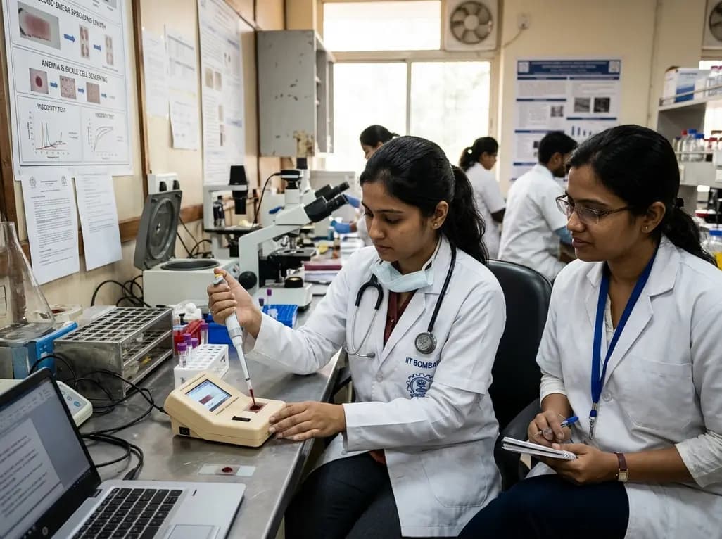
Researchers at IIT Bombay have engineered an automated, cost-effective point-of-care device that diagnoses anaemia and sickle cell disease from a single drop of blood. By measuring blood smear spreading length, this portable tool assesses blood viscosity to offer rapid screening in resource-limited healthcare centers.
Today
A population-based study of 20,928 children reveals that small vulnerable newborns face sex-divergent mental capital outcomes at ages 6-7: term SGA girls show higher behavioral difficulties and lower resilience, while preterm non-SGA boys experience increased behavioral problems.
Today
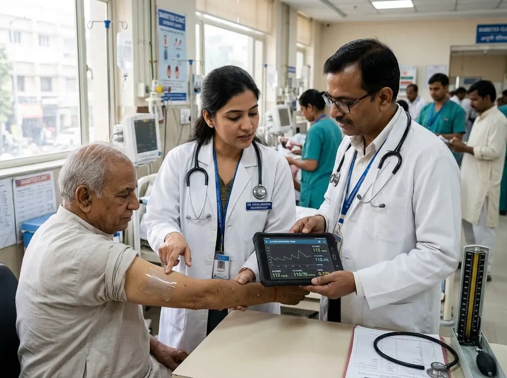
A breakthrough ultrathin multimodal sensor decouples physical hemodynamic monitoring from disposable electrochemical calibration, achieving long-term wearable glucose prediction with 81.36% accuracy and a 9.12% MARD. This offers a robust, drift-free solution for outpatient metabolic surveillance.
Today

A comparative review of tension band wiring and intramedullary screw fixation in thumb metacarpophalangeal arthrodesis, evaluating union rates, hardware complications, and clinical outcomes.
Today
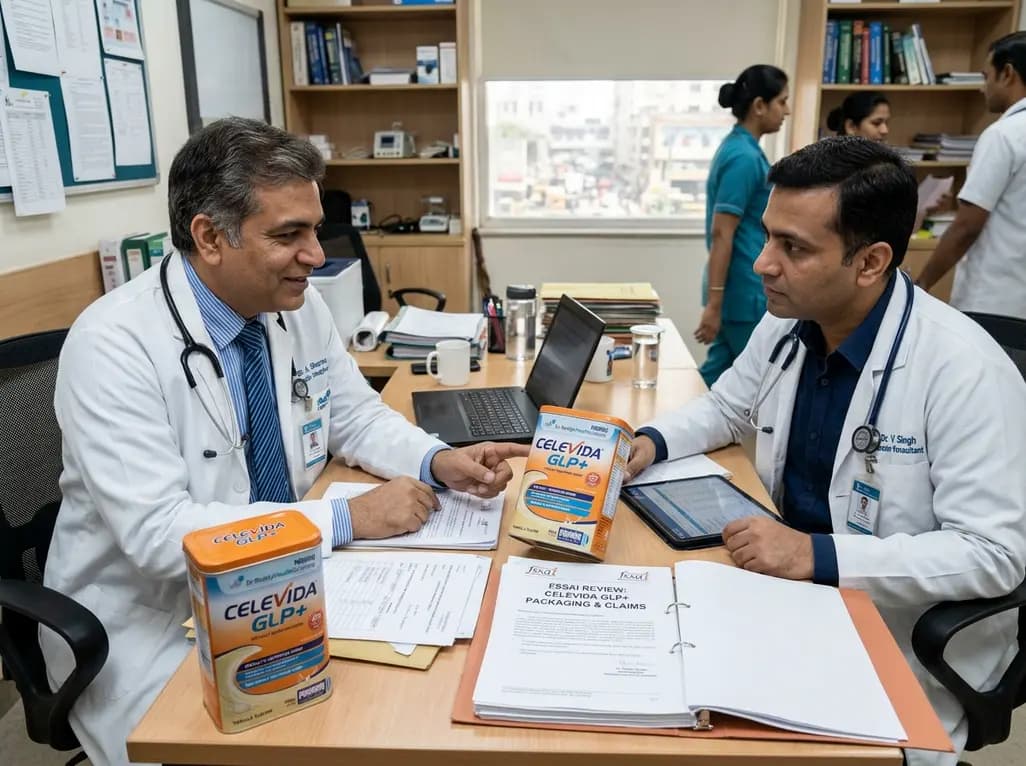
Following regulatory scrutiny from FSSAI regarding promotional representations and lean muscle preservation claims, Dr. Reddy's Laboratories and Nestlé Health Science are currently reviewing the branding and marketing strategy for their joint nutritional supplement Celevida GLP+.
Today
