.webp&w=384&q=75)

Loading, please wait...
Loading, please wait...

Surgical management of anterior skull base tumors has witnessed a transformative paradigm shift over the past two decades. Historically, surgeons relied on open craniofacial resection to eradicate sinonasal tumors abutting the cranial vault. However, the modern endoscopic endonasal transcribriform approach provides an effective, minimally invasive corridor to achieve complete tumor clearance with negative oncologic margins. Malignancies arising in the ethmoid roof and superior nasal cavity frequently threaten the cribriform plate and dura. Consequently, specialized surgical teams must master exact anatomical nuances to optimize oncologic radicality while minimizing neurological morbidity. This specialized operative route combines wide intranasal debulking with targeted craniectomy, delivering exceptional visualization of critical neural structures.
Successful skull base surgery demands a profound understanding of ventral cranial anatomy. The cribriform plate serves as a delicate osseous boundary separating the sinonasal corridor from the olfactory grooves. Tumors such as adenoid cystic carcinoma, olfactory neuroblastoma, and adenocarcinoma readily traverse these thin bony structures. Therefore, the endoscopic endonasal transcribriform approach targets this fragile interface directly from below. Surgeons systematically resect the bilateral ethmoid labyrinths, middle turbinates, and superior nasal septum. This broad exposure reveals the entire anterior skull base from the posterior frontal sinus table to the planum sphenoidale. Furthermore, the fovea ethmoidalis and lamina papyracea define the critical lateral borders. Establishing this unobstructed corridor ensures direct, perpendicular visualization without requiring frontal lobe retraction. Consequently, teams can execute meticulous osteotomies and protect neighboring neurovascular conduits safely.
Appropriate patient selection remains paramount when contemplating an extended endonasal resection. Clinicians must perform comprehensive high-resolution computed tomography to assess osseous erosion of the fovea ethmoidalis and cribriform plate. In addition, contrast-enhanced magnetic resonance imaging provides crucial detail regarding intracranial extension and dural enhancement. Specifically, dynamic imaging distinguishes inflammatory sinus obstruction from frank malignant invasion. Perineural spread along the olfactory fibers or branches of the trigeminal nerve requires particular attention during evaluation. Surgeons must evaluate the lateral extent of the malignancy relative to the orbit. When a neoplasm invades the periorbita, orbital fat, or external facial skin, an exclusively endonasal approach becomes contraindicated. In such scenarios, multidisciplinary teams must pivot toward combined craniofacial or open approaches to guarantee negative surgical margins.
The operative procedure begins with extensive bilateral sphenoethmoidectomies and posterior septectomy to maximize working angles. First, surgeons ligate the anterior and posterior ethmoidal arteries along the frontoethmoidal suture. This early devascularization substantially mitigates intraoperative bleeding throughout subsequent dissection. Next, the surgical team performs rapid debulking of the intranasal tumor mass to delineate the primary site of origin. In cases with orbital abutment, surgeons carefully expose and preserve the intact periorbita. Subsequently, the surgical team utilizes high-speed diamond burrs to perform a precise endoscopic craniectomy along the cribriform plate. Surgeons drill the bony boundaries around the tumor margin until they visualize healthy dura circumferentially. Then, the team incises the dura widely, transects the olfactory rootlets, and resects the involved falx cerebri to obtain negative histological margins.
Extensive resection of the cribriform plate and dura creates a high-flow cerebrospinal fluid defect. Therefore, robust skull base reconstruction is vital to prevent postoperative meningitis and persistent cerebrospinal fluid leakage. Surgeons universally prefer vascularized flaps, most notably the pedicled nasoseptal flap. However, when tumor invades the superior septal mucosa, surgeons harvest alternate regional vascularized tissues or free fascia lata grafts. Surgical teams employ a watertight, multi-layered repair strategy to secure the skull base. First, clinicians place an inlay graft of collagen matrix or fascia lata beneath the intracranial dural edges. Second, they apply an onlay fascial layer across the bony defect to distribute pressure evenly. Finally, surgeons rotate the vascularized flap directly over the reconstruction site, reinforcing the construct with surgical sealant and bioresorbable nasal packing.
Meticulous postoperative care ensures uneventful healing of the reconstructed skull base. Patients remain on strict bed rest with head-of-bed elevation for forty-eight hours to mitigate intracranial pressure spikes. Clinicians strictly avoid high positive-pressure ventilation and continuous positive airway pressure devices during the immediate recovery period. In addition, serial neurological examinations detect subtle changes in mental status or early signs of tension pneumocephalus. Routine nasal saline irrigation commences after pack removal to prevent mucosal crusting and promote rapid remucosalization. Histopathologic assessment of all peripheral and deep frozen margins directs adjuvant therapy protocols. Because sinonasal malignancies exhibit high propensity for local recurrence, postoperative adjuvant radiotherapy or concurrent chemoradiation typically follows within six to eight weeks.
Although endoscopic techniques reduce surgical morbidity, major complications can occasionally occur during complex skull base resections. Arterial injury to the ethmoidal arteries or internal carotid artery represents a life-threatening emergency demanding prompt endonasal tamponade or endovascular intervention. Furthermore, unaddressed dural dehiscence can lead to delayed cerebrospinal fluid leaks, tension pneumocephalus, and ascending bacterial meningitis. Surgeons must maintain extreme vigilance during lateral dissection to avoid inadvertent intraorbital penetration and resultant retrobulbar hematoma. Long-term surveillance requires rigorous endoscopic inspection coupled with serial magnetic resonance imaging every three to six months. Because tumors such as adenoid cystic carcinoma present delayed recurrences, multidisciplinary teams must follow these patients indefinitely. Collaborative efforts between otolaryngologists, neurosurgeons, and radiation oncologists consistently yield optimal local control and excellent functional outcomes.
Surgeons utilize this approach primarily for sinonasal tumors invading the cribriform plate, fovea ethmoidalis, and anterior cranial fossa dura. Common pathologies include olfactory neuroblastoma, adenoid cystic carcinoma, adenocarcinoma, and sinonasal undifferentiated carcinoma. It provides direct visualization for en-bloc dural resection while avoiding external facial incisions and brain retraction.
Gross invasion of the external nasal skin, massive soft tissue infiltration of the anterior facial soft tissues, and extensive lateral orbital involvement contraindicate this endoscopic approach. Furthermore, extensive brain parenchymal invasion or encasement of major intracranial arteries necessitates open transcranial or combined craniofacial surgical approaches instead.
Surgeons reconstruct large defects using a multi-layered technique comprising inlay and onlay fascial grafts followed by a vascularized flap. The vascularized nasoseptal flap represents the primary choice when uninvolved by tumor. Alternatively, clinicians utilize pericranial flaps, temporoparietal fascia flaps, or tensor fascia lata grafts with fibrin sealants.
Disclaimer: This content is for informational and educational purposes only... Refer to the latest local and national guidelines for clinical practice.
References
1. Singh SK et al. Surgical Nuances of Managing Sinonasal Malignancy by Endoscopic Endonasal Transcribriform Approach. Neurol India. 2026 May 01. doi: 10.4103/neurol-india.Neurol-India-D-25-00598. PMID: 42087631.
2. Majmundar N, Kamal NH, Reddy RK, Eloy JA, Liu JK. Limitations of the endoscopic endonasal transcribriform approach. J Neurosurg Sci. 2018 Jun;62(3):287-296. doi: 10.23736/S0390-5616.18.04348-5.
3. Mendelson ZS, Sheikh A, Patel SK, Eloy JA, Liu JK. Endoscopic Endonasal Transcribriform Approach for Anterior Skull Base Lesions Involving the Cribriform Plate: Indications, Techniques, and Results in 24 Patients. J Neurol Surg B Skull Base. 2016;77(Suppl 1):S1-S146.

Read summarized clinical updates, watch expert medical content, and earn CME certifications right from your smartphone.
Open your smartphone camera to scan and install.





Sinonasal malignancies invading the cribriform plate require precise surgical clearance. Discover how the endoscopic endonasal transcribriform approach offers complete tumor resection with negative margins, multi-layered skull base reconstruction, and minimal morbidity in carefully selected patients.
Today
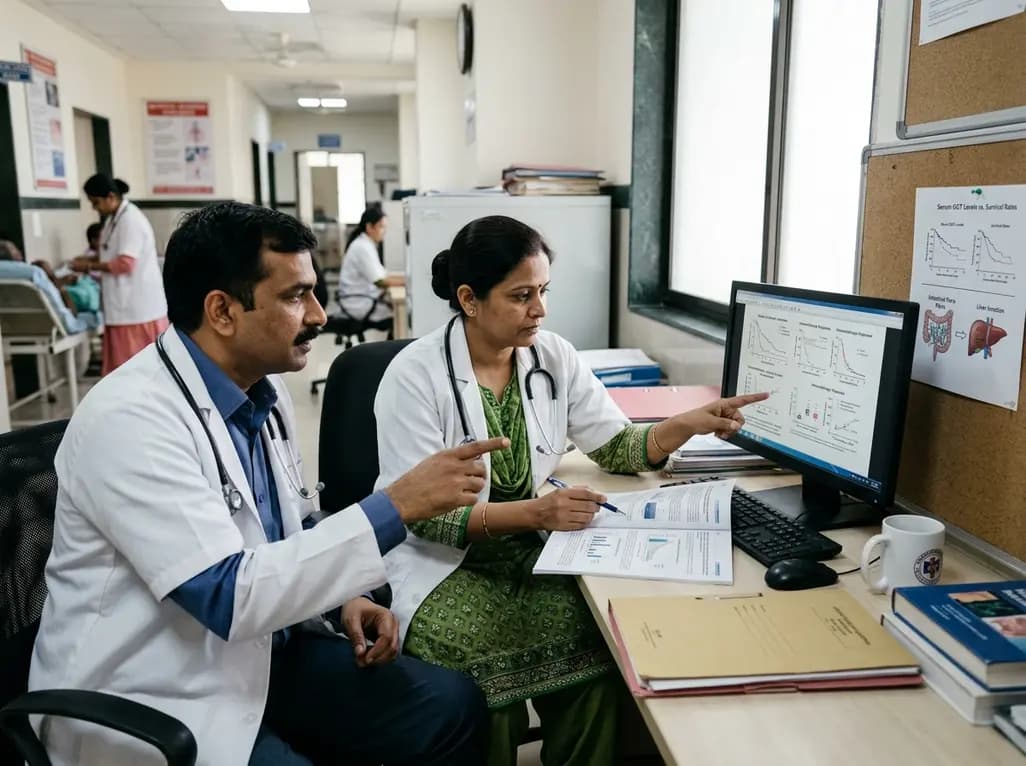
Recent research reveals that subclinical cholestasis, marked by elevated serum gamma-glutamyl transferase, reflects gut dysbiosis and predicts resistance to immune checkpoint blockade. Bile acid remodeling and mucosal MAdCAM-1 loss impair antitumor immunity, highlighting novel therapeutic targets.
Today
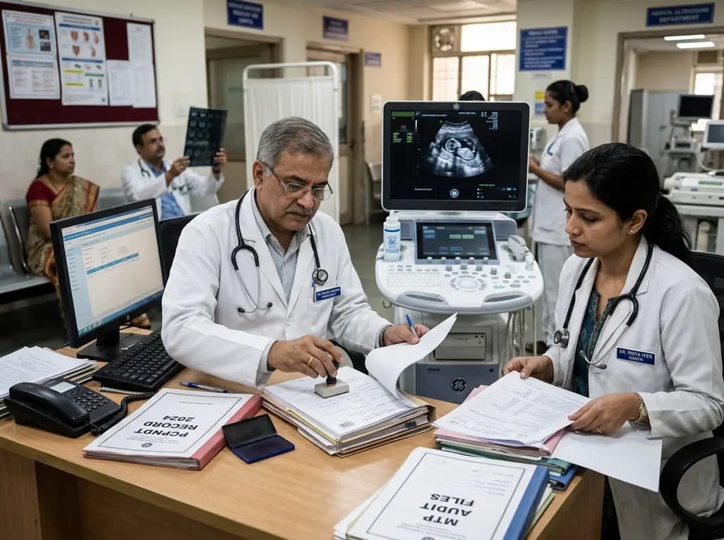
Maharashtra has initiated the appointment of two dedicated special public prosecutors in every district alongside a joint police-health SOP to fast-track PCPNDT and MTP cases. Medical imaging centers, radiologists, and obstetricians must strengthen statutory documentation and audit preparedness.
Today
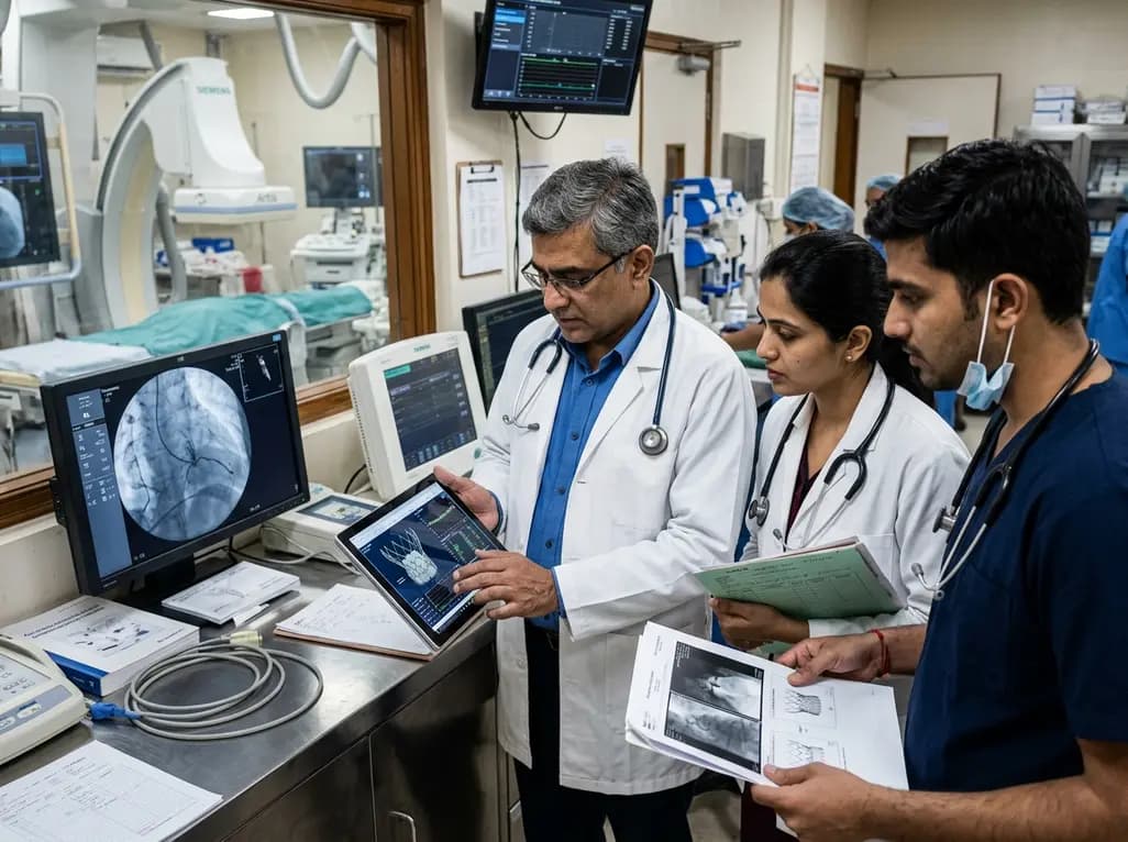
The US FDA has released an early safety alert regarding Edwards Lifesciences Commander M delivery catheters used in transcatheter mitral valve replacement. Reports of balloon bursting during deployment led to one death and one injury, prompting a recall of specific lots and urgent clinical inventory reviews.
Today
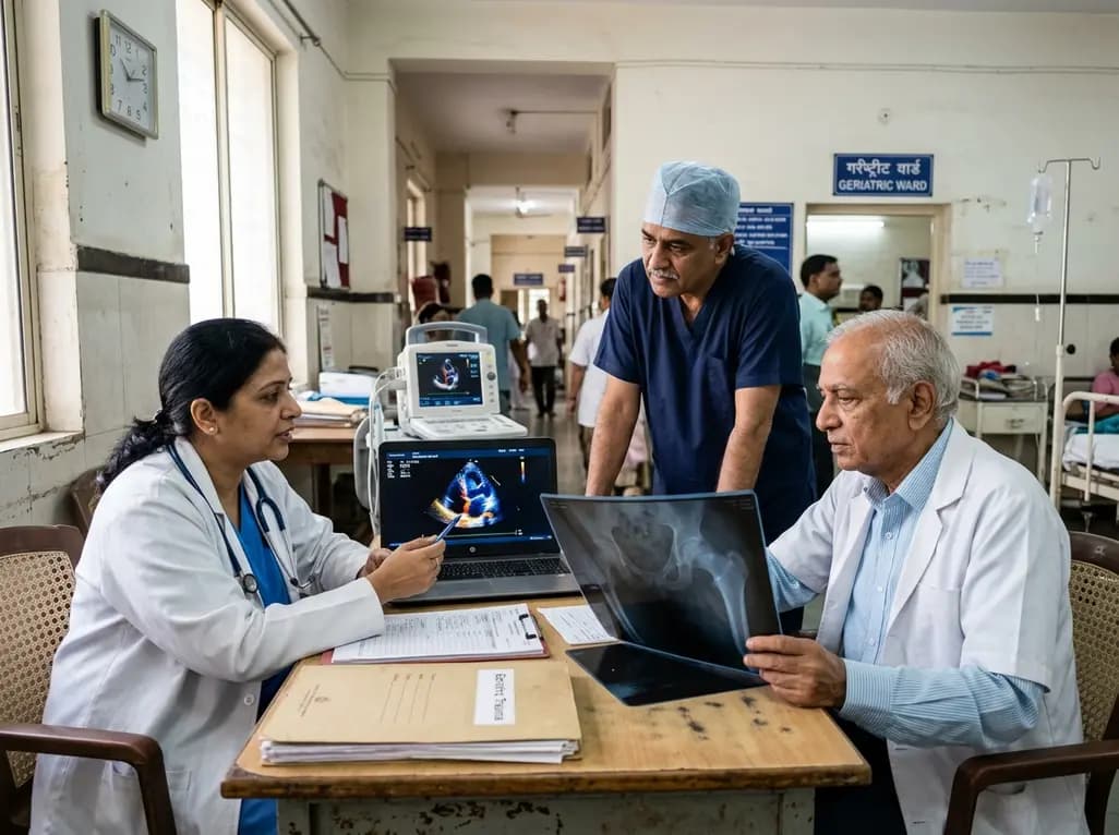
A meta-analysis evaluates the impact of preoperative transthoracic echocardiography on surgical delay and hospital stay in geriatric hip fracture patients. Evidence emphasizes balancing cardiac workups against operative urgency and adhering to clinical guidelines.
Today

An exploratory pilot study evaluates metoprolol pharmacokinetics across cisgender males, cisgender females, and transgender women on gender-affirming hormone therapy. Findings reveal distinct drug disposition trends, emphasizing the critical need for genotype-stratified research in transgender cardiovascular care.
Today
