

Loading, please wait...
Loading, please wait...

Obstructive sleep apnea remains a widespread disorder that triggers severe cardiovascular and metabolic consequences. While polysomnography identifies apneic events, it fails to visualize pharyngeal collapse in real time. Recent technological developments now overcome this longstanding barrier. Specifically, dynamic upper-airway MRI offers clinicians a non-invasive window into airway mechanics without ionizing radiation. Conventional scanning speeds previously caused substantial motion blur during sleep. However, an innovative self-supervised variational manifold learning framework reconstructs under-sampled spiral data with high fidelity during natural sleep. Consequently, sleep specialists and surgeons can now observe true physiological collapse patterns with remarkable spatial precision.
Upper airway collapse during sleep involves dynamic interactions between anatomical narrowing and pharyngeal muscle relaxation. Currently, otolaryngologists often utilize drug-induced sleep endoscopy to locate obstructive sites before surgical intervention. Although endoscopy provides direct visualization, pharmacological sedation fundamentally alters upper airway collapsibility. Sedatives suppress natural neuromuscular reflexes, potentially producing artificial collapse patterns. Furthermore, endoscopes view only one airway segment at a time.
Magnetic resonance imaging provides superior multiplanar soft tissue contrast, yet standard sequences require prolonged acquisition times. Because sleep-related obstruction occurs rapidly within hundreds of milliseconds, conventional scanners generate severe motion artifacts. Moreover, loud acoustic noise disrupts authentic slumber. In previous trials, compressed sensing and parallel imaging attempted to accelerate data collection. Nevertheless, these classical techniques struggled to preserve temporal sharpness across multiple contiguous slices without fully sampled calibration data. Thus, evaluating spontaneous airway collapse during natural sleep demanded a completely autonomous reconstruction strategy.
To resolve dynamic obstruction without prior training datasets, researchers created a self-supervised variational manifold learning algorithm. This framework capitalizes on prospectively under-sampled spiral k-space trajectories. Spiral sampling offers superior scanning efficiency while minimizing flow and motion sensitivity. Instead of relying on vast pre-existing anatomical databases, the framework jointly optimizes a convolutional neural network generator alongside compact latent vectors directly from patient measurements.
Consequently, the network learns a patient-specific temporal manifold without an explicit motion model. The algorithm accurately captures deformation across respiratory cycles. In addition, an adaptive learning rate guided by validation loss delivers principled early stopping without requiring ground truth. This algorithmic constraint prevents overfitting while preserving delicate anatomical boundaries. Most importantly, the technique functions purely in a self-supervised manner. Because clinicians cannot capture fully sampled, motion-free images during active airway collapse, this self-supervised capability represents a vital technical breakthrough for physiological sleep evaluation.
Investigators validated this imaging pipeline across eight patients with obstructive sleep apnea during natural sleep and four healthy volunteers performing the Müller maneuver. Remarkably, the reconstruction captured eleven concurrent axial slices from the velopharynx to the hypopharynx. The imaging protocol achieved a temporal resolution of 183 milliseconds per frame alongside substantial acceleration. Therefore, physicians could inspect simultaneous structural collapse across the upper aerodigestive tract.
Comparative evaluations confirmed marked technical superiority over existing methods. Three expert raters evaluated image quality against parallel imaging, compressed sensing, view sharing, and manifold baselines. Specifically, the proposed method achieved statistically superior ratings across aliasing, spatial blurring, and temporal blurring categories. Reconstruction artifacts occurred in merely six percent of the evaluated slices. Furthermore, these minor artifacts never obscured critical airway boundaries. Real-time dynamic imaging also matched synchronized physiological monitors, closely aligning with respiratory effort curves and oxygen desaturation dips. In single imaging sessions, the system reliably highlighted up to thirteen distinct obstructive episodes.
Pharyngeal obstruction in sleep apnea rarely occurs at a solitary anatomical level. Instead, many individuals present with complex, multilevel collapsibility involving both soft palate and tongue base structures. The manifold learning framework successfully delineated concurrent collapse events across consecutive cross-sectional views. For instance, the reconstructed datasets showed complete concentric retropalatal collapse alongside lateral pharyngeal wall collapse at the velopharyngeal transition. Concurrently, the imaging depicted partial tongue-base displacement and retroglossal narrowing within the same apneic event.
Accurately distinguishing these dynamic collapse configurations carries profound clinical significance. While continuous positive airway pressure remains first-line therapy, poor compliance frequently prompts surgical referral. Surgical interventions such as hypoglossal nerve stimulation require precise patient selection. Specifically, surgeons must exclude patients exhibiting complete concentric collapse at the retropalatal level, as neurostimulation rarely succeeds in this cohort. Because dynamic magnetic resonance imaging visualizes authentic collapse patterns without sedation, it equips clinicians to select optimal candidates for surgical implants and personalized mandibular advancement splints.
Integrating self-supervised dynamic magnetic resonance imaging into daily practice could transform sleep apnea treatment paradigms. Traditional polysomnography quantifies oxygen desaturations and arousal indices, yet it provides no spatial map of airway obstruction. Dynamic magnetic resonance imaging bridges this diagnostic deficit by providing comprehensive anatomical phenotyping. Moreover, imaging during unmedicated natural sleep avoids the respiratory depression and altered neuromuscular tone associated with drug-induced endoscopy.
Nevertheless, health systems must address several technical and workflow demands before widespread clinical integration. First, radiology suites must embed reconstruction algorithms directly into scanner consoles, enabling fast reconstruction times for immediate reporting. Second, scanners should incorporate quiet acoustic sequences to prevent waking sleeping patients. In addition, future clinical investigations must confirm whether MRI-guided interventions produce superior long-term clinical outcomes compared to standard care. Automated segmentation powered by deep neural networks will likely accelerate volumetric measurements of airway caliber. Ultimately, real-time sleep MRI establishes an objective foundation for precision therapy, guiding targeted surgeries and non-surgical device selection.
Drug-induced sleep endoscopy requires pharmacological sedation, which often exaggerates muscular collapse and alters spontaneous breathing patterns. Furthermore, endoscopes view only one airway segment at a time. In contrast, dynamic upper-airway MRI evaluates the entire pharynx simultaneously across eleven contiguous axial slices. This non-invasive approach captures natural physiological collapse patterns during authentic unmedicated sleep without altering baseline pharyngeal muscle tone or requiring invasive instrumentation.
Standard deep learning techniques require extensive databases of fully sampled, artifact-free reference images for network training. However, obtaining fully sampled cine images of dynamic airway collapse during natural sleep remains physically impossible due to rapid physiological movement. Self-supervised manifold learning solves this obstacle by optimizing neural network generators directly from patient-specific, under-sampled spiral k-space data. Consequently, the model reconstructs crisp, multi-slice temporal dynamics without requiring any external training ground truth.
Selecting appropriate surgical interventions requires accurate anatomical localization of obstructive sites. For example, hypoglossal nerve stimulation therapy fails in patients exhibiting complete concentric collapse at the retropalatal level. Dynamic sleep MRI provides volumetric visualization of simultaneous multi-level collapse, including lateral wall, tongue base, and velopharyngeal movements. Therefore, surgeons can pinpoint precise obstruction patterns, rule out unsuitable surgical candidates, and customize targeted surgical procedures to optimize long-term clinical success.
Disclaimer: This content is for informational and educational purposes only. It is not intended to be a substitute for professional medical advice, diagnosis, or treatment. Always seek the advice of your physician or other qualified health provider with any questions you may have regarding a medical condition. Refer to the latest local and national guidelines for clinical practice.
References

Read summarized clinical updates, watch expert medical content, and earn CME certifications right from your smartphone.
Open your smartphone camera to scan and install.





Self-supervised spiral variational manifold learning enables high-speed dynamic upper-airway MRI during natural sleep in obstructive sleep apnea patients. Reconstructing 11 concurrent slices at 183 ms temporal resolution, this novel approach resolves multi-level airway collapse to enhance surgical planning.
Today
A landmark toxicology study demonstrates that airborne VOC dermal uptake contributes substantially to total systemic chemical burdens, highlighting unique epidermal cellular changes and challenging conventional inhalation-focused exposure paradigms.
Today

A nationwide cohort study published in JAMA Pediatrics reveals that maternal exposure to wildfire fine particulate matter, especially in late pregnancy, is linked to an elevated risk of childhood autism. Clinicians should evaluate ambient air quality risks and provide proactive protective counseling to mothers.
Yesterday

With hypertension affecting over 300 million adults in India and household monitor penetration remaining under 5%, local manufacturing expansion of validated oscillometric devices marks a pivotal shift. This overview explores clinical protocols, diagnostic thresholds, and strategies to improve hypertension control.
Today
Atraumatic subcapital femoral neck fracture can emerge as the initial manifestation of osteonecrosis of the femoral head. A comprehensive systematic review highlights key diagnostic features, the indispensable role of MRI, and critical considerations when choosing between arthroplasty and internal fixation.
Today
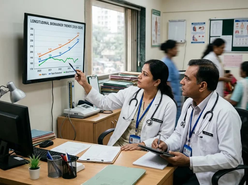
Annual health checks only offer static snapshots. Real-world evidence from 2.1 million insured Indians reveals that tracking dynamic biomarker trends over time enables clinicians to spot evolving cardiometabolic risks early, reverse borderline pathology, and deliver tailored preventive interventions.
Yesterday
