

Loading, please wait...
Loading, please wait...

Osteonecrosis of the femoral head typically manifests with progressive groin ache, restricted motion, and eventual collapse of the articular surface. However, an atraumatic femoral neck fracture can occasionally emerge as the initial clinical presentation of underlying bone ischemia. Because clinicians routinely associate subcapital fractures with significant falls in osteoporotic patients or severe high-energy impacts, an unprovoked fracture pattern creates considerable diagnostic uncertainty. Consequently, healthcare providers must evaluate spontaneous hip symptoms with heightened suspicion. When clinicians recognize ischemic necrosis prior to surgical intervention, they can avoid catastrophic fixation failures. Therefore, evaluating underlying biological factors remains essential for optimizing functional outcomes.
Osteonecrosis originates when microvascular impairment disrupts blood perfusion to the femoral head. Consequently, prolonged hypoxia induces osteocyte apoptosis and structural trabecular breakdown in weight-bearing zones. Over time, normal physiological stresses trigger microfractures that fail to repair due to deficient osteogenesis. Although subchondral impaction represents the most recognized complication of avascular necrosis, ischemic weakness frequently extends distal to the femoral neck. Furthermore, common predisposing factors such as corticosteroid therapy, chronic alcohol excess, hemoglobinopathies, and systemic autoimmune diseases accelerate regional skeletal degradation. In addition, localized osteoclastic bone resorption creates focal lucencies that destabilize the subcapital junction. When physiological load crosses this severely compromised bony zone, a complete fracture occurs without mechanical trauma. Therefore, clinicians should understand that these fractures represent a late biological failure of necrotic bone rather than an isolated traumatic insult. Recognizing this distinct pathological cascade helps surgical teams anticipate mechanical instability and poor bone quality during reconstructive planning.
Patients experiencing this atypical presentation generally complain of insidious groin or anterior thigh pain that gradually worsens before functional collapse occurs. However, because these individuals experience no preceding fall or sports injury, primary care providers often attribute initial discomfort to lumbar spine pathology, sciatica, or soft tissue strain. Furthermore, physical examination demonstrates painful limitations in internal rotation and an antalgic gait. Once complete osseous disruption develops, patients experience sudden inability to bear weight along with subtle leg shortening. In addition, diagnostic delay remains prevalent because early plain radiographs often show minimal cortical abnormalities or subtle subcapital radiolucencies. When radiologists interpret these findings simply as primary osteoporotic insufficiency fractures, they overlook the underlying necrosis. Consequently, surgical teams may choose inappropriate fixation devices that fail in necrotic bone. Therefore, medical teams must maintain heightened vigilance whenever patients report spontaneous hip pain accompanied by risk factors for avascular necrosis.
Standard pelvis and hip radiographs serve as the mandatory initial screening modality for acute hip pain. However, plain films frequently demonstrate poor sensitivity for early osteonecrosis and non-displaced fracture lines. Consequently, magnetic resonance imaging provides the gold standard for establishing a definitive diagnosis and evaluating fracture configurations. MRI scans clearly demonstrate low-signal geographic bands on T1-weighted sequences alongside surrounding bone marrow edema. Furthermore, T2-weighted images frequently display the characteristic double-line sign that demarcates devitalized tissue from vascular repair zones. In addition, high-resolution computed tomography helps delineate subcapital cortical breaches and assesses three-dimensional trabecular integrity. Therefore, clinicians must promptly request advanced cross-sectional imaging when radiographs appear equivocal or atypical. Importantly, magnetic resonance evaluation should always encompass the contralateral hip joint, because bilateral avascular necrosis occurs in a high percentage of patients. Consequently, early comprehensive imaging prevents missed contralateral lesions and directs timely joint-preserving interventions.
Choosing an appropriate surgical strategy demands thorough consideration of underlying bone viability and biomechanical longevity. Historically, orthopedic surgeons attempted closed reduction and internal fixation with cannulated screws for subcapital fractures in younger individuals. However, systematic clinical evidence reveals that internal fixation in necrotic femoral heads carries exceptionally high failure rates. Because dead trabeculae cannot achieve biological osseous union or support implant purchase, fixation screws frequently migrate, cut out, or collapse. Furthermore, subsequent femoral head fragmentation accelerates degenerative osteoarthritis and forces early revision procedures. In contrast, hip arthroplasty provides significantly greater reliability by resecting devitalized tissue and restoring immediate joint mechanics. Consequently, total hip arthroplasty represents the gold standard reconstructive treatment for the vast majority of symptomatic patients. Nevertheless, surgeons must carefully individualize implant selection based on age, physiological status, and anatomical requirements. Therefore, recognizing underlying necrosis fundamentally transforms surgical decision-making from preservation to reliable prosthetic reconstruction.
Optimizing recovery for patients with complex hip necrosis requires coordinated teamwork across surgical, medical, and rehabilitation specialties. Firstly, physicians must investigate and mitigate systemic risk factors, such as discontinuing non-essential corticosteroids and managing metabolic comorbidities. Furthermore, counseling regarding alcohol cessation and smoking cessation promotes systemic bone healing and implant longevity. In addition, physical therapists must institute early mobilization protocols that protect reconstructive components while restoring functional gait mechanics. Diligent long-term surveillance remains equally crucial, particularly for monitoring the asymptomatic contralateral hip with periodic magnetic resonance imaging. Clinicians must educate patients regarding early warning symptoms to facilitate timely joint-preserving measures on the opposite side. Moreover, structured clinical follow-up tracks implant fixation and overall quality of life over extended intervals. Ultimately, combining precise surgical reconstruction with aggressive medical optimization yields the best long-term outcomes for patients facing this challenging clinical condition.
An atraumatic femoral neck fracture develops when prolonged ischemic compromise destroys bone microarchitecture in the proximal femur. Continuous physiological weight-bearing forces overwhelm this weakened, necrotic trabecular framework, producing progressive microfractures that eventually cause complete bone separation without trauma. In addition, predisposing factors such as systemic steroid use and chronic alcohol intake impair bone repair mechanisms. Consequently, severe structural degradation precipitates spontaneous fracture under everyday biomechanical stress.
Magnetic resonance imaging offers exceptional sensitivity for detecting occult osteonecrosis that standard radiographs regularly miss. Specifically, MRI visualizes characteristic bone marrow edema, pathognomonic low-signal demarcation lines, and the classic double-line sign. In addition, confirming ischemic necrosis prevents surgeons from mistakenly attempting internal fixation on devitalized femoral bone. Furthermore, MRI scans systematically screen the contralateral hip, enabling clinicians to identify silent bilateral necrosis and implement timely joint-preserving care.
Total hip arthroplasty offers the most durable and reliable surgical outcome for patients suffering from this condition. Internal fixation exhibits frequent hardware failure and nonunion because ischemic bone lacks the biological healing potential required for screw stability. Consequently, replacing the damaged joint with a prosthetic construct completely eliminates nonunion risk. In addition, arthroplasty facilitates rapid postoperative weight-bearing, reduces revision rates, and quickly restores functional mobility.
Disclaimer: This content is for informational and educational purposes only and does not constitute medical advice. Consult qualified healthcare professionals for diagnosis and treatment. Refer to the latest local and national guidelines for clinical practice.
References

Read summarized clinical updates, watch expert medical content, and earn CME certifications right from your smartphone.
Open your smartphone camera to scan and install.





Atraumatic subcapital femoral neck fracture can emerge as the initial manifestation of osteonecrosis of the femoral head. A comprehensive systematic review highlights key diagnostic features, the indispensable role of MRI, and critical considerations when choosing between arthroplasty and internal fixation.
Today
A landmark toxicology study demonstrates that airborne VOC dermal uptake contributes substantially to total systemic chemical burdens, highlighting unique epidermal cellular changes and challenging conventional inhalation-focused exposure paradigms.
Today

A nationwide cohort study published in JAMA Pediatrics reveals that maternal exposure to wildfire fine particulate matter, especially in late pregnancy, is linked to an elevated risk of childhood autism. Clinicians should evaluate ambient air quality risks and provide proactive protective counseling to mothers.
Yesterday
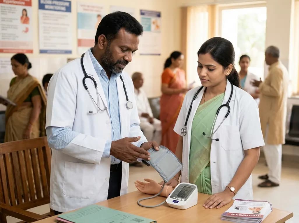
With hypertension affecting over 300 million adults in India and household monitor penetration remaining under 5%, local manufacturing expansion of validated oscillometric devices marks a pivotal shift. This overview explores clinical protocols, diagnostic thresholds, and strategies to improve hypertension control.
Today
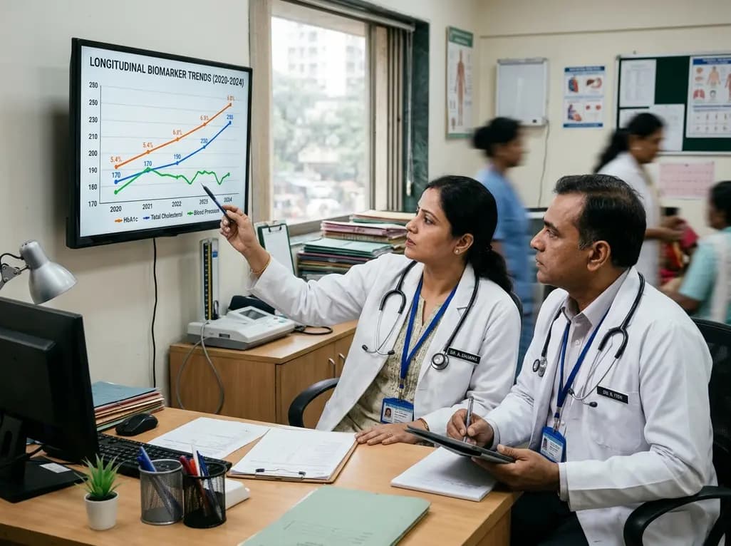
Annual health checks only offer static snapshots. Real-world evidence from 2.1 million insured Indians reveals that tracking dynamic biomarker trends over time enables clinicians to spot evolving cardiometabolic risks early, reverse borderline pathology, and deliver tailored preventive interventions.
Yesterday
