.webp&w=384&q=75)

Loading, please wait...
Loading, please wait...
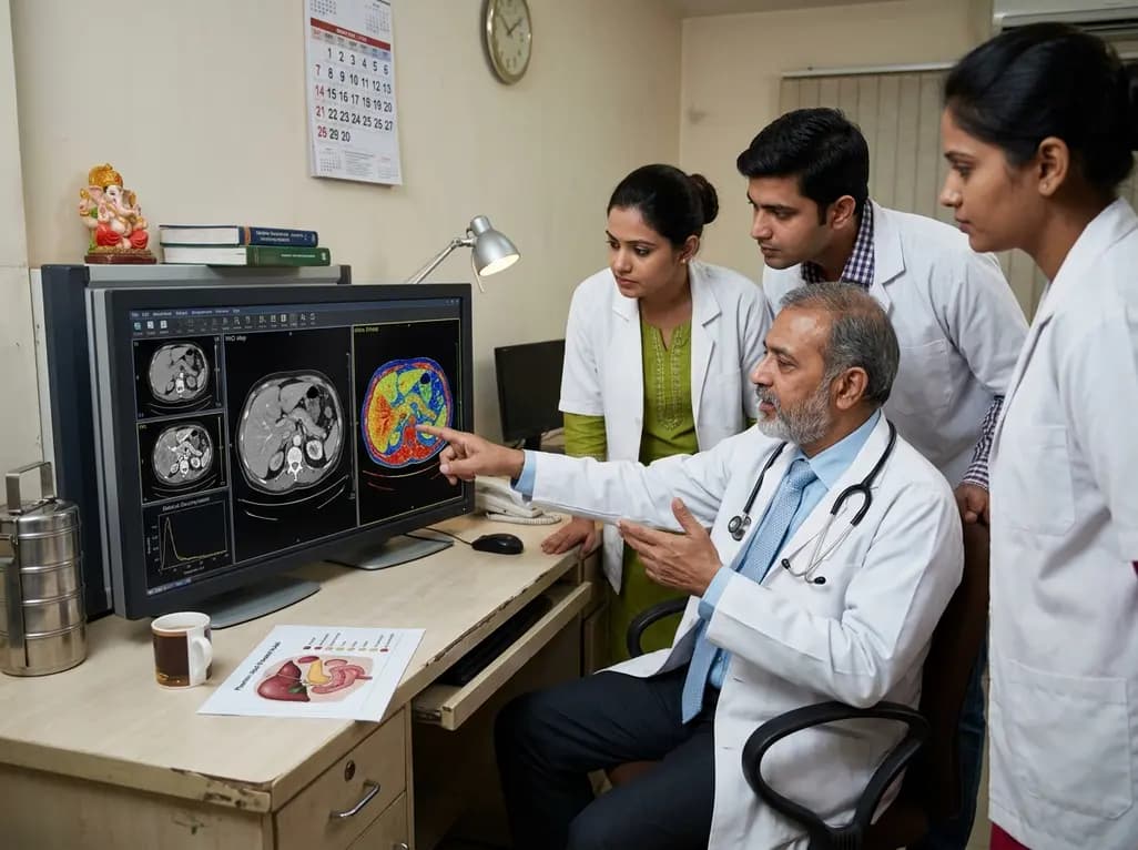
Dual-layer spectral computed tomography (DLCT) has revolutionized abdominal diagnostics by providing more than just anatomical maps. It allows for the simultaneous acquisition of conventional images and spectral data, enabling the creation of virtual non-contrast (VNC) images and precise iodine quantification. This technological leap is particularly beneficial for evaluating vascularity and characterizing lesions without the need for additional, non-enhanced scans. However, clinical experts have long noted that tissue composition can influence the reliability of these reconstructions. Specifically, the presence of lipids within abdominal organs presents a significant challenge to the material decomposition algorithms that undergird dual-layer spectral CT accuracy. When lipids are present in high concentrations, such as in fatty liver disease or certain adrenal lesions, the expected spectral signature of tissues is altered. Consequently, this can lead to errors in both attenuation measurements on VNC images and the estimation of iodine density. Understanding these nuances is essential for radiologists in India, where metabolic conditions like non-alcoholic fatty liver disease (NAFLD) are increasingly prevalent, necessitating highly accurate quantitative imaging to guide patient management and treatment decisions.
The core of dual-layer spectral CT accuracy lies in its ability to differentiate materials based on their energy-dependent attenuation profiles. Specifically, the detector layers capture low-energy and high-energy photons simultaneously, allowing the system to solve for the photoelectric effect and Compton scattering. Furthermore, iodine and water are the primary materials used in these decomposition models. When lipids are introduced into the equation, their unique attenuation properties, which differ significantly from both water and iodine, can disrupt the mathematical separation. Because lipids have a lower effective atomic number than water, they can mimic or mask the attenuation characteristics of other materials during the post-processing phase. Therefore, as the concentration of lipids increases, the algorithm may incorrectly attribute a portion of the attenuation to the wrong material category. This phenomenon results in a systematic shift in the Hounsfield units (HU) observed on VNC images. Moreover, it leads to an underestimation of iodine density, as the presence of fat effectively dilutes the spectral signal that the system uses to quantify contrast agent concentration. These findings emphasize that spectral CT is not a "one-size-fits-all" solution and requires careful calibration based on the patient's internal milieu.
Recent phantom studies have provided quantitative insights into how varying lipid levels affect imaging outcomes. By utilizing solutions with lipid concentrations ranging from 0% to 40%, researchers were able to simulate the diverse fatty environments found in human organs. Specifically, the study evaluated tubes containing contrast agents alongside non-contrast controls within a water-filled phantom. The results clearly demonstrated that as the lipid content rose, the VNC estimation error increased proportionally. Notably, the difference in attenuation between virtual and true non-contrast images became more pronounced, suggesting that clinicians must exercise caution when interpreting VNC values in patients with high-grade steatosis. Additionally, the accuracy of iodine density measurements suffered under these conditions, with greater underestimation observed as the lipid fraction grew. Such errors are not merely academic; they can potentially lead to the mischaracterization of an adrenal adenoma as a malignant lesion or result in the inaccurate quantification of extracellular volume (ECV) in the liver. Consequently, these results provide a critical benchmark for identifying the limitations of current spectral decomposition techniques and highlight the necessity for optimized scanning protocols that can mitigate these lipid-induced discrepancies.
One of the most actionable findings from recent research is the significant role of tube voltage in maintaining dual-layer spectral CT accuracy. The study compared the performance of 120 kVp and 140 kVp settings, revealing a distinct advantage for the higher voltage. Specifically, scans performed at 140 kVp exhibited smaller VNC estimation errors and less severe underestimation of iodine density compared to those acquired at 120 kVp. This improvement is likely due to the superior energy separation and increased photon flux achieved at higher voltages, which enhances the signal-to-noise ratio in the high-energy detector layer. By providing a clearer spectral signal, 140 kVp scanning allows the material decomposition algorithm to better differentiate between lipids, iodine, and water. Consequently, for patients suspected of having high lipid deposition, such as those with hepatic steatosis or lipid-rich incidentalomas, opting for a higher voltage may be a superior clinical strategy. This approach not only improves the reliability of quantitative data but also ensures that the benefits of spectral CT, such as radiation dose reduction through VNC, are not compromised by inaccuracies. Radiologists should therefore consider adjusting their default abdominal protocols to favor 140 kVp when quantitative spectral analysis is the primary goal.
The practical implications of these findings are far-reaching for daily radiological practice. In the context of adrenal imaging, differentiating between lipid-rich adenomas and non-adenomatous lesions often relies on precise attenuation thresholds. If VNC images overestimate attenuation due to lipid interference, a benign adenoma might incorrectly appear indeterminate, triggering unnecessary follow-up imaging or biopsy. Furthermore, in the assessment of fatty liver disease, the ability to accurately quantify both fat and iodine is becoming increasingly important for monitoring disease progression and therapeutic response. Specifically, the underestimation of iodine density could lead to false negatives when evaluating liver perfusion or extracellular volume, which are key markers of fibrosis. Therefore, clinicians must integrate the knowledge of lipid-induced errors into their diagnostic workflow. By understanding that 140 kVp scans provide better dual-layer spectral CT accuracy, centers can refine their standard operating procedures to enhance diagnostic confidence. Ultimately, these advancements contribute to the growing field of precision medicine, allowing for more personalized and accurate non-invasive assessments of complex abdominal pathologies, which is particularly relevant in the diverse and high-volume clinical environment of modern India.
As spectral imaging technology continues to evolve, the integration of these phantom study findings will be crucial for the development of the next generation of algorithms. Manufacturers are already looking into more sophisticated material decomposition models that can explicitly account for a third or fourth material, such as fat, to improve dual-layer spectral CT accuracy. However, until such software updates are universally available, the onus remains on the radiologist to optimize hardware parameters. The recommendation for high-voltage scanning serves as a robust interim solution that provides immediate benefits in measurement precision. Additionally, future research should focus on validating these phantom results in vivo, exploring how different patient body habitus and organ-specific lipid distributions might further influence spectral data. By bridging the gap between phantom research and clinical application, the medical community can ensure that quantitative CT becomes a truly reliable tool for assessing tissue composition. In conclusion, while lipids do pose a challenge to spectral decomposition, the strategic use of 140 kVp tube voltage offers a clear path toward minimizing errors and maximizing the diagnostic potential of DLCT in the abdominal region.
Lipids interfere with VNC reconstruction by altering the expected energy-dependent attenuation profile of the tissue. Since material decomposition algorithms typically rely on water and iodine as base materials, the unique attenuation properties of lipids can lead to mathematical errors. As lipid concentration increases, the system may fail to perfectly subtract the iodine signal, resulting in a virtual non-contrast attenuation value that differs significantly from the true non-contrast measurements, potentially leading to diagnostic misinterpretations.
Higher tube voltage settings, such as 140 kVp, provide better dual-layer spectral CT accuracy because they generate a broader spectrum of X-ray energies. This increases the photon count reaching the high-energy detector layer and improves the energy separation between the two layers. Consequently, the material decomposition algorithm can more effectively distinguish between the spectral signatures of lipids and iodine, leading to reduced estimation errors and more consistent iodine density quantification even in fatty tissues.
The primary clinical implication is the prevention of diagnostic errors. In adrenal imaging, overestimation of VNC attenuation could mask the low-density characteristic of lipid-rich adenomas, leading to more indeterminate findings. For extracellular volume (ECV) quantification in the liver, underestimation of iodine density could result in inaccurate fibrosis staging. Utilizing 140 kVp protocols ensures more reliable quantitative data, which is essential for characterizing these lesions accurately and avoiding unnecessary invasive procedures for benign conditions.
Disclaimer: This content is for informational and educational purposes only. It does not constitute medical advice or a professional relationship between the reader and the author or publisher. While every effort has been made to ensure accuracy, medical knowledge is constantly evolving. Refer to the latest local and national guidelines for clinical practice.
References
Yamane S et al. The effect of lipid content on virtual non-contrast images and iodine density estimation from dual-layer spectral computed tomography: A phantom study. Radiography (Lond). 2026 Jul 09. doi: undefined. PMID: 42424720.
Sauter A et al. Accuracy of iodine quantification in dual-layer spectral CT: Influence of iterative reconstruction, patient habitus and tube parameters. Eur J Radiol. 2018 May;102:112-119. doi: 10.1016/j.ejrad.2018.03.009. PMID: 29685549.
Gröber S et al. Accuracy of iodine quantification using dual energy CT in latest generation dual source and dual layer CT. Eur Radiol. 2026 Feb 26. doi: 10.1007/s00330-017-4785-0.

Read summarized clinical updates, watch expert medical content, and earn CME certifications right from your smartphone.
Open your smartphone camera to scan and install.





A new phantom study reveals how lipid content in abdominal organs can interfere with dual-layer spectral CT accuracy, affecting virtual non-contrast (VNC) attenuation and iodine density. Researchers found that using a higher tube voltage of 140 kVp significantly improves measurement precision.
3 months ago
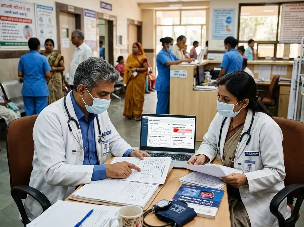
Co-administering antiseizure medications and DOACs frequently causes pharmacokinetic interactions, subtherapeutic drug levels, and thromboembolic failures. This review highlights enzyme induction risks, unpredictable outcomes with levetiracetam, and the vital role of therapeutic drug monitoring.
Today

Demanding exhaustive hormonal profiling can inadvertently discourage clinical studies involving females. Methodological standards must align with the specific scientific question to expand, not restrict, equitable evidence for women.
Today
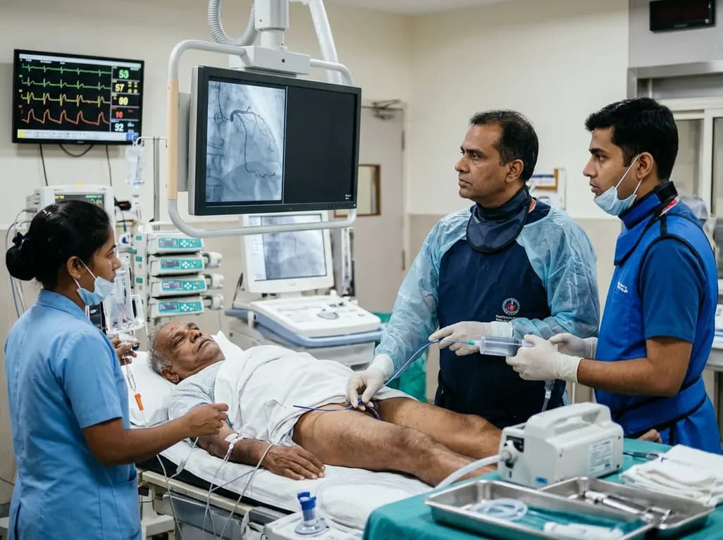
An elderly man with partially repaired Tetralogy of Fallot presented with STEMI caused by coronary embolism rather than atherosclerosis. Successful treatment with sustained mechanical aspiration thrombectomy underscores the vital need for high clinical vigilance in adult congenital heart disease.
Today
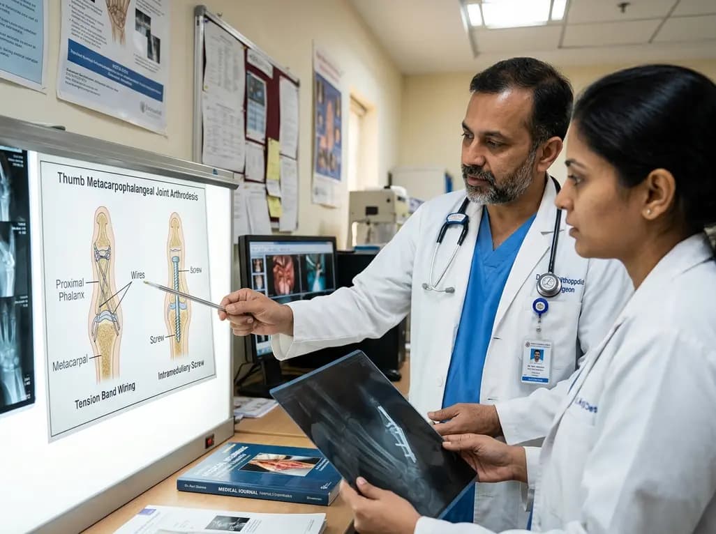
A comparative review of tension band wiring and intramedullary screw fixation in thumb metacarpophalangeal arthrodesis, evaluating union rates, hardware complications, and clinical outcomes.
Today

Following regulatory scrutiny from FSSAI regarding promotional representations and lean muscle preservation claims, Dr. Reddy's Laboratories and Nestlé Health Science are currently reviewing the branding and marketing strategy for their joint nutritional supplement Celevida GLP+.
Today
