

Loading, please wait...
Loading, please wait...
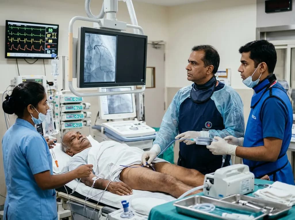
Acute myocardial infarction typically results from atherosclerotic plaque rupture and subsequent occlusive thrombosis. However, coronary embolism represents an uncommon yet critical nonatherosclerotic etiology that interventional cardiologists encounter during emergency primary catheterization. Adult congenital heart disease creates distinct anatomical and physiological vulnerabilities that predispose patients to thromboembolic complications. Specifically, residual intracardiac shunts, progressive atrial chamber dilatation, and surgically scarred myocardial tissue foster significant blood stasis. When elderly individuals with congenital malformations develop acute coronary syndromes, clinicians frequently assume conventional atherosclerotic vascular disease. Consequently, this assumption can delay targeted diagnostic evaluation and misguide acute revascularization strategies. Tetralogy of Fallot repairs performed decades ago frequently left residual hemodynamic defects, including persistent ventricular septal communications and pulmonary regurgitation. Furthermore, secondary electrophysiological remodeling often triggers sustained atrial arrhythmias in these aging patients. These combined mechanical and electrical abnormalities dramatically amplify the risk of systemic embolization. Therefore, acute chest pain protocols in specialized centers must incorporate thromboembolic differentials whenever congenital repairs coexist with cardiac arrhythmias. Identifying nonatherosclerotic coronary occlusion early fundamentally transforms invasive procedural planning. As a result, interventional operators can select targeted thrombectomy techniques while avoiding unnecessary coronary stenting in non-diseased epicardial vessels.
A recent clinical investigation highlighted a 77-year-old male with partially repaired Tetralogy of Fallot presenting with acute inferior ST-elevation myocardial infarction. The patient had undergone palliative surgical intervention during childhood, leaving a persistent residual ventricular septal defect with bidirectional shunting. In addition, the patient developed new-onset atrial fibrillation, which severely disrupted left and right atrial transport mechanics. He arrived at the emergency department experiencing severe retrosternal chest pressure and classic electrocardiographic ST-segment elevations across inferior leads. Interventional cardiologists initiated emergency cardiac catheterization to restore epicardial coronary flow. Angiography revealed an abrupt total occlusion in the distal right coronary artery containing an exceptionally high thrombus burden. Remarkably, the remaining coronary tree exhibited smooth luminal borders without significant angiographic atherosclerosis. This stark discrepancy between severe focal thrombotic occlusion and completely pristine coronary vessels strongly suggested an embolic origin. Moreover, bedside echocardiography demonstrated bidirectional flow across the residual septal defect alongside marked biatrial enlargement. Clinicians recognized that either left atrial thrombosis or paradoxical migration through the ventricular defect caused this acute vessel occlusion. Thus, the medical team quickly adapted their revascularization plan to address large-volume thrombus extraction rather than atherosclerotic plaque modification.
Understanding the interplay between intracardiac shunts and atrial arrhythmogenesis clarifies the genesis of coronary embolism. In healthy hearts, competent cardiac valves and intact septal barriers prevent peripheral venous thrombi from entering systemic arterial circulation. However, residual ventricular septal defects create viable anatomical channels for bidirectional or right-to-left paradoxical migration. Elevated right ventricular pressures, common after congenital pulmonary valve repairs, intermittently reverse trans-septal pressure gradients during respiration or physical strain. Consequently, deep venous thrombi can bypass the pulmonary capillary filter and directly enter the left ventricle and ascending aorta. Alternatively, new-onset atrial fibrillation promotes spontaneous blood stasis within dilated atria, directly generating left-sided mural thrombi. Regardless of the exact upstream origin, dislodged thrombotic fragments preferentially enter epicardial vessels with favorable take-off angles and flow dynamics. The right coronary artery frequently receives these migratory emboli due to its anterior aortic origin and systolic flow patterns. Furthermore, the absence of underlying atherosclerotic plaques means the distal vessel architecture remains fragile and susceptible to sudden mechanical blockage. Therefore, managing clinicians must recognize that systemic anticoagulation, rather than antiplatelet monotherapy, addresses the primary source of recurrent intravascular events.
Managing extensive coronary thrombi in pristine epicardial vessels presents severe interventional challenges. Routine manual syringe aspiration catheters frequently fail when encountering dense, organized fibrin-rich emboli. Furthermore, aggressive balloon predilatation or direct stent deployment risks distal embolization, extensive microvascular obstruction, and the dreaded no-reflow phenomenon. To overcome these hazards, operators successfully deployed sustained mechanical aspiration thrombectomy using continuous computer-controlled vacuum suction. Unlike transient manual suction, continuous mechanical aspiration provides uniform, high-flow vacuum extraction through large-lumen aspiration catheters. The catheter engages the proximal thrombus interface and continuously draws the entire clot into an external collection canister. In this case, sustained aspiration successfully retrieved the massive thrombus burden without inducing distal fragmentation or epicardial dissection. Angiographic imaging immediately confirmed complete restoration of normal coronary perfusion with TIMI 3 flow. Additionally, the underlying arterial segment displayed perfectly intact endothelium, which eliminated any justification for intracoronary stent implantation. The patient experienced prompt resolution of ischemic chest pain and immediate electrocardiographic ST-segment normalization. Accordingly, avoiding permanent metallic stents preserved natural vessel vasomotion and spared the patient from long-term dual antiplatelet bleeding hazards.
This landmark case offers vital diagnostic and therapeutic lessons for physicians caring for adult congenital heart disease cohorts. First, interventional cardiologists must maintain high suspicion for coronary embolism whenever acute coronary syndromes occur alongside intracardiac shunts. When coronary angiography shows abrupt cutoff without diffuse atherosclerosis, clinicians should immediately refrain from reflexive stent placement. Second, transesophageal echocardiography and saline contrast bubble studies remain essential to delineate intracardiac shunt dynamics precisely. In addition, ambulatory rhythm monitors should evaluate occult atrial arrhythmias, given their high prevalence in surgically repaired hearts. From a therapeutic standpoint, long-term secondary prevention requires oral anticoagulation rather than standard post-STEMI dual antiplatelet therapy. Anticoagulants directly address the primary source of thromboembolism by preventing fibrin propagation in dilated cardiac chambers. Furthermore, multidisciplinary adult congenital heart disease teams must evaluate patients for potential percutaneous or surgical defect closure. Closing significant residual shunts substantially reduces lifelong risk of stroke and paradoxical systemic embolism. Ultimately, combining innovative catheter-based thrombectomy with tailored anticoagulation regimens ensures optimal survival and preserves myocardial function in complex congenital survivors.
Clinicians diagnose coronary embolism using established clinical and angiographic criteria. Coronary angiography typically demonstrates abrupt vessel cutoff with heavy thrombus burden without underlying atherosclerotic lesions. Additionally, clinicians should confirm an embolic source, such as intracardiac shunts, mural thrombi, or atrial fibrillation. Transesophageal echocardiography and systemic vascular imaging help verify paradoxical or direct embolic pathways. Therefore, comprehensive multi-modality evaluation remains essential to distinguish paradoxical coronary emboli from typical plaque rupture events.
Sustained mechanical aspiration thrombectomy provides continuous, vacuum-assisted negative pressure to extract large thrombi safely from epicardial arteries. Unlike routine manual syringe aspiration, sustained mechanical devices evacuate substantial thrombotic burdens without excessive vessel trauma. Consequently, this intervention restores epicardial flow rapidly without requiring balloon angioplasty or stent placement. In cases involving pristine coronary anatomy, this mechanical strategy prevents unnecessary stenting, lowers lifelong bleeding risks, and markedly preserves microvascular perfusion.
Long-term management requires targeted systemic anticoagulation rather than prolonged dual antiplatelet therapy. Because the underlying etiology stems from arrhythmia and intracardiac shunting rather than plaque rupture, oral anticoagulants effectively suppress further thrombus formation. Furthermore, clinicians must evaluate the hemodynamic significance of residual shunts and correct atrial tachyarrhythmias. Regular multidisciplinary surveillance ensures timely identification of recurrent thromboembolic risks, thereby preventing future cerebrovascular accidents and recurrent myocardial infarction in this vulnerable population.
Disclaimer: This content is for informational and educational purposes only and is intended solely for healthcare professionals. It does not constitute medical advice, diagnosis, or treatment recommendations. Clinical decisions should always be made by qualified healthcare providers based on individual patient evaluations and applicable clinical standards. Refer to the latest local and national guidelines for clinical practice.
References

Read summarized clinical updates, watch expert medical content, and earn CME certifications right from your smartphone.
Open your smartphone camera to scan and install.





An elderly man with partially repaired Tetralogy of Fallot presented with STEMI caused by coronary embolism rather than atherosclerosis. Successful treatment with sustained mechanical aspiration thrombectomy underscores the vital need for high clinical vigilance in adult congenital heart disease.
Today

Demanding exhaustive hormonal profiling can inadvertently discourage clinical studies involving females. Methodological standards must align with the specific scientific question to expand, not restrict, equitable evidence for women.
Today

A recent study demonstrates that spinal oxygen sensors do not require mitochondrial electron transport to trigger sympathetic excitation during acute hypoxia, revealing new insights into how mitochondrial oxidative stress modulates autonomic tone in cardiovascular and neurological diseases.
Today

A novel calcium polyphosphate coacervate enables injectable bone implants with in situ hierarchical porosity via phase separation and gas foaming, significantly enhancing cell proliferation, energy metabolism, and osteogenic differentiation for minimally invasive bone defect repair.
Today

Following regulatory scrutiny from FSSAI regarding promotional representations and lean muscle preservation claims, Dr. Reddy's Laboratories and Nestlé Health Science are currently reviewing the branding and marketing strategy for their joint nutritional supplement Celevida GLP+.
Today
