

Loading, please wait...
Loading, please wait...

Complex cranial wound infections after trauma or craniotomy present severe reconstructive challenges. Patients frequently present with chronic suppurative drainage, hardware extrusion, and progressive calvarial osteomyelitis. When infection destroys the anterior cranial vault, surgeons must confront exposed dura alongside denuded bone. Traditional reconstructive algorithms emphasize free tissue transfer or extensive local transposition flaps to achieve soft tissue obliteration. However, morbidities associated with lengthy microvascular operations may pose substantial systemic risks in fragile patients. Consequently, clinicians increasingly appreciate recipient-bed-based salvage approaches. In selected scenarios, direct dural skin grafting represents an effective and pragmatic alternative to burdensome microvascular reconstruction. This technique requires an intact, vascularized dural envelope following complete removal of all compromised tissues. Furthermore, achieving reliable coverage depends entirely on establishing a healthy granulation bed free of residual colonization. Chronic inflammation often induces fibrous thickening and neovascularization of the dura mater. As a result, the dura develops sufficient microvascular architecture to sustain split-thickness skin grafts. Therefore, reconstructive teams must tailor their interventions to the precise tissue biology of the recipient site rather than instinctively escalating surgical complexity.
Successful management of calvarial osteomyelitis fundamentally demands uncompromising source control before attempting any structural closure. Retained synthetic hardware and devitalized bone harbor resilient bacterial biofilms that resist systemic antimicrobial therapy. Therefore, neurosurgical and plastic surgical teams must collaborate closely to resect all infected calvarium back to bleeding, viable margins. In chronic implant infections, surgeons must completely remove extruded titanium plates, fixation screws, and necrotic bone fragments. Incomplete debridement inevitably invites recurrent suppuration, flap necrosis, and life-threatening intracranial infections such as subdural empyema or meningitis. During this radical resectional phase, surgeons must meticulously evaluate and preserve the underlying dural membrane. Because the dura functions as an impermeable physical barrier safeguarding the brain parenchyma, preventing dural lacerations remains paramount. When chronic infection induces peridural scarring, careful blunt dissection separates the infected calvarium without violating underlying cerebrospinal fluid pathways. Additionally, intraoperative microbial cultures guide targeted parenteral antibiotic regimens to sterilize adjacent soft tissues. Once surgeons achieve complete infectious debridement, they expose a clean wound floor consisting of viable peripheral bone and intact dura. Consequently, this definitive clearance creates an ideal substrate for tailored soft tissue coverage.
Applying skin grafts directly onto cranial dura was once considered hazardous or biologically improbable. However, modern clinical experience reveals that direct dural skin grafting succeeds reliably when surgeons respect underlying vascular physiology. Intact cranial dura possesses a rich vascular supply derived from meningeal arteries and dense periosteal microcapillary networks. Furthermore, chronic osteomyelitis stimulates local angiogenic pathways, converting fragile dura into a robust, fibrotic, and hypervascular granulation bed. This vascular bed readily supports split-thickness skin grafts through standard imbibition and subsequent inosculation. In contrast, bare cortical bone lacking periosteum cannot support graft take and inevitably requires vascularized soft tissue flaps. Thus, a hybrid reconstruction strategy logically emerges after extensive cranial debridement. Surgeons deploy vascularized local tissue to cover exposed calvarial margins while reserving split-thickness skin grafts for residual dural exposures. Split-thickness autografts offer distinct advantages over composite tissue transfers, including minimal donor site morbidity, rapid healing, and minimal operative duration. Furthermore, avoiding bulky musculocutaneous flaps preserves the regional aesthetic profile of the forehead. Consequently, recognizing the viable biological capacity of vascularized dura expands surgical alternatives during high-risk cranial salvage operations.
To execute this hybrid reconstructive approach, surgeons frequently employ local pedicled flaps in combination with advanced biomaterials. Bilateral supratrochlear-based forehead flaps provide well-vascularized tissue ideal for advancing over viable exposed calvarial rims. These axial flaps supply durable cutaneous and subcuticular coverage with reliable arterial inflow. However, mobilizing local flaps occasionally leaves central residual defects directly over the dura. For these specific dural zones, split-thickness skin grafting provides rapid, tension-free closure. In traditional grafting, surgeons place multiple transfixing sutures to secure the graft onto the wound bed. Placing sutures into or through the dura mater, however, introduces unacceptable risks of dural tears, intracranial hemorrhage, and cerebrospinal fluid leaks. To eliminate this danger, reconstructive teams apply topical fibrin sealant directly beneath the split-thickness skin graft. Fibrin glue achieves immediate hemostasis, prevents seroma or hematoma accumulation, and eliminates mechanical dead space. Furthermore, the adhesive matrix promotes intimate graft-to-bed apposition without requiring traumatic penetrating sutures. This refined application significantly simplifies the procedure while protecting delicate neural structures beneath. As a result, the graft achieves rapid vascular integration without endangering the intracranial vault.
Postoperative surveillance in hybrid cranial reconstructions demonstrates excellent functional durability and minimal patient morbidity. At midterm follow-up, well-vascularized forehead flaps remain viable, and skin grafts over fibrotic dura demonstrate complete epithelial integration. Patients maintain stable wound coverage without evidence of chronic infection recurrence, graft breakdown, or secondary dural fistula formation. Furthermore, avoiding extensive free-tissue transfers significantly reduces overall anesthesia time, donor-site disability, and postoperative hospital length of stay. This simplified approach demonstrates the value of adhering to sound surgical principles rather than reflexively choosing complex microvascular operations. In patients with multi-morbid conditions or previous surgical failures, simplicity frequently yields superior safety and reliability. Nevertheless, surgical candidates must meet specific prerequisites: complete hardware eradication, healthy debrided margins, and an intact, non-leaking dural membrane. If cerebrospinal fluid leakage or severe dural destruction occurs, surgeons must instead pursue formal vascularized flap coverage and watertight dural repair. Consequently, careful case selection remains the cornerstone of successful outcomes. Overall, this targeted hybrid strategy provides reconstructive surgeons with a practical, reproducible salvage pathway for challenging anterior cranial vault infections.
Yes, a split-thickness skin graft can survive directly on cranial dura provided the dural surface is intact, vascularized, and uninfected. Chronic calvarial osteomyelitis often stimulates fibrous thickening and prominent capillary proliferation across the outer dural layer. This hypervascular granulation tissue provides excellent nutrient imbibition and inosculation for the graft. However, the dura must remain entirely watertight without active cerebrospinal fluid leakage, because continuous fluid egress inevitably prevents graft adherence.
Surgeons prefer fibrin sealant over sutures because penetrating dural sutures pose severe hazards, including dural tears, intracranial hemorrhage, and cerebrospinal fluid leaks. Fibrin glue establishes rapid, uniform adherence across the entire wound bed, eliminating microscopic dead spaces where seromas or hematomas could form. Additionally, the adhesive matrix stabilizes the delicate graft without mechanical shearing. Consequently, fibrin sealant promotes uninterrupted plasmatic imbibition while maintaining intracranial barrier integrity and minimizing surgical trauma.
Free flap coverage is mandatory when radical debridement leaves exposed, non-vascularized cortical bone without periosteum or when significant dural tears permit cerebrospinal fluid leakage. Skin grafts cannot survive over avascular bone or across large dead spaces. Furthermore, extensive frontal defects involving the frontal sinus, irradiated wound beds, or compromised soft tissues demand bulky, well-vascularized tissue transfer. In such severe circumstances, microvascular free flaps provide the robust perfusion required to obliterate cavities and eliminate recalcitrant infections.
Disclaimer: This content is for informational and educational purposes only... Refer to the latest local and national guidelines for clinical practice.
References

Read summarized clinical updates, watch expert medical content, and earn CME certifications right from your smartphone.
Open your smartphone camera to scan and install.





Direct dural skin grafting offers a reliable, low-morbidity salvage strategy after radical debridement of frontal calvarial osteomyelitis. Combining bilateral forehead flaps for exposed bone and skin grafts with fibrin sealant over vascular dura ensures durable reconstruction without complex free tissue transfer.
Today

A nationwide UK Cystic Fibrosis Registry study demonstrates that higher updated mean HbA1c levels correlate with a graded, significant increase in all-cause mortality among individuals with cystic fibrosis-related diabetes, reinforcing the critical need for proactive glycemic monitoring and intervention.
Today

A KGMU study reveals over half of women with breast abnormalities fail to undergo diagnostic confirmation, leading to advanced-stage detection. Discover the socio-cultural barriers, clinical risks of incomplete resection, and community-level solutions needed to bridge the critical screening-to-diagnosis gap in India.
Today

A comparative non-contact study reveals that high-power radiofrequency delivery with the QDOT MICRO catheter induces an artifactual contact force rise. Electrophysiologists must understand this power-dependent thermal and electrical phenomenon to avoid inadvertent catheter withdrawal and ineffective ablation.
Yesterday
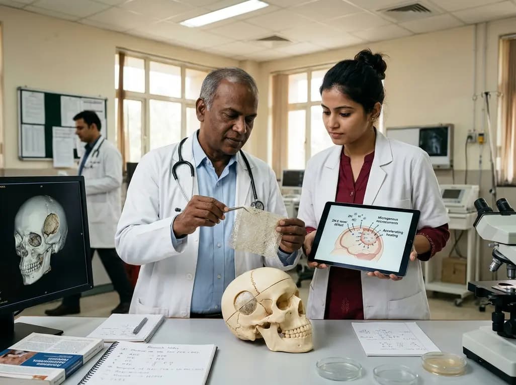
Researchers have engineered a self-powered Zn/C micro-galvanic electrospun membrane that couples sustained zinc release with endogenous microcurrents. This bioelectric scaffold stimulates M2 macrophage polarization and accelerates bone deposition to achieve robust cranial defect regeneration.
Today
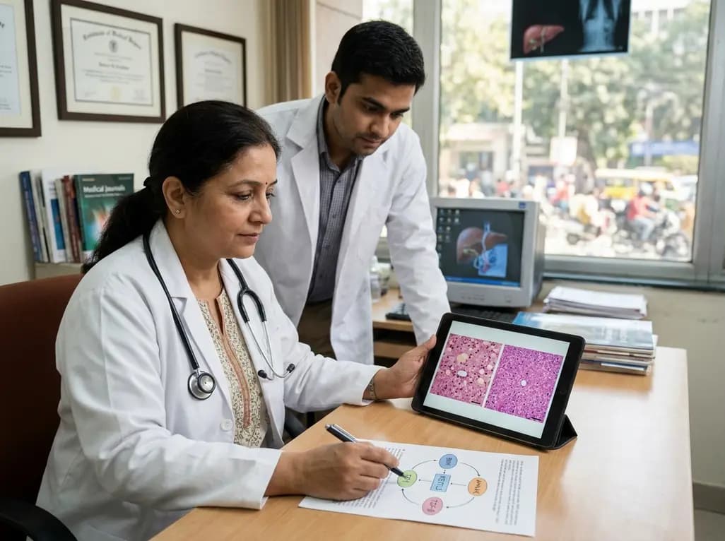
Preclinical research demonstrates that BMI1 overexpression alleviates metabolic syndrome-related liver injury. By triggering ubiquitin-mediated METTL3 degradation, BMI1 prevents AHNAK m6A methylation and suppresses fibrogenic TGF-β/SMAD signaling, significantly improving hepatic steatosis and insulin resistance.
Today
