.webp&w=384&q=75)

Loading, please wait...
Loading, please wait...
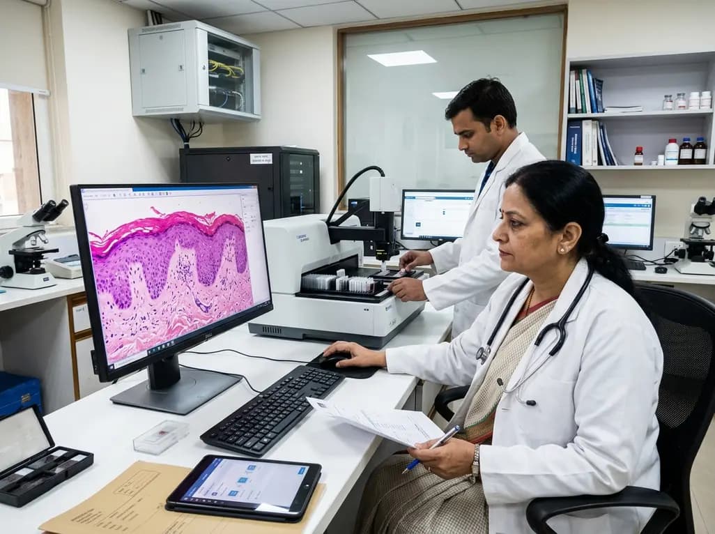
Transitioning from traditional light microscopy to whole slide imaging marks a transformative evolution in cutaneous pathology. Establishing an efficient digital dermatopathology workflow requires a comprehensive understanding of hardware capabilities, software integration, and laboratory logistics. While glass slide interpretation has anchored histopathologic diagnosis for decades, modern clinical demands require agile teleconsultation, rapid turnaround, and scalable data storage. Consequently, dermatopathology practices must redesign operational pathways from initial tissue accessioning to final electronic sign-out. This operational guide explores the fundamental pillars required to execute a dependable digital workflow. Practitioners will gain actionable insights into selecting central management platforms, designing scanning operations, maintaining diagnostic concordance, and fulfilling stringent regulatory requirements.
The image management system functions as the technological epicenter of the digital laboratory. This software environment connects physical whole slide scanners with the primary laboratory information system. Therefore, a robust image platform must seamlessly synchronize case metadata, clinical histories, gross pathology photographs, and high-resolution whole slide images. When pathologists open a requisition, the software automatically loads corresponding slides without manual barcode entry.
Furthermore, modern platforms incorporate bidirectional communication interfaces that automate diagnostic worklists and status updates. Pathologists can annotate microscopic lesions, order deeper levels, and request immunohistochemical stains directly within the image viewer. In addition, the image management system serves as the foundational launchpad for advanced artificial intelligence algorithms. These computational tools assist clinicians with epidermal measurement, margin assessment, and mitotic figure identification. However, institutions must ensure that the software architecture supports fast pan-and-zoom functionality and low latency. Clinicians rapidly lose diagnostic efficiency when digital slides exhibit loading delays. Implementing scalable vendor-neutral platforms also prevents vendor lock-in, enabling laboratories to integrate varied scanner hardware over time. Ultimately, an intuitive, responsive interface drives clinician adoption and protects diagnostic throughput across diverse clinical environments.
Selecting the appropriate scanning model represents a pivotal decision in operational design. Laboratories typically choose between inline scanning and retrospective batch scanning workflows. Inline scanning digitizes glass slides immediately after coverslipping and microtome sectioning. Consequently, pathologists receive digital cases concurrently with physical slide completion, which minimizes turnaround delays for urgent skin biopsies.
Moreover, inline workflows streamline routine operations because histotechnologists detect scanning errors, tissue folds, and out-of-focus areas before distributing cases. This immediate feedback loop reduces re-scan delays and prevents diagnostic interruptions for the signing pathologist. However, inline processing requires dedicated scanning personnel throughout the workday, alongside high-throughput scanners capable of rapid continuous loading. In contrast, retrospective scanning models digitize slides after initial glass review or during overnight laboratory shifts. Academic institutions and commercial laboratories often adopt this strategy when establishing archival repositories or preparing consultation sets. While retrospective scanning decreases daytime hardware bottlenecks, it provides fewer real-time efficiencies for routine primary sign-out. Therefore, high-volume cutaneous pathology centers increasingly favor inline models to maximize rapid clinical turnaround. Clinicians must evaluate biopsy volume, staffing resources, and physical lab space before committing to a specific scanning paradigm.
Before deploying whole slide imaging for primary cutaneous diagnosis, laboratories must complete rigorous clinical validation. Standardized validation protocols confirm that digital slide interpretations demonstrate diagnostic concordance with traditional light microscopy. According to professional accreditation guidelines, validation sets should encompass at least sixty representative routine cases spanning inflammatory skin conditions, melanocytic neoplasms, and epithelial malignancies.
Additionally, validation studies require a washout period of at least two weeks between glass and digital slide evaluations. This mandatory separation prevents recall bias and ensures true intraobserver concordance assessments. Pathologists must evaluate all diagnostic parameters, including architectural patterns, nuclear atypia, subtle solar elastosis, and melanin pigment distribution. Any discordant diagnoses require formal categorization into major or minor clinical variances. Furthermore, validation must encompass the complete digital ecosystem rather than isolated hardware units. Clinicians must validate specific monitor resolutions, network bandwidths, and remote viewing environments. When laboratories introduce new scanner models, updated image management software, or novel staining techniques, technical teams must perform targeted re-validation. Maintaining detailed documentation of these validation trials guarantees clinical safety, satisfies accreditation inspectors, and builds diagnostic confidence across the pathology team.
Implementing digital pathology requires substantial capital expenditure alongside ongoing operational funding. Upfront investments include high-throughput slide scanners, enterprise image servers, high-resolution diagnostic monitors, and software licenses. Additionally, laboratories must budget for increased network bandwidth and secure cloud or on-premises data storage. High-resolution whole slide images consume substantial storage capacity, generating several gigabytes per complex biopsy case.
However, organizations realize meaningful return on investment through tangible operational efficiencies and long-term cost offsets. Digital platforms eliminate physical slide courier expenses, reduce lost slide incidents, and accelerate internal specialist consultations. Pathologists can review complex melanocytic lesions remotely across regional hospital networks without mailing physical glass trays. Consequently, overall turnaround times decrease, improving patient satisfaction and physician collaboration. Furthermore, digitized archives eliminate off-site physical storage lease costs and retrieve historical biopsies instantaneously. Laboratories can also monetize specialized computational pathology tools and digital second-opinion services. To maintain financial sustainability, healthcare institutions should adopt tiered storage architectures, transitioning older cases to cold archive tiers. Strategic economic planning ensures that the long-term clinical benefits comfortably justify the initial capital investments.
Digital dermatopathology operations must adhere strictly to established clinical quality assurance and regulatory frameworks. In the United States, regulatory oversight involves compliance with the Clinical Laboratory Improvement Amendments, College of American Pathologists standards, and Food and Drug Administration clearances. Accreditation inspectors specifically audit patient identification integrity, ensuring that barcode metadata matches slide labels perfectly across all software interfaces.
Moreover, laboratories must enforce robust quality control protocols for every digitized specimen. Histotechnologists must inspect digital images for out-of-focus fields, scanning lines, tissue folding, and missed tissue fragments. Establishing daily calibration procedures for scanner optics and monitor color profiles maintains uniform image fidelity. Laboratories must also document standard operating procedures for system downtime, data recovery, and cybersecurity safeguards. Additionally, regulatory bodies require defined competency assessments for all participating pathologists and technical staff. Clinicians must demonstrate proficiency in digital navigation, measurement calibrations, and viewing software functions prior to primary sign-out. When laboratories utilize mix-and-match configurations combining non-bundled scanners, viewers, and displays, local medical directors assume responsibility for end-to-end system validation. Adhering to these rigorous quality frameworks ensures unwavering diagnostic reliability and patient safety.
An Image Management System integrates with laboratory information systems using standardized communication protocols such as HL7 and DICOM. This bidirectional connection synchronizes patient demographic data, specimen accession numbers, and clinical requisitions directly with whole slide images. Pathologists view organized case folders without re-entering identifiers, minimizing manual clerical errors. Furthermore, when pathologists complete their digital diagnostic reports, the integrated system automatically updates case sign-out status across hospital records.
Inline scanning digitizes glass slides immediately after coverslipping, allowing technical staff to resolve quality defects before case distribution. This approach supports rapid primary sign-out and minimizes diagnostic turnaround times for acute clinical biopsies. Conversely, retrospective scanning digitizes slides after light microscopic review or during off-peak hours. While retrospective scanning avoids daytime hardware bottlenecks, it serves primarily for archival storage, educational repositories, and secondary remote consultations rather than immediate clinical workflows.
Laboratories validate whole slide imaging by evaluating at least sixty routine pathology cases covering diverse tissue types and disease spectra. Pathologists review glass and digital slides separated by a mandatory two-week washout interval to prevent recall bias. The validation confirms high intraobserver concordance across architectural and cytological features. Technical teams must document diagnostic concordance rates, investigate minor or major variances, and validate viewing monitors and software environments thoroughly before clinical sign-out.
Disclaimer: This content is for informational and educational purposes only and does not constitute medical or regulatory advice. Digital pathology systems and diagnostic workflows require formal validation according to institutional, regional, and national accreditation standards. Refer to the latest local and national guidelines for clinical practice.
References

Read summarized clinical updates, watch expert medical content, and earn CME certifications right from your smartphone.
Open your smartphone camera to scan and install.





A comprehensive playbook on navigating the digital dermatopathology workflow from slide scanning and image management system integration to validation and regulatory compliance.
last month
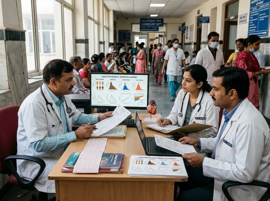
Atherosclerotic cardiovascular disease guidelines often rely on European-derived lipoprotein(a) cutoffs. However, baseline Lp(a) levels vary widely across ancestries, including South Asians and Africans. Clinicians must interpret Lp(a) values within overall absolute cardiovascular risk.
Today

Maternal anaemia contributes to nearly half of pregnancy-related fatalities across India. To prevent complications, Uttar Pradesh has added single-dose parenteral iron to its essential medicines list. This clinical transition aims to restore haemoglobin rapidly and protect high-risk mothers across rural facilities.
Yesterday
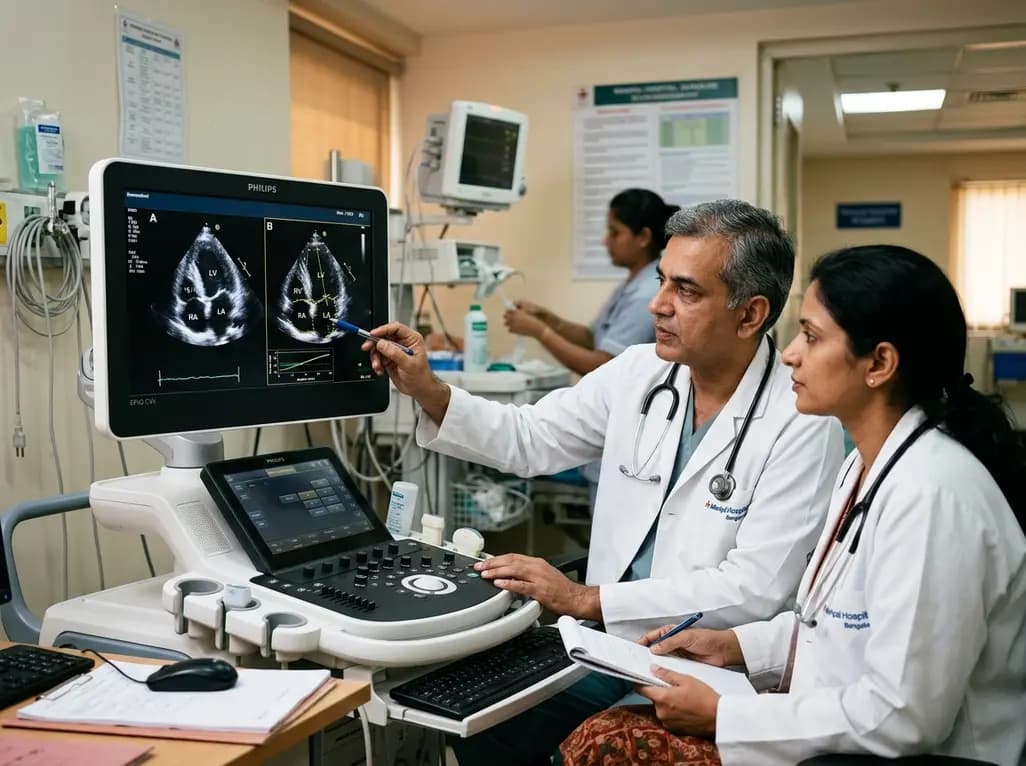
A multi-expert study shows pairwise deep learning accurately assesses echocardiographic rotational alignment in apical four-chamber views, outperforming pointwise regression. This technique improves automated image quality control and standardizes functional cardiac assessment.
Today

A longitudinal study of 1,055 chronic low back pain patients reveals that higher patient activation fosters constructive pain medication attitudes, reduces disability, and decreases short- and long-term opioid use through collaborative, multimodal care.
Today

A landmark cross-sectional study in Asian adults with type 2 diabetes identifies novel urinary metabolites linked to mild cognitive impairment. Elevated urinary sucrose and altered metabolites highlight non-invasive pathways for early cognitive risk stratification and targeted metabolic management.
Yesterday
