

Loading, please wait...
Loading, please wait...

In modern stereotactic radiosurgery, CyberKnife quality assurance serves as a critical pillar for safe patient care. Delivering ablative radiation doses demands rigorous geometric fidelity to protect surrounding critical structures. Historically, radiation oncologists and medical physicists have relied on meticulous verification protocols to confirm spatial alignment. Among these periodic tests, laser-radiation field coincidence remains essential because clinicians rely on visual lasers during patient setup and positional reference verification. However, tiny mechanical misalignments can introduce spatial uncertainties that compromise therapeutic margins. Consequently, medical physics teams must establish reliable routines to verify beam geometry with submillimeter accuracy. Platforms like the CyberKnife M6 deliver extremely steep dose gradients around intracranial lesions and spinal targets. Therefore, any undiscovered geometric shift between the optical guidance lasers and the therapeutic radiation beam can result in geographical miss or normal-tissue complications. Regulatory standards and professional physics guidelines demand strict compliance with submillimeter tolerances. As clinical workloads expand across radiation therapy centers, departments face increasing pressure to execute these daily and monthly checks rapidly. Thus, investigating faster, highly reproducible calibration approaches represents an urgent clinical priority.
Traditionally, medical physicists use radiochromic film to verify laser and radiation field coincidence. In typical practice, the physicist positions a radiographic film at the nominal calibration distance and manually marks laser crosshair intersections with a fine pen. Next, the robotic arm delivers a radiation beam through selected collimators onto the film. Although radiochromic film offers superior spatial resolution, the overall process entails notable practical drawbacks. Manual pen marking inherently introduces subjective human error and inter-observer variability. Furthermore, film processing requires substantial post-exposure waiting intervals to allow complete dye polymerization before scanning. Medical physicists must then digitize the exposed films and carry out image processing steps. Consequently, film-based verification consumes considerable clinical resources and creates significant workflow delays. Additionally, recurring expenditures on radiochromic films increase departmental operational costs over time. In busy radiation therapy facilities, these time-intensive steps often restrict testing frequency. Therefore, while radiochromic film historically served as the gold standard, its reliance on manual handling hinders daily implementation. Radiation oncology teams consequently seek robust digital alternatives that eliminate manual marking errors and deliver immediate analytical feedback.
To address the operational challenges of film, researchers recently introduced a fully digitized acquisition and evaluation pipeline. This innovative workflow integrates a specialized multileaf collimator quality assurance tool with the QA StereoChecker digital imaging platform. Instead of relying on manual pen marks, the protocol utilizes the central metal fiducial marker to define the laser reference position objectively. Subsequently, the QA StereoChecker flat-panel sensor captures high-resolution digital images directly under beam irradiation. The acquired images transfer immediately into automated analytical software known as CKQA for rapid geometric calculation. This setup entirely bypasses chemical or post-irradiation film development. Moreover, automated beam analysis algorithms instantly identify both the radiation field center and the reference marker location. As a result, the digital platform extracts beam profile parameters and calculates coordinate offsets within seconds. Clinicians can test multiple fixed collimators, ranging from five millimeters to forty millimeters, in a single efficient session. Furthermore, the standardized mounting mechanism guarantees consistent positioning between successive measurements. By combining reproducible hardware referencing with automated digital image acquisition, this approach establishes a standardized, filmless framework for routine validation.
A comprehensive experimental investigation recently compared this digital framework against three clinically realistic film-based configurations. The researchers designated manual film marking as method a and collimator tool marker film referencing as method b. Meanwhile, method c employed the fully digital QA StereoChecker workflow, and method d utilized film placed directly on the digital detector assembly. Across all ten collimator sizes, every tested protocol successfully maintained mean spatial deviations within submillimeter limits. However, the experimental results revealed striking performance hierarchies among the approaches. The mean deviations ranked consistently in descending order from method a to method b, followed by method d, and finally method c. Specifically, the fully digital workflow achieved the highest precision, yielding a mean deviation of only 0.116 millimeters and a root mean square error of 0.155 millimeters. In contrast, manual film marking exhibited the greatest geometric variability and largest positional deviations. Dataset comparisons confirmed statistically significant differences between manual marking and the standardized digital approach. Additionally, within the digital datasets, the automated CKQA software produced significantly lower deviation metrics than manual ImageJ processing.
These clinical findings provide compelling evidence for updating routine quality control practices in stereotactic radiosurgery centers. Transitioning to a digital, software-driven workflow significantly elevates quality assurance efficiency while strengthening treatment safety. Because the digital pipeline removes manual drafting and film development, physics staff can complete routine coincidence evaluations rapidly. Consequently, radiotherapy facilities can transition from burdensome monthly checks to frequent, highly reproducible quality assessments. Furthermore, automated software eliminates individual subjective bias during center determination, ensuring uniform geometric tracking across longitudinal datasets. In developing regions, such as oncology facilities across India where patient volumes remain exceptionally high, streamlined digital quality assurance directly translates into increased machine uptime. Linear accelerators and robotic radiosurgery platforms can spend less non-productive time in offline testing and more time delivering vital cancer treatments. In addition, the consistent submillimeter precision reassures radiation oncologists when delivering single-fraction radiosurgery to critical cranial and extracranial targets. Ultimately, embracing automated digital verification frameworks strengthens radiation protection, minimizes human calculation errors, and reinforces clinical precision.
The digital workflow significantly enhances stereotactic quality assurance by substituting physical film with a high-resolution electronic detector array. This technological transition eliminates lengthy chemical or optical development intervals, cuts recurring film expenditure, and prevents subjective manual marking errors. In addition, automated software immediately processes the captured beam images. Consequently, medical physicists obtain precise, submillimeter spatial measurements in minutes, enabling departments to perform routine geometric checks far more frequently and reliably.
Stereotactic radiosurgery delivers ablative, highly concentrated radiation doses to target volumes adjacent to radiosensitive organs. Clinicians frequently use positioning lasers to confirm patient alignment before initiating beam delivery. If the optical laser crosshairs deviate from the actual therapeutic radiation field center, the robotic system risks underdosing tumor margins or delivering unintended high radiation to normal tissues. Therefore, maintaining strict submillimeter coincidence ensures that visual positioning references faithfully reflect true radiation delivery geometry.
Automated analysis algorithms like CKQA utilize standardized mathematical routines to detect beam edges, radiation centers, and reference fiducials without human intervention. In contrast, manual tools like ImageJ require individual operators to define profile lines and select threshold levels manually, introducing inter-observer variation. Clinical testing demonstrates that CKQA yields significantly lower spatial deviation and root mean square error values than manual tools, guaranteeing greater measurement reproducibility across routine linear accelerator testing cycles.
Disclaimer: This content is for informational and educational purposes only... Refer to the latest local and national guidelines for clinical practice.
References
Hu L et al. Implementation and preliminary evaluation of a practical digital workflow for routine laser-radiation field coincidence quality assurance on the CyberKnife M6. Precis Radiat Oncol. 2026 Oct 24. doi: 10.1002/pro6.70103. PMID: 42927071.
Dieterich S, Cavedon C, Chuang CF, et al. Report of AAPM TG 135: Quality assurance for robotic radiosurgery. Med Phys. 2011;38(6):2914-2936.
Schmidhalter D, Henzen D, Malthaner M, Fix MK, Manser P. Evaluation of a digital detector array for daily quality assurance of a robotic radiosurgery system. J Appl Clin Med Phys. 2017;18(4):114-123.

Read summarized clinical updates, watch expert medical content, and earn CME certifications right from your smartphone.
Open your smartphone camera to scan and install.





A novel digital CyberKnife quality assurance workflow combining the QA StereoChecker and automated CKQA software achieves submillimeter laser-radiation field coincidence precision of 0.116 mm, outperforming conventional film-based techniques and streamlining routine medical physics verification.
Today
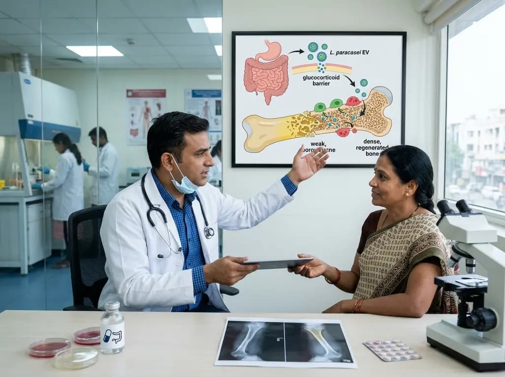
A novel oral nanoplatform using Lacticaseibacillus paracasei-derived extracellular vesicles delivers alendronate, reversing glucocorticoid-induced bone loss and restoring gut-immune-bone homeostasis.
Today
A rare presentation of cervical villoglandular adenocarcinoma metastasizing to the ovary mimicked a borderline ovarian tumor. Multimodal testing with immunohistochemistry and HPV16 DNA confirmed metastatic origin, guiding optimal surgical and chemoradiotherapy intervention.
Today

A landmark study details how an enterprise pediatric remote patient management model expanded from a single cardiology pilot to 16 diverse specialty programs by combining centralized digital operations with specialty clinical leadership.
Yesterday

A comparative review of tension band wiring and intramedullary screw fixation in thumb metacarpophalangeal arthrodesis, evaluating union rates, hardware complications, and clinical outcomes.
Yesterday
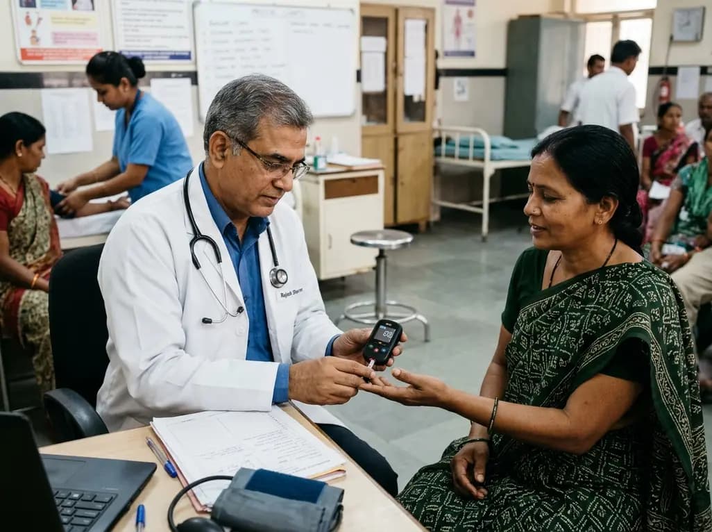
A real-world trial evaluating a near-infrared spectroscopy noninvasive glucose monitoring device in adults with type 2 diabetes demonstrated a combined MAD/MARD of 16.3 across 2,616 paired readings, highlighting its clinical feasibility.
Today
