

Loading, please wait...
Loading, please wait...

Chronic diabetes mellitus frequently provokes severe complications across the musculoskeletal framework. Clinicians increasingly recognize diabetic musculoskeletal degeneration as a debilitating comorbidity characterized by skeletal muscle atrophy, bone mineral loss, and persistent physical frailty. Consequently, prolonged exposure to glucolipotoxicity drives profound metabolic stress within peripheral muscle tissue. High circulating glucose levels and lipid overload impair tissue microcirculation and induce premature cellular senescence. Therefore, muscle satellite cells gradually lose their endogenous regenerative capacity over time. In addition, chronic low-grade inflammation accelerates the degradation of structural myofibrillar proteins. Patients subsequently experience diminished muscle mass, declining contractile strength, and reduced functional independence. Standard glycemic management often fails to reverse this established myogenic deficit. Furthermore, conventional pharmacological therapies cannot repair cellular exhaustion in chronically damaged satellite cells. Pathologists observe significant disruptions in muscle architectural integrity and associated bone mineralization. Accordingly, this coupled deterioration underscores the necessity for comprehensive musculoskeletal protection. Researchers now emphasize therapeutic approaches that simultaneously arrest metabolic damage and stimulate endogenous tissue renewal. Therefore, understanding the intricate interplay between cellular senescence, lipotoxicity, and regenerative arrest is vital for developing effective clinical interventions.
Recent investigations reveal that ferroptosis acts as a principal driver of myocyte depletion during metabolic stress. Ferroptosis represents an iron-dependent, non-apoptotic form of programmed cell death driven by toxic lipid peroxidation. In glucolipotoxic environments, intracellular iron homeostasis encounters severe deregulation. Consequently, unbound ferrous ions catalyze the generation of lethal reactive oxygen species via Fenton chemistry. In addition, chronic nutrient surplus rapidly depletes intracellular glutathione pools and inactivates glutathione peroxidase 4. As a result, damaged myoblasts undergo extensive membrane lipid peroxidative rupture. Moreover, this persistent ferroptotic cascade severely impairs muscle progenitor viability and blunts myogenic differentiation. Traditional antioxidants provide only transient relief because they cannot halt underlying iron-mediated lipid cascades. Similarly, synthetic ferroptosis inhibitors fail to re-establish myogenic regenerative competency on their own. Thus, single-target interventions remain inadequate for comprehensive tissue restoration. Chronic cellular loss additionally promotes local macrophage infiltration and pro-inflammatory polarization. Subsequently, inflamed M1 macrophages secrete destructive cytokines that further amplify tissue devastation. Therefore, interrupting ferroptosis while concurrently restoring the regenerative microenvironment represents an essential therapeutic objective. Coordinated bioengineering strategies provide a robust foundation for counteracting progressive metabolic sarcopenia in vulnerable diabetic populations.
To overcome these biochemical challenges, bioengineers designed a sophisticated cellular manufacturing platform. Mesenchymal stem cells derived from bone marrow demonstrate remarkable secretome plasticity under specific environmental cues. Specifically, investigators integrated the stem cell screen-enrich-combine biomaterials circulating system, known as SECCS. This advanced three-dimensional culture platform incorporates porous beta-tricalcium phosphate granules and intermittent media circulation. Furthermore, researchers conditioned the stem cells with fullerol, a potent water-soluble polyhydroxylated fullerene antioxidant. Nano-priming with fullerol profoundly enhances the cytoprotective payload within secreted extracellular vesicles. Meanwhile, the dynamic three-dimensional bioreactor mimics native physiological fluid dynamics and mechanical forces. Consequently, fullerol-primed SECCS extracellular vesicles, termed F-SECCS-EVs, acquire unique biomolecular packaging. In comparative assessments, standard two-dimensional vesicles and unprimed vesicles exhibited limited biological efficacy. In contrast, F-SECCS-EVs delivered superior bioavailability and markedly elevated membrane cargo stability. Furthermore, these primed vesicles demonstrated powerful tropism toward stressed myoblasts and skeletal tissues. Thus, multimodal bioengineering effectively transforms conventional stem cell vesicles into targeted nanotherapeutic agents. Such innovations bridge the critical gap between raw biomaterials and effective cell-free clinical therapeutics for metabolic disorders. Accordingly, this bioengineering breakthrough provides scalable manufacturing options for regenerative medicine.
Comprehensive transcriptomic analyses revealed the precise molecular mechanisms governing F-SECCS-EV therapeutic actions. Notably, researchers identified the forkhead box O3 transcription factor, FOXO3, as a pivotal mediator. In glucolipotoxicity-challenged myoblasts, these engineered vesicles robustly stimulate FOXO3 signaling pathways. Consequently, activated FOXO3 coordinates cellular antioxidant defenses while simultaneously regulating myogenic transcription networks. Furthermore, elevated FOXO3 expression upregulates systemic detoxifying enzymes and stabilizes cellular iron balance. As a result, treated myoblasts successfully withstand severe oxidative challenges and suppress ferroptotic death. Moreover, the vesicles restore crucial myogenic markers, including MyoD and myogenin, within impaired satellite cells. This functional rescue enhances progenitor cell proliferation and promotes mature multinucleated myotube fusion. In addition, downstream signaling actively modulates the local immune microenvironment. Specifically, the vesicles reprogram pro-inflammatory M1 macrophages toward a pro-regenerative M2 phenotype. Thus, coordinated immune modulation mitigates chronic inflammation while facilitating structural matrix repair. Simultaneously, renewed satellite cells initiate rapid muscle fiber reconstruction without triggering aberrant fibrotic responses. Collectively, these molecular events establish a self-reinforcing regenerative cycle in metabolically stressed muscle beds.
In vivo preclinical evaluations demonstrated substantial therapeutic benefits in diabetic murine models. Specifically, systemic administration of F-SECCS-EVs produced dramatic functional and structural recoveries. Animals receiving these engineered vesicles exhibited enhanced motor coordination, increased running endurance, and superior limb grip strength. Furthermore, histological analysis confirmed a marked expansion of skeletal myofiber cross-sectional area. In contrast, free fullerol or conventional two-dimensional vesicles produced only negligible functional recovery. Importantly, the therapeutic benefits extended beyond skeletal muscle tissue into adjacent skeletal structures. Treated animals exhibited favorable microarchitectural remodeling and preserved bone mineral density within the tibia. Consequently, this dual osteomyogenic preservation highlights the holistic power of stem cell-derived vesicular therapy. For practicing clinicians, these findings highlight a promising cell-free paradigm for diabetic frailty. In addition, cell-free vesicles circumvent the tumorigenic and immunogenic risks inherent to live stem cell transplantation. Therefore, bioengineered extracellular vesicle therapy represents a viable future strategy for complex musculoskeletal complications. Moreover, standardized production protocols will accelerate phase-one clinical investigations in metabolic medicine. Continued clinical translation may soon transform the therapeutic outlook for debilitating diabetic sarcopenia across healthcare systems.
Diabetic muscle wasting stems from sustained glucolipotoxicity, chronic low-grade inflammation, and accelerated microvascular impairment. Elevated circulating glucose and free fatty acids overwhelm cellular defenses and trigger catastrophic lipid peroxidation. Consequently, satellite cells suffer ferroptotic programmed cell death and functional exhaustion. Furthermore, impaired insulin signaling suppresses essential protein synthesis while elevating muscle proteolysis. These converging pathophysiological disturbances progressively decrease skeletal muscle cross-sectional area, compromise physical endurance, and diminish overall contractile force.
Fullerol is a water-soluble polyhydroxylated carbon nanoparticle exhibiting exceptional free-radical scavenging capacity. Priming bone marrow stem cells with fullerol under dynamic three-dimensional culture optimizes vesicular cargo packaging. Consequently, the resulting extracellular vesicles carry enriched antioxidant mediators, protective microRNAs, and regulatory proteins. Moreover, these engineered vesicles demonstrate enhanced biophysical stability and targeted uptake by recipient cells. They effectively neutralize toxic lipid radicals, shield vulnerable organelles, and promote sustained tissue restoration.
Ferroptosis represents a unique iron-dependent cell death mechanism that directly destroys lipid membranes in stressed myocytes. Chronic metabolic disorders generate high oxidative stress, rendering muscle progenitor cells exceptionally vulnerable to ferroptotic injury. Standard anti-inflammatory therapies cannot counteract this iron-catalyzed lipid degradation. Therefore, directly suppressing ferroptosis preserves satellite cell populations and prevents catastrophic cellular attrition. Once ferroptosis is controlled, progenitor cells regain their intrinsic capacity to proliferate, differentiate, and regenerate damaged skeletal myofibers.
Disclaimer: This content is for informational and educational purposes only and does not constitute medical advice. Refer to the latest local and national guidelines for clinical practice.
References
Wang Z et al. Multimodal Programming of Extracellular Vesicles via Nano-Primed SECCS-Based 3D Microenvironments for Coordinated Ferroptosis Suppression and Myogenic Regeneration in Diabetes. Adv Healthc Mater. 2026 Oct 18. doi: 10.1002/adhm.71868. PMID: 42893935.
Zhang H et al. Mesenchymal stem cell-derived extracellular vesicles for disease therapy by regulating ferroptosis: focus on diabetes mellitus and diabetic complications. Stem Cell Res Ther. 2025;16(1):412.
Chen Y et al. Fullerol Attenuates Diabetic Muscle Atrophy and Ferroptosis-Associated Injury: Intervention Timing Influences Myogenesis. Antioxidants (Basel). 2026;15(10):1245.

Read summarized clinical updates, watch expert medical content, and earn CME certifications right from your smartphone.
Open your smartphone camera to scan and install.





A breakthrough study demonstrates that fullerol-primed extracellular vesicles generated in a 3D SECCS bioreactor attenuate ferroptosis and restore myogenic regeneration in diabetes via the FOXO3 pathway, offering a cell-free therapeutic strategy for diabetic musculoskeletal degeneration.
Today

A recent maternal mortality case in Kota has drawn national scrutiny toward operational deficits in emergency transport networks. With low ambulance utilization among high-risk pregnant women, delayed inter-facility transfers, and preventable clinical complications, urgent systemic reforms are vital for patient safety.
Today

A study reveals that the MYLK c.4079G>A, p.(Gly1360Asp) variant is linked to high-penetrance heritable thoracic aortic disease and early-onset aortic dissection, showing a founder effect in Finnish cohorts. Findings emphasize early cascade screening and vigilant surveillance.
Yesterday
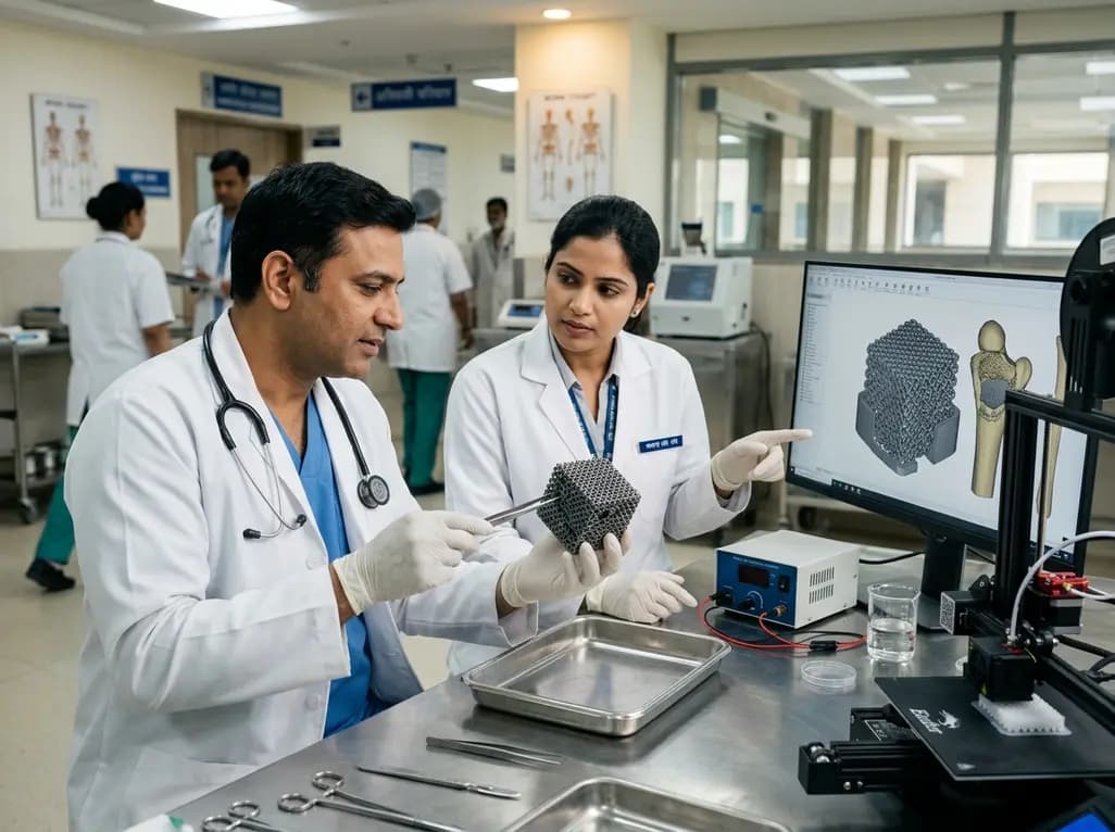
A novel sacrificial-template technique combines FDM polymer 3D printing and micro-arc oxidation to fabricate orientation-controlled porous titanium scaffolds. These bioactive constructs offer tailored stiffness, exceptional multidirectional load resistance, and robust osseointegration for bone defect repair.
Today
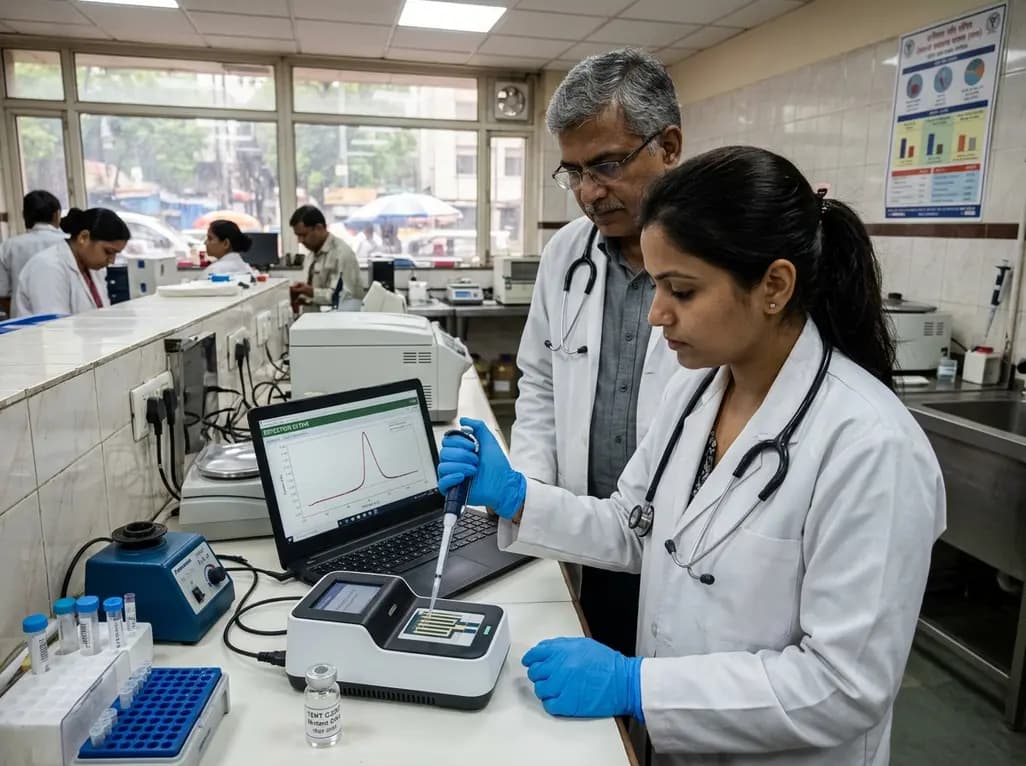
A novel label-free electrochemical biosensor utilizing magnetic nanocomposites enables ultrasensitive TERT C228T mutation detection (LOD: 0.67 pM) for rapid risk stratification in papillary thyroid cancer, offering promising point-of-care utility for endocrine oncology.
Today
