

Loading, please wait...
Loading, please wait...

Continuous hemodynamic tracking remains fundamental for managing critically ill patients presenting with cardiovascular instability. In conventional intensive care units, clinicians primarily rely on indwelling arterial catheters to guide inotrope administration and fluid resuscitation. Although indwelling arterial lines provide the clinical gold standard for real-time arterial tension tracking, they introduce significant iatrogenic hazards. Patients frequently experience severe complications, including catheter-associated bloodstream infections, local hematomas, vasospasm, and peripheral arterial thrombosis. Furthermore, arterial cannulation demands substantial clinical expertise and sterile maintenance, which substantially strains nursing resources in crowded tertiary facilities. Consequently, researchers actively seek noninvasive alternatives that provide comparable precision without compromising patient safety. Advanced cuffless blood pressure monitoring offers a revolutionary paradigm shift away from repeated invasive arterial punctures. Standard noninvasive oscillometric arm cuffs only deliver intermittent snapshot recordings. Unfortunately, intermittent cuff inflation cannot detect sudden hypotensive collapses, and frequent cycling causes tissue edema and petechial skin injury. Therefore, developing continuous, reliable, and sensor-based arterial pressure tracking addresses an urgent unmet requirement across acute care environments globally. By eliminating cannulation risks, bedside clinicians can preserve vascular access while maintaining vigilant surveillance over vulnerable patients. This transition supports proactive clinical decision-making during acute hemodynamic destabilization.
To overcome the limitations of invasive lines, investigators developed an innovative framework using single-sensor photoplethysmography data. Photoplethysmography optical sensors capture peripheral microvascular blood volume pulsations during each cardiac cycle. Traditionally, machine learning engineers extract hundreds of mathematical morphological indicators from arterial pulse waveforms. However, incorporating excessively complex feature matrices often causes algorithmic overfitting and requires unsustainable computational processing. In this prospective intensive care unit study, researchers recorded synchronized ten-minute continuous waveforms from one hundred critically ill patients. The team initially isolated 201 candidate digital pulse features. Subsequently, they deployed a hybrid feature selection strategy combining RReliefF algorithms with intrinsic model-specific rankings. This systematic mathematical reduction refined the candidate pool to an optimized subset of only thirty core features. Consequently, the team retained approximately fifteen percent of the initial feature inventory without degrading predictive fidelity. They subsequently trained multiple supervised regression architectures, including random forest, support vector regression, and extreme gradient boosting algorithms. Among these mathematical models, the extreme gradient boosting framework achieved an exceptional balance of speed, compactness, and analytical accuracy. Furthermore, this compact feature configuration drastically lowers processing latency, making real-time bedside execution feasible on resource-constrained monitoring devices. As a result, bedside systems can deliver instantaneous blood pressure feedback without demanding expensive dedicated graphics processing units.
Rigorous clinical validation remains mandatory before any artificial intelligence diagnostic technology enters bedside critical care practice. Therefore, the investigative team implemented patient-wise testing alongside nested cross-validation to prevent data leakage and evaluate real-world generalizability. Across heterogeneous intensive care cohorts, the thirty-feature extreme gradient boosting model demonstrated extraordinary fidelity. Specifically, the compact model achieved mean absolute errors of 8.61 mm Hg for systolic blood pressure, 3.50 mm Hg for mean arterial pressure, and 2.62 mm Hg for diastolic blood pressure. Moreover, the mean differences for mean arterial pressure and diastolic blood pressure reached minus 0.09 mm Hg and minus 1.07 mm Hg, respectively. These precise metric outputs comfortably met the strict numerical accuracy criteria established by the Association for the Advancement of Medical Instrumentation. Furthermore, the model achieved a prestigious Grade A evaluation under the British Hypertension Society international grading standards. While systolic estimation exhibited slightly wider statistical dispersion due to systemic arterial compliance changes, the algorithm maintained robust hemodynamic tracking across dynamic physiological fluctuations. Ultimately, these clinical outcomes confirm that reduced feature sets can replicate continuous arterial line precision while markedly reducing hardware requirements. In addition, meeting these stringent international benchmarks represents a major milestone toward formal clinical adoption in critical care units.
Many machine learning algorithms in critical care medicine suffer from the opaque black-box problem. Clinicians often hesitate to trust automated numerical recommendations when the underlying physiological mechanisms remain completely hidden. In contrast, this computational framework prioritizes mechanistic interpretability by directly linking mathematical parameters to human cardiovascular physiology. Feature importance analyses revealed distinct, clinically logical physiological clusters across systolic, diastolic, and mean arterial estimations. For example, diastolic and mean arterial estimations correlated strongly with wave reflection timing and peripheral arterial stiffness. Meanwhile, systolic estimates depended heavily on pulse arrival intervals, systolic ejection timing, and low-frequency vasomotor fluctuations. Additionally, autonomic regulatory variations registered through photoplethysmographic contour alterations mirrored sympathetic vascular tone shifts. By validating these biological associations, the investigators demonstrated that machine learning algorithms can mirror established hemodynamic principles rather than capturing spurious signal artifacts. Thus, transparent algorithmic reasoning builds vital clinician confidence, encouraging safe deployment across demanding intensive care environments. Furthermore, physiological interpretability enables intensivists to cross-examine algorithmic predictions against expected bedside pathophysiology during complex circulatory collapse scenarios. Consequently, healthcare providers can confidently differentiate between genuine hemodynamic deterioration and technical signal distortion.
The successful development of single-sensor optical hemodynamic estimation offers transformative potential for Indian healthcare infrastructure. In India, public and private intensive care units face high patient volumes alongside variable nurse-to-patient staffing ratios. Invasive arterial line placement requires continuous transducer calibration, expensive disposable pressure transducer kits, and vigilant catheter maintenance to prevent catastrophic nosocomial infections. Consequently, an accurate, single-sensor photoplethysmography platform can dramatically reduce equipment costs while protecting vulnerable patients from invasive vascular line sepsis. Furthermore, resource-constrained district hospitals and emergency departments can implement this noninvasive technology to monitor deteriorating patients during critical inter-facility transfers. Because standard pulse oximetry hardware exists universally across Indian wards, deploying lightweight machine learning software could democratize continuous hemodynamic surveillance beyond tier-one tertiary hospitals. Ultimately, integrating physiologically explainable artificial intelligence into everyday monitoring systems empowers Indian physicians to detect shock states earlier, initiate prompt therapeutic interventions, and substantially improve critical patient survival rates. In addition, simplifying hemodynamic monitoring workflows significantly alleviates administrative workloads for intensive care nursing teams. Therefore, this noninvasive optical approach presents a practical, scalable opportunity to elevate critical care monitoring standards across India.
Single-sensor photoplethysmography captures peripheral microvascular blood volume pulsations using light absorption changes across vascular tissue beds. An optimized machine learning algorithm extracts key morphological characteristics from each pulse wave, including systolic ejection duration, pulse reflection peaks, and vascular notch timing. By correlating these physiological wave dimensions with systemic vascular resistance and arterial compliance, the model accurately calculates continuous systolic, diastolic, and mean arterial pressures without requiring an inflated arm cuff.
While machine learning models provide remarkable noninvasive accuracy, they currently complement rather than fully replace indwelling arterial lines. Invasive catheters remain indispensable for frequent arterial blood gas sampling and immediate beat-to-beat titration during severe vasopressor-dependent shock. However, for hemodynamically stabilizing patients, intermediate critical care wards, and resource-limited settings, validated photoplethysmographic algorithms provide continuous, reliable surveillance while successfully eliminating dangerous catheter-related infections, bleeding risks, and thrombosis complications.
Explainable artificial intelligence allows clinicians to understand the physiological basis supporting algorithmic predictions instead of blindly relying on opaque black-box calculations. In acute critical care, intensivists must verify that arterial pressure estimates reflect genuine cardiovascular properties like arterial stiffness, vasomotor tone, and stroke volume timing. Understanding these biological relationships ensures clinical trust, helps identify potential sensor motion artifacts, and facilitates safe, reliable clinical decision-making during life-threatening patient emergencies.
Disclaimer: This content is for informational and educational purposes only... Refer to the latest local and national guidelines for clinical practice.
References

Read summarized clinical updates, watch expert medical content, and earn CME certifications right from your smartphone.
Open your smartphone camera to scan and install.





A prospective machine learning study demonstrates that an interpretable XGBoost model using 30 photoplethysmography features accurately estimates continuous blood pressure in intensive care units, achieving Grade A British Hypertension Society standards and reducing reliance on invasive arterial catheters.
Today

A qualitative study on the Dutch HPV vaccination catch-up campaign reveals that young adult hesitancy stems from information gaps and accessibility barriers rather than rampant misinformation. Clinicians can leverage these social listening insights to improve vaccine communication and address patient concerns.
Today
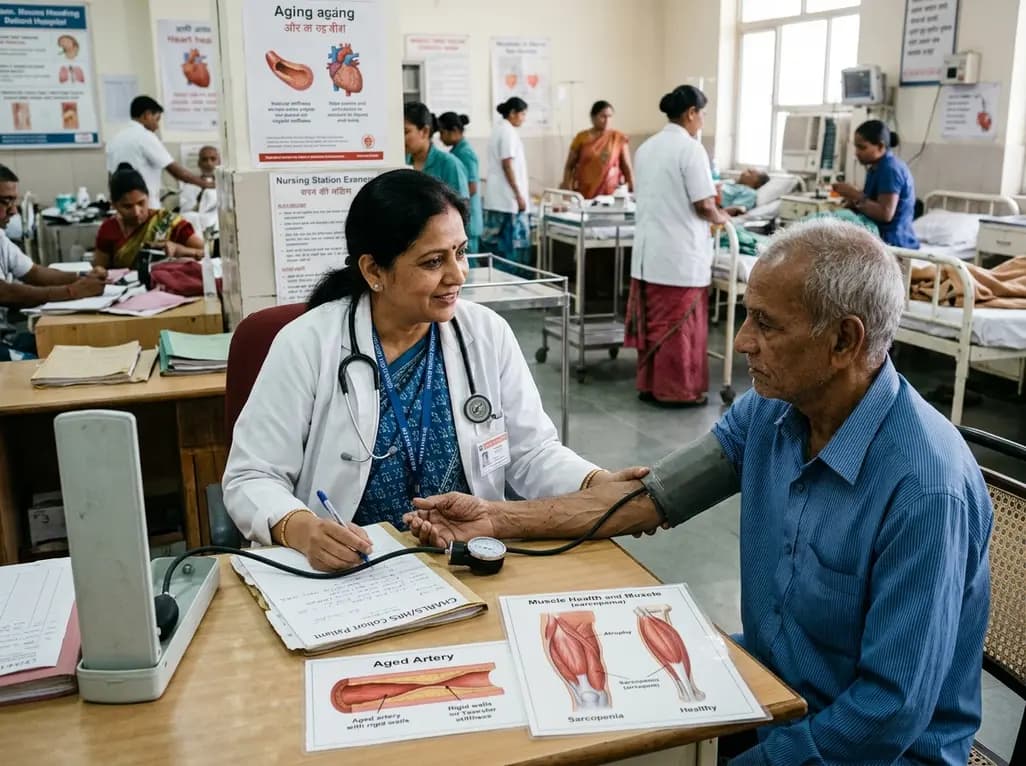
A multicenter longitudinal study of the CHARLS and HRS cohorts demonstrates that elevated cumulative pulse pressure and high-stable trajectories significantly increase incident sarcopenia risk in older adults, particularly in those with heart disease, linking arterial stiffness directly to progressive muscle loss.
Today

A 491-patient cohort study in Sudan reveals critical insights into war trauma reconstructive surgery, analyzing injury patterns, soft-tissue procedures, and a 25.5% complication rate in resource-limited conflict zones.
Today
A secondary analysis of the Living Healthier with Diabetes trial reveals that positive baseline technology attitudes significantly improve engagement with digital diabetes self-management apps, leading to enhanced self-care behaviors and better long-term glycemic control in adults with type 2 diabetes.
Today
