

Loading, please wait...
Loading, please wait...

Carotid artery disease remains a major cause of ischemic stroke worldwide. When clinicians choose carotid revascularization to restore cerebral perfusion, they must carefully evaluate plaque characteristics. Plaque composition significantly influences procedural success and long-term durability. While heavy calcification has long raised technical concerns, its precise morphological configuration matters most. Recent evidence indicates that luminal calcified nodules act as severe mechanical impediments during endovascular procedures. Consequently, clinicians require detailed preoperative computed tomography angiography evaluations to determine the safest therapeutic pathway for each patient.
Atherosclerotic calcification does not behave uniformly throughout the arterial tree. Historically, clinicians quantified calcification by volume or circumferential arc alone. However, geometric distribution within the vascular wall plays a decisive role during carotid revascularization. Intramural calcification situated along the outer wall preserves a compliant inner lumen. In contrast, calcified nodules that protrude directly into the arterial lumen create rigid, unyielding obstructions. These projecting structures disrupt laminar blood flow and elevate shear stress. Furthermore, they resist mechanical dilatation when interventionalists deploy endovascular devices. Stent struts cannot embed uniformly into dense mineralized projections. As a result, incomplete stent expansion and strut malapposition frequently follow. Therefore, identifying luminal nodular morphology on computed tomography angiography provides vital insights that overall calcium burden cannot capture.
A landmark investigation led by Hiroki Uchida and colleagues thoroughly examined this clinical dilemma. The investigators retrospectively analyzed 92 consecutive patients undergoing carotid revascularization with either carotid artery stenting or carotid endarterectomy. They categorized preoperative computed tomography angiography findings into three distinct anatomical patterns. Group 1 displayed no or spotty calcification, whereas Group 2 exhibited outer calcification without luminal disruption. Group 3 featured calcified nodules directly abutting the contrast-enhanced vascular lumen. The researchers defined a primary composite endpoint comprising technical failure, perioperative ischemic complications, or post-procedural restenosis. Notably, the composite endpoint occurred in only 4.8% of patients in Groups 1 and 2 combined. In sharp contrast, 55.6% of patients in Group 3 developed the adverse endpoint, demonstrating a striking difference between smooth and nodular morphologies.
The study demonstrated an exceptionally high hazard when operators treated calcified nodules with carotid artery stenting. Among patients undergoing stenting, those with smooth or outer calcification experienced complications in only 7.9% of cases. Conversely, all five stenting patients displaying the nodular pattern reached the adverse composite endpoint, yielding a 100% failure rate. When self-expanding stents encounter non-compliant mineral masses, the metal scaffolds cannot achieve adequate radial expansion. In addition, aggressive post-dilatation against calcified nodules risks arterial rupture or plaque fragmentation. Microscopic debris can bypass distal protection filters, triggering cerebral microemboli and neurological deficits. Furthermore, malapposed struts serve as niduses for platelet aggregation and accelerated in-stent restenosis. Consequently, endovascular approaches carry substantial risks when rigid nodules protrude directly into the lumen.
In multivariable regression modeling controlling for baseline risks, the luminal nodular configuration remained an independent predictor of failure, with an adjusted odds ratio of 49.05. Meanwhile, carotid endarterectomy demonstrated a markedly protective profile, reducing adverse events with an adjusted odds ratio of 0.045. Open surgical endarterectomy allows direct mechanical en bloc excision of the atherosclerotic core. During endarterectomy, the operating surgeon carefully identifies the subintimal plane and extracts both the lipid debris and rigid calcium nodules together. Thus, surgical resection leaves behind an open, smooth arterial bed free of metallic scaffolding. Because the surgeon removes the mineral protrusion completely, hemodynamic turbulence resolves immediately. These findings clearly indicate that surgical endarterectomy provides superior procedural reliability and patient safety when challenging calcified nodules exist.
These findings provide clear guidance for multidisciplinary neurovascular teams managing complex carotid stenosis. Before selecting carotid revascularization, clinicians must scrutinize thin-slice computed tomography angiography images in multiplanar reformatted views. Radiologists and neurointerventionalists should systematically report whether calcification lies on the outer wall or projects into the lumen. If cross-sectional imaging reveals an intraluminal calcified nodule, clinicians should favor carotid endarterectomy whenever the patient can safely tolerate open surgery. If prohibitive anatomical or medical risks mandate stenting, the team must anticipate severe technical hurdles. In such scenarios, interventionalists should evaluate advanced plaque modification tools or specialized stent architectures. In conclusion, integrating morphologic assessment into routine preoperative workflows improves case selection, avoids avoidable complications, and optimizes stroke prevention.
A calcified nodule on computed tomography angiography represents a distinct, heavily mineralized atherosclerotic mass that projects inward and directly abuts the contrast-filled arterial lumen. Unlike outer wall calcification, these irregular luminal protrusions disrupt blood flow, resist mechanical dilation, and create significant friction during endovascular device delivery.
Carotid artery stenting frequently fails in these lesions because rigid calcium prevents full stent expansion and apposition. Aggressive balloon dilatation risks arterial dissection or plaque disruption. Furthermore, malapposed stent struts promote acute thrombus formation and accelerated neointimal hyperplasia, ultimately producing technical failure, embolic stroke, or recurrent restenosis.
Clinicians should strongly consider carotid endarterectomy whenever cross-sectional imaging demonstrates luminal calcified nodules in surgically eligible patients. Open surgical resection allows complete en bloc removal of calcified plaques and restores a clean vascular wall, thereby avoiding the severe stent-expansion failures and restenosis risks associated with endovascular stenting.
Disclaimer: This content is for informational and educational purposes only. It is not intended to provide medical advice, diagnosis, or treatment recommendations. Refer to the latest local and national guidelines for clinical practice.
References
Uchida H et al. CTA-defined calcified nodules as a predictor of adverse outcomes following carotid revascularization. Neurosurg Rev. 2026 Nov 10. doi: 10.1007/s10143-026-04565-7. PMID: 43003963.
Brott TG, Halperin JL, Abbara S, et al. 2011 ASA/ACCF/AHA/AANN/AANS/ACR/ASNR/CNS/SAIP/SCAI/SIR/SNIS/SVM/SVS guideline on the management of patients with extracranial carotid and vertebral artery disease. Circulation. 2011;124(4):e54-e130.
Bonati LH, Dobson J, Featherstone RL, et al. Long-term outcomes after stenting versus endarterectomy for treatment of carotid artery stenosis: the Carotid Open Stent Trial (ICST). Lancet Neurol. 2015;14(6):577-586.

Read summarized clinical updates, watch expert medical content, and earn CME certifications right from your smartphone.
Open your smartphone camera to scan and install.





A new study reveals that CTA-defined calcified nodules directly abutting the lumen dramatically increase complications and restenosis after carotid revascularization, particularly in carotid artery stenting, highlighting the protective advantage of carotid endarterectomy in high-risk anatomical phenotypes.
Today

Ambient digital scribes promise effortless medical documentation, but a critical sociotechnical commentary reveals that AI alters rather than eliminates charting work. Discover how shifting cognitive burdens, oversight risks, and workflow redesign impact clinical safety and healthcare practice.
Today
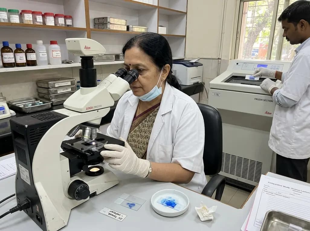
A new comparative study demonstrates that intraoperative imprint cytology achieves 96.6% diagnostic accuracy in ovarian tumours, outperforming frozen section in sensitivity for borderline lesions and offering an accessible, rapid alternative for resource-constrained surgical centres.
Today
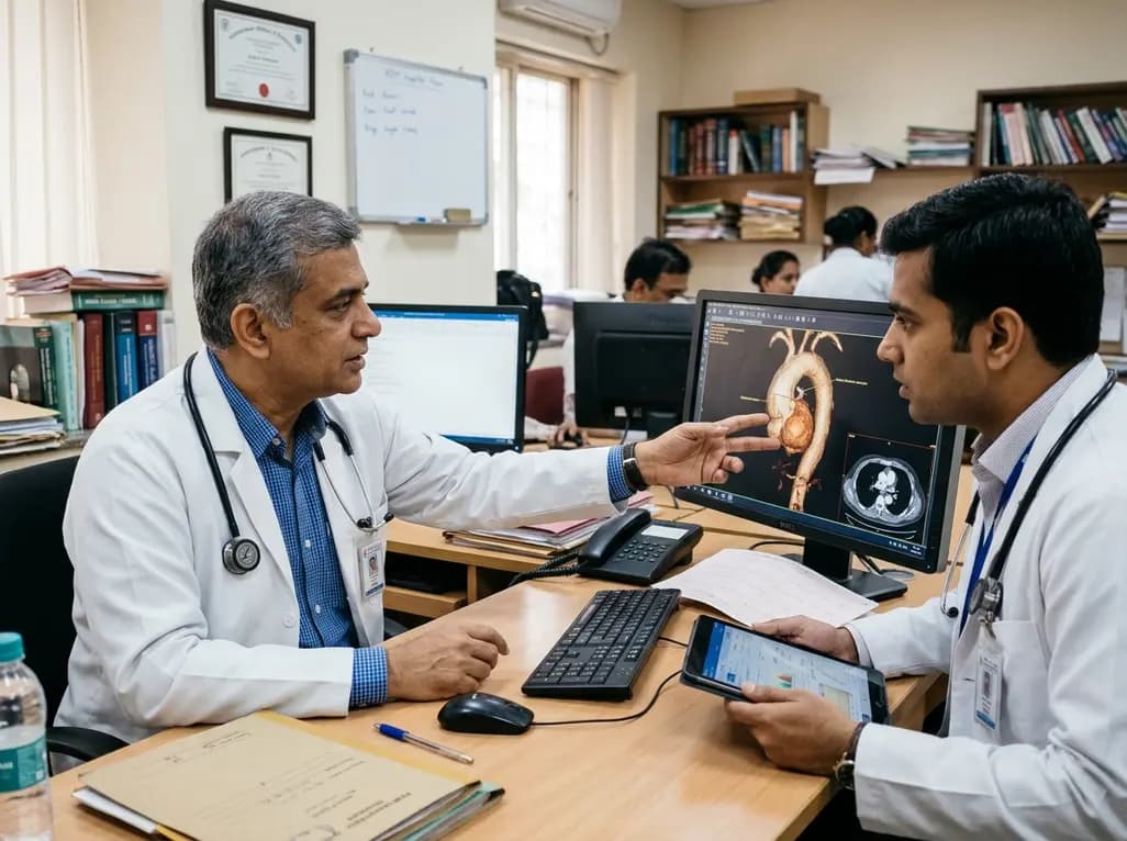
Thoracic aortic aneurysm lacks etiologic medical therapy. A new study reveals that targeting specific αV integrins with Cilengitide attenuates TGF-β and mechanotransduction pathways, effectively limiting pro-fibrotic remodeling and aortic bulb dilation in both syndromic and sporadic disease models.
Today
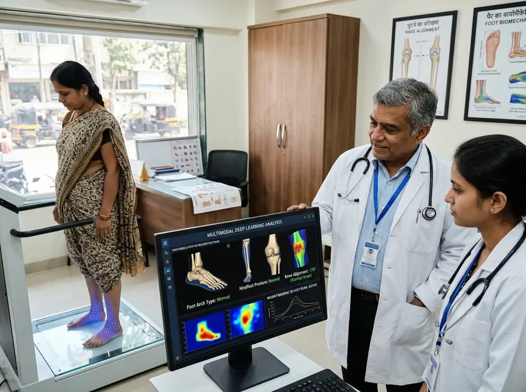
A multimodal deep learning framework utilizing weight-bearing 3D foot scans accurately screens lower-limb alignment, including arch type, hindfoot posture, and knee alignment, offering an accessible, radiation-free alternative to radiography for early biomechanical triage.
Today
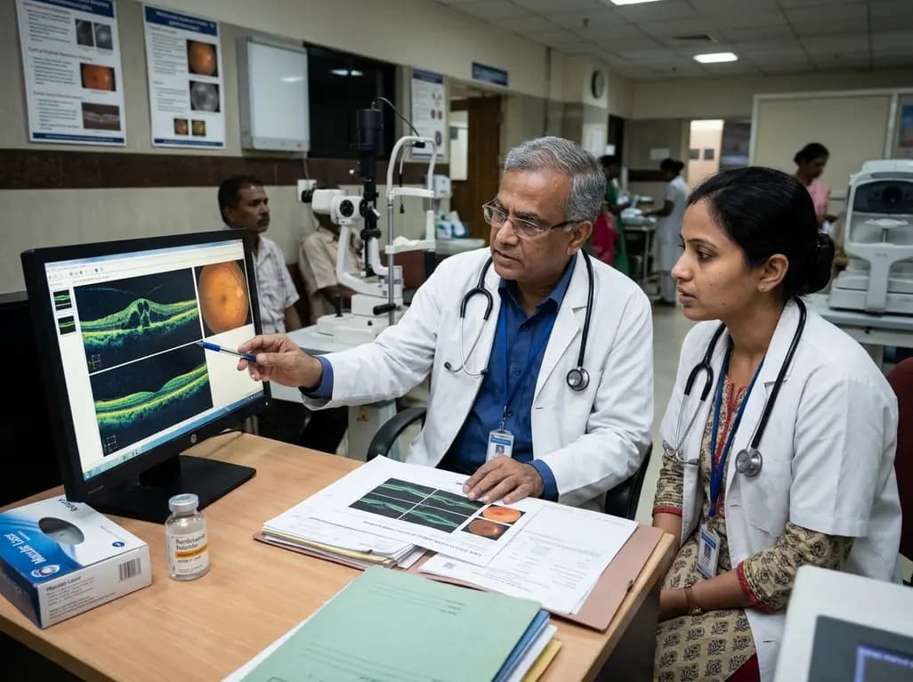
A real-world retrospective study demonstrates that combining intravitreal ranibizumab with focal/grid macular laser significantly improves visual acuity and central macular thickness compared to laser monotherapy in patients with center-involved diabetic macular edema.
Today
