

Loading, please wait...
Loading, please wait...

Temporomandibular disorders encompass a broad spectrum of clinical conditions affecting the masticatory musculature and the temporomandibular joint. Consequently, clinicians frequently observe progressive osseous remodeling and degenerative changes in the mandibular condyle. Recent advances in quantitative computed tomography now allow detailed characterization of condylar trabecular bone texture. This objective imaging approach reveals subtle microstructural shifts that conventional radiographic visual examination often overlooks.
The mandibular condyle undergoes constant functional adaptation in response to biomechanical stresses. However, sustained mechanical overloading or articular inflammation disrupts normal bone homeostatic balance. As a result, the condylar subchondral microarchitecture experiences progressive remodeling, matrix resorption, and focal sclerosis. Evaluating condylar trabecular bone texture provides vital insights into these complex biological transformations. When pathological loading exceeds normal physiological tolerance, trabecular struts alter their thickness, spatial alignment, and mineral distribution.
Standard radiological imaging effectively identifies macroscopic damage, such as gross osteophytes or severe cortical erosions. Nevertheless, macroscopic assessment cannot capture earlier, microscopic reorganizations within the trabecular compartment. Therefore, researchers developed computational texture metrics to bridge this diagnostic void. By extracting mathematical values from gray-level pixel distributions, quantitative texture analysis reveals subtle structural heterogeneity across the condyle. Furthermore, these digital markers uncover microarchitectural degradation well before extensive cortical collapse develops. Understanding these fine osseous patterns helps dental specialists and maxillofacial radiologists comprehend the natural evolution of temporomandibular joint degenerative disease. Ultimately, quantifying trabecular characteristics deepens our grasp of joint biomechanics and functional adaptation.
Computed tomography and cone-beam computed tomography represent standard diagnostic modalities for visualizing osseous temporomandibular structures. For example, clinicians routinely rely on these scans to identify cortical flattening, marginal osteophytes, and subchondral cystic lesions. However, conventional radiological analysis relies heavily on subjective visual inspection. Consequently, diagnostic assessments often suffer from inter-observer variability and limited sensitivity during early disease stages.
Moreover, two-dimensional visual assessments cannot reliably measure the internal density gradients of cancellous bone. While visual grading confirms established degenerative joint disease, it provides minimal quantitative data regarding early bone quality. In addition, bone remodelling occurs dynamically along lines of mechanical stress before cortical deformities appear. Thus, qualitative visual reporting fails to detect the earliest phases of subchondral osteopenia or localized microfractures. Clinicians require more precise, objective computational metrics to monitor joint stability and treatment response. Integrating quantitative imaging algorithms into diagnostic protocols solves this persistent limitation. By extracting sub-visual parameters, advanced image processing transforms routine computed tomography from a purely qualitative viewer into an objective analytical platform.
To investigate trabecular microarchitecture, investigators screened 250 computed tomography scans and enrolled 116 eligible subjects in a rigorous study. Specifically, the researchers established three clinical cohorts: temporomandibular disorder with subchondral cysts, temporomandibular disorder without cysts, and healthy asymptomatic controls. Sixty patients presented with subchondral cysts, while sixteen had disorder symptoms without cyst formation, and forty individuals served as asymptomatic controls.
Furthermore, examiners manually segmented regions of interest on three central sagittal slices of each mandibular condyle. In patients presenting with subchondral cysts, specialists positioned the region of interest immediately adjacent to the cystic perimeter. Conversely, in condyles without cystic degeneration, examiners placed the region of interest within superior subchondral trabecular bone. The team subsequently extracted eleven distinct grey-level co-occurrence matrix features from these designated regions. These mathematical parameters quantify spatial relationships, contrast variations, and local pixel dependencies throughout the trabecular grid. Finally, investigators applied the Kruskal-Wallis test alongside Dunn's post hoc testing. To ensure methodological precision, they incorporated false discovery rate adjustments while controlling for patient age and biological sex.
The analytical findings revealed substantial microarchitectural differences among the evaluated clinical cohorts. After false discovery rate correction and multivariable adjustment for confounding factors, four texture metrics demonstrated statistically significant differences. Specifically, Correlation, Sum of Squares, Sum Variance, and Sum Entropy varied significantly across the study groups. Receiver operating characteristic analyses confirmed that these four metrics possessed notable discriminatory capacity for differentiating pathological condyles.
Particularly, the peri-cystic trabecular bone in condyles with subchondral cysts demonstrated marked spatial disruption. Subchondral cysts represent focal areas of bone marrow breakdown accompanied by elevated intra-articular hydraulic pressures. Consequently, the bone surrounding these cystic cavities undergoes intense secondary remodelling, producing marked local heterogeneity. The elevated Sum Variance and Sum Entropy values directly reflect this architectural chaos and disorganized trabecular alignment. In contrast, asymptomatic control condyles maintained regular, uniform pixel arrangements indicative of cohesive trabecular scaffolding. Patients with temporomandibular disorders without cysts showed intermediate structural values. Hence, these quantitative metrics reliably track the severity gradient of degenerative bone alterations.
The implementation of quantitative computed tomography texture analysis offers immense promise for contemporary dental and maxillofacial practice. Currently, practitioners frequently struggle to predict which patients with temporomandibular disorders will develop progressive condylar degeneration. However, objective radiomic parameters can identify vulnerable trabecular bone before extensive cortical breakdown becomes irreversible. Therefore, incorporating these texture algorithms into clinical workflows enhances diagnostic precision and risk stratification.
Furthermore, quantitative imaging tools provide objective biomarkers for monitoring therapeutic outcomes. For instance, clinicians prescribing occlusal appliances, physical therapy, or targeted pharmacotherapy can evaluate whether interventions successfully arrest trabecular deterioration. In surgical cases, identifying peri-cystic bone compromise assists maxillofacial surgeons in determining structural stability prior to arthroplasty or joint reconstruction. Additionally, automated texture software can seamlessly integrate into digital hospital networks to generate automated bone health scores. Such computational aids eliminate subjective operator bias and standardize radiographic interpretations across clinical settings. As digital health technologies expand throughout modern healthcare systems, radiomic texture quantification will undeniably empower clinicians to deliver personalized, proactive management for temporomandibular joint diseases.
Standard computed tomography relies on visual examination to identify macroscopic osseous abnormalities such as cortical erosions, flattening, and marginal osteophytes. In contrast, texture analysis employs computational algorithms, including grey-level co-occurrence matrix features, to measure mathematical patterns and spatial pixel arrangements within cancellous bone. Consequently, this advanced methodology captures subtle microstructural trabecular variations and bone heterogeneity that remain completely undetectable to the human eye during routine subjective visual evaluations.
Subchondral cysts signify advanced degenerative joint disease and severe localized mechanical overload within the temporomandibular joint. Specifically, these fluid-filled cavities develop following microtrauma, microvascular impairment, or bone marrow necrosis beneath the articular cartilage. As joint forces concentrate abnormally, surrounding peri-cystic trabecular bone undergoes intense secondary remodelling and structural disruption. Therefore, detecting cystic changes and adjacent bone degradation helps clinicians identify patients facing higher risks of progressive condylar collapse.
Yes, radiomic texture metrics offer valuable quantitative biomarkers that significantly enhance clinical decision-making for temporomandibular disorders. By assessing subchondral microarchitecture objectively, clinicians can stratify disease severity, anticipate joint instability, and personalize therapeutic interventions. Furthermore, longitudinal texture monitoring allows dental practitioners to evaluate the structural efficacy of splints, pharmacotherapies, or surgical treatments. Thus, integrating automated radiomics into clinical workflows enables earlier, targeted therapies before irreversible condylar destruction occurs.
Disclaimer: This content is for informational and educational purposes only. It is not intended to substitute for professional medical advice, diagnosis, or treatment. Always seek the advice of your physician or other qualified healthcare provider with any questions you may have regarding a medical condition. Refer to the latest local and national guidelines for clinical practice.
References

Read summarized clinical updates, watch expert medical content, and earn CME certifications right from your smartphone.
Open your smartphone camera to scan and install.





A quantitative CT study shows that condylar trabecular bone texture analysis detects subtle microstructural changes in temporomandibular disorders. Significant texture features differentiate TMD patients with and without subchondral cysts from healthy controls, highlighting altered peri-cystic trabecular bone.
Today

The TREM-AB open-label pilot study evaluated a wearable passive nonlinear vibration absorber for essential tremor. The Nonlinear Energy Sink bracelet reduced clinical Fahn-Tolosa-Marin tremor scores by 32.1% and electrophysiological tremor power by 49.2% with no adverse effects reported.
Today

A population-based study of 20,928 children reveals that small vulnerable newborns face sex-divergent mental capital outcomes at ages 6-7: term SGA girls show higher behavioral difficulties and lower resilience, while preterm non-SGA boys experience increased behavioral problems.
Today
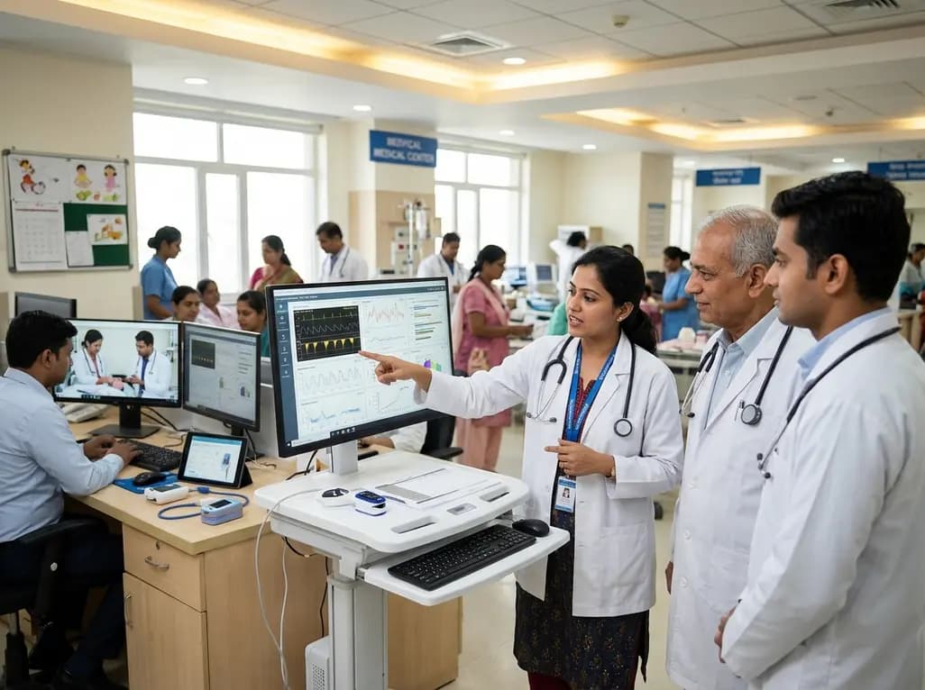
A landmark study details how an enterprise pediatric remote patient management model expanded from a single cardiology pilot to 16 diverse specialty programs by combining centralized digital operations with specialty clinical leadership.
Today

A comparative review of tension band wiring and intramedullary screw fixation in thumb metacarpophalangeal arthrodesis, evaluating union rates, hardware complications, and clinical outcomes.
Today

A breakthrough ultrathin multimodal sensor decouples physical hemodynamic monitoring from disposable electrochemical calibration, achieving long-term wearable glucose prediction with 81.36% accuracy and a 9.12% MARD. This offers a robust, drift-free solution for outpatient metabolic surveillance.
Today
