.webp&w=384&q=75)

Loading, please wait...
Loading, please wait...
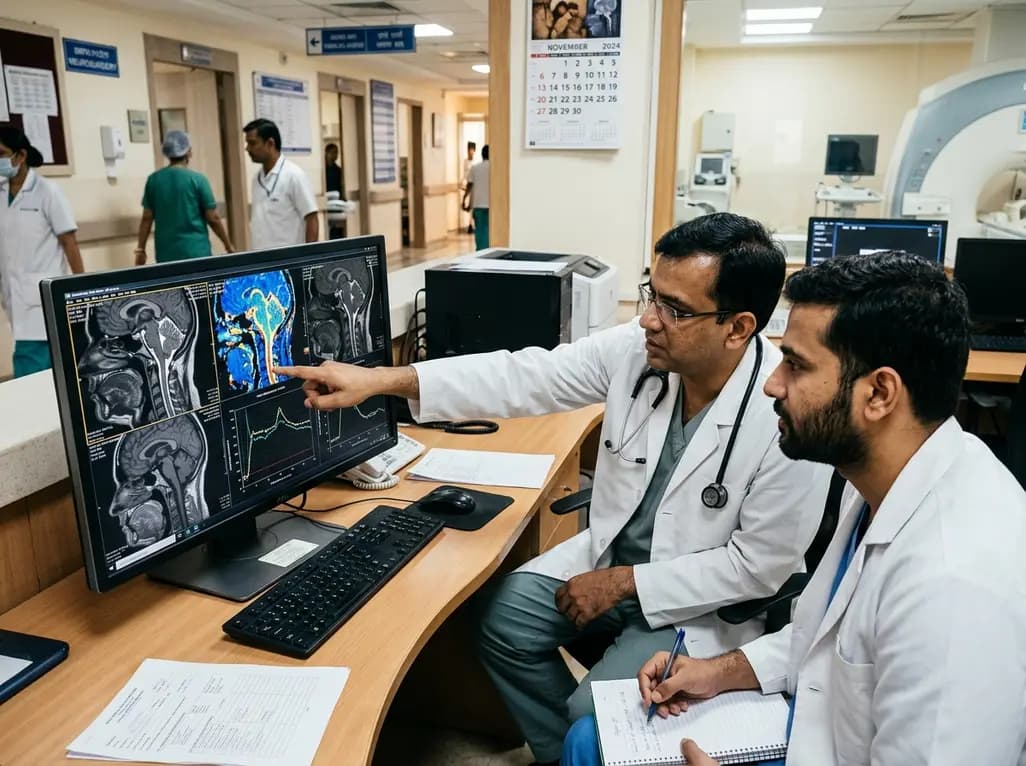
Chiari malformation type I presents a complex neurosurgical conundrum characterized by tonsillar herniation through the foramen magnum. Clinicians conventionally define this condition by cerebellar tonsillar descent exceeding five millimeters below the basion-opisthion line. However, tonsillar displacement magnitude correlates poorly with neurological symptom severity. Furthermore, static anatomical measurements fail to predict surgical outcomes reliably. Posterior fossa decompression surgery provides clinical relief in roughly seventy-five percent of symptomatic patients. Nevertheless, surgical decompression entails inherent morbidity, including cerebrospinal fluid leaks, pseudomeningocele formation, and persistent neuropathic pain. Therefore, neurosurgeons require dependable physiological biomarkers to stratify operative candidates accurately. Cerebrospinal fluid dynamics and pulsatile neural tissue kinematics provide substantial physiological insights into craniocervical pathophysiology. Because static anatomical neuroimaging cannot capture these hemodynamic fluctuations, dynamic magnetic resonance techniques have emerged as essential investigative modalities. Consequently, evaluating pulsatile biophysical parameters directly addresses diagnostic limitations, allowing clinicians to tailor treatment algorithms for vulnerable individuals.
Physiological cardiac contraction drives dynamic craniospinal biomechanics. During systole, rapid expansion of intracranial arterial blood volumes displaces cerebrospinal fluid caudally into the compliant spinal subarachnoid space. Subsequently, diastole allows cranial fluid rebound. In healthy physiology, the foramen magnum accommodates these cyclic hydrodynamic shifts effortlessly. Conversely, tonsillar herniation restricts fluid transit and elevates local impedance. As a result, cardiac pulsations transfer excessive kinetic forces directly into cerebellar and brainstem parenchyma. Novel neuroimaging sequences accurately quantify these pathological interactions. Specifically, phase-contrast magnetic resonance imaging measures directional cerebrospinal fluid velocity and stroke volume across the craniocervical junction. In addition, displacement encoding with stimulated echoes cine imaging quantifies tissue motion and mechanical strain with sub-millimeter precision. These non-invasive modalities transform clinical assessments by replacing subjective anatomical appraisals with objective kinetic data. Thus, clinicians gain unparalleled access to the functional biomechanics governing symptomatic craniocervical junction compromise.
A prospective investigation evaluated one hundred and eight patients with Chiari malformation type I, tracking sixty-one individuals through posterior fossa decompression surgery. Investigators utilized phase-contrast magnetic resonance imaging alongside cine displacement encoding sequences before and after surgical intervention. Strikingly, decompression surgery produced profound physiological restorations across both fluid and solid compartments. Cerebrospinal fluid stroke volume across the foramen magnum increased by 28.9 percent, confirming substantial canal decompression. Furthermore, brainstem tissue displacement decreased significantly by 17.3 percent postoperatively. Cerebellar motion demonstrated even more dramatic improvements, exhibiting a 45.2 percent decrease in mechanical displacement. Patients presenting with the greatest preoperative cerebellar motion and the lowest initial cerebrospinal fluid flow experienced the most significant dynamic normalization. Therefore, baseline biomechanical disturbances strongly correlate with therapeutic response. These empirical measurements demonstrate that successful surgery restores regional hydrodynamic compliance, isolating neural tissue from excessive cardiac-induced kinetic stress.
Neurosurgical patient selection has historically relied upon clinical symptomatology combined with static sagittal imaging. Unfortunately, this paradigm exposes non-responders to unnecessary surgical risks while delaying essential decompression in patients with subtle tonsillar descent. Dynamic neuroimaging provides objective functional criteria to bridge this persistent diagnostic gap. For example, patients exhibiting severe tonsillar ectopia without significant fluid impedance rarely benefit from aggressive osseous and dural decompression. Conversely, individuals with marginal tonsillar descent who experience severe cerebrospinal fluid dampening and pronounced brain motion show marked clinical recovery following intervention. Hence, functional biomechanical indices distinguish genuine craniocervical stenosis from benign anatomical variants. Additionally, quantitative kinetic parameters enable clinicians to monitor postoperative healing objectively. If a patient experiences recurrent suboccipital headaches, repeat dynamic imaging readily differentiates mechanical restenosis from chronic central sensitization or occipital neuralgia. Integrating physiological metrics therefore optimizes surgical decision-making and protects patients from ineffective interventions.
Posterior fossa decompression strategies vary widely, ranging from simple suboccipital craniectomy to extensive duraplasty and tonsillar reduction. The ultimate objective remains the restoration of unrestricted cerebrospinal fluid passage and normalized neural kinematics. However, aggressive intradural dissection increases perioperative risks, such as aseptic meningitis and surgical site infection. Quantitative biomechanical findings inform surgical extent significantly. When preoperative imaging reveals profound flow obstruction, comprehensive decompression incorporating duraplasty reliably restores spinal fluid stroke volume. In contrast, patients with mild kinetic disturbances might achieve adequate decompression through bone-only approaches, minimizing arachnoid scarring. Moreover, intraoperative ultrasonography complements these magnetic resonance parameters by offering real-time verification of pulsatile tonsillar movements. Postoperative cine sequences validate surgical success by documenting normalized cerebellar and brainstem motion. Consequently, surgical teams can standardize technique selection, balancing risk profiles against dynamic restorative potential to secure optimal patient safety.
In Indian clinical environments, the diagnosis of craniocervical junction abnormalities involves distinct epidemiological and infrastructural considerations. For instance, congenital anomalies such as basilar invagination and atlantoaxial dislocation frequently coexist with Chiari malformations across Indian patient cohorts. Consequently, determining whether primary neural compression or craniovertebral instability drives clinical morbidity represents a pivotal challenge. Incorporating dynamic magnetic resonance sequences allows tertiary Indian neurosurgical centers to differentiate between isolated hydrodynamic compromise and complex spinal instability. Although advanced sequences require specialized radiofrequency coils and post-processing software, modern high-field scanners in major public and private hospitals can readily implement these protocols. Furthermore, objective imaging prevents unwarranted surgical interventions, thereby reducing direct healthcare expenditures for economically vulnerable families. Establishing standardized dynamic craniocervical imaging protocols across Indian medical institutions will substantially improve operative selection, refine surgical outcomes, and elevate tertiary neurosurgical care nationwide.
Phase-contrast magnetic resonance imaging measures the velocity and directional flow of cerebrospinal fluid during the cardiac cycle. By quantifying stroke volume and peak systolic velocities across the foramen magnum, this non-invasive technique detects flow obstruction and validates hydrodynamic restoration after decompression surgery with superior diagnostic accuracy.
Surgical decompression enlarges the crowded subarachnoid space at the craniovertebral junction, substantially reducing hydrodynamic outflow resistance. Consequently, cyclic arterial pulse waves dissipate freely through cerebrospinal fluid pathways rather than transmitting excessive mechanical force directly into brainstem and cerebellar tissue, markedly reducing brain motion.
Yes, dynamic magnetic resonance imaging reliably stratifies surgical candidates. Research confirms that patients displaying pronounced preoperative cerebellar motion and severely diminished cerebrospinal fluid stroke volume experience the greatest biomechanical restoration postoperatively. These quantitative metrics guide clinicians in identifying individuals most likely to achieve symptomatic improvement.
Disclaimer: This content is for informational and educational purposes only and should not be considered medical advice. Always consult a qualified healthcare professional regarding any medical condition or before making any healthcare decisions. Refer to the latest local and national guidelines for clinical practice.
References
McIlvain G et al. Measurement of CSF flow and brain motion in Chiari malformation type I subjects undergoing posterior fossa decompression surgery. J Neurosurg. 2025 Jul 01. doi: 10.3171/2024.11.JNS241509. PMID: 40053923.
Mohsenian S, Ibrahimy A, Al Samman MMF, Oshinski JN, Bhadelia RA, Loth F. Association between resistance to cerebrospinal fluid flow and cardiac-induced brain tissue motion for Chiari malformation type I. Neuroradiology. 2023;65(10):1535-1543. doi: 10.1007/s00234-023-03204-x.
Bhadelia RA, Ibrahimy A, Al Samman MM, Ebrahimzadeh SA, Zhao Y, Loth F. Transient decrease in cerebrospinal fluid motion is related to cough-associated headache in Chiari I malformation. World Neurosurg. 2024;189:e709-e717. doi: 10.1016/j.wneu.2024.06.126.

Read summarized clinical updates, watch expert medical content, and earn CME certifications right from your smartphone.
Open your smartphone camera to scan and install.





A landmark study evaluated CSF flow and brain motion in Chiari malformation type I patients undergoing decompression. Advanced MRI metrics revealed significant dynamic improvements, providing objective physiological markers to refine patient selection and surgical outcomes.
last month

As monsoon seasons elevate arboviral disease threats, Tamil Nadu has initiated an ICMR-monitored evaluation of the tetravalent dengue vaccine in Tenkasi district. Clinicians emphasize clinical monitoring, public prevention, and high-quality early pediatric care to reduce disease morbidity across endemic regions.
Today

A comparative cohort study evaluates vNOTES vs TLH hysterectomy for benign conditions, demonstrating shorter operative duration, faster return to daily activities, and equivalent female sexual function scores without increased dyspareunia.
Today

Review how tailored exercise programs benefit frail older patients with heart failure. Explore pathophysiological mechanisms, individualized training prescriptions, and strategies to overcome adherence barriers in clinical practice.
Today

This comparative study evaluates posterior labral tears with and without glenohumeral instability, highlighting distinct demographic patterns, MRI underestimation in stable shoulders, and successful arthroscopic repair outcomes.
Today

A clinical study demonstrates that early intervention with tocilizumab, either as monotherapy or bridged with corticosteroids, achieves significant proptosis reduction and resolves active inflammation in treatment-naive thyroid eye disease.
Today
