.webp&w=384&q=75)

Loading, please wait...
Loading, please wait...
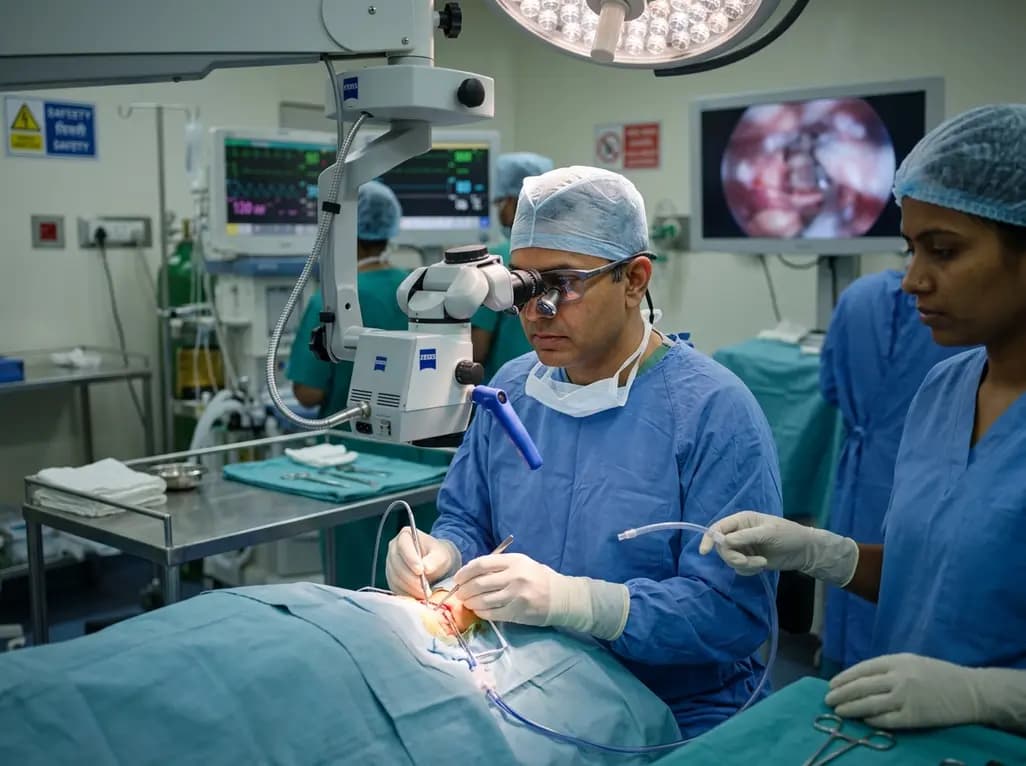
Degenerative cervical spine disorders frequently lead to progressive spinal cord compression. Cervical spondylotic myelopathy remains a leading cause of spinal cord dysfunction in aging populations worldwide. While surgical intervention effectively halts neurological deterioration, surgeons continually strive to minimize structural disruption. Recent surgical advances highlight cervical microendoscopic laminotomy as a promising, motion-preserving decompression technique.
Cervical spondylotic myelopathy arises from degenerative changes in the intervertebral discs, facet joints, and supporting ligaments. Consequently, these structural alterations narrow the spinal canal and compress the cervical spinal cord. Patients typically experience progressive clumsiness of the hands, gait unsteadiness, sensory disturbances, and motor weakness. When conservative therapy fails, decompression surgery becomes necessary to prevent irreversible neurological damage.
Historically, posterior decompression strategies have relied on extensive approaches such as expansive laminoplasty or segmental partial laminectomy. Although these conventional surgeries successfully relieve neural compression, they often disrupt posterior muscular attachments and ligamentous complexes. Therefore, patients frequently suffer from persistent axial neck pain, significant stiffness, and reduced cervical range of motion. Furthermore, wide muscle stripping compromises cervical dynamic stability and alters sagittal alignment.
In elderly patients, post-surgical neck morbidity poses a substantial barrier to functional independence. Because aging spines already exhibit diminished flexibility and muscle sarcopenia, extensive posterior exposure exacerbates long-term axial disability. As a result, spinal surgeons increasingly seek minimally invasive techniques that provide adequate neural decompression while preserving physiological biomechanics. Minimizing muscular detachment directly translates to better recovery trajectories and improved patient-reported outcomes.
To address the limitations of open posterior procedures, surgeons developed cervical microendoscopic laminotomy. This minimally invasive technique utilizes tubular retractor systems and high-definition endoscopes or microscopes. Consequently, surgeons access the lamina through small paramedian incisions without extensively detaching the posterior paraspinal musculature.
During the procedure, the surgeon creates a precise laminotomy window to resect the compressive ligamentum flavum and offending bony osteophytes. Furthermore, the microendoscopic approach enables bilateral neural decompression from a unilateral approach by undercutting the base of the spinous process. In contrast, segmental partial laminectomy requires broader midline exposure and extensive resection of posterior bony elements. While segmental partial laminectomy achieves robust central decompression, it compromises posterior tension bands and facet joint integrity.
Additionally, microendoscopic visualization grants exceptional illumination and magnified clarity of the thecal sac and exiting nerve roots. Surgeons can safely execute targeted bone drilling under continuous visual guidance, reducing accidental dural tears. Because the microendoscopic technique avoids extensive periosteal stripping, it significantly preserves local blood supply to the musculature. Therefore, this method creates a biologically favorable environment that promotes rapid soft tissue healing and diminishes postoperative muscular atrophy.
Preserving cervical kinematics represents a vital goal in modern decompressive spine surgery. Recent comparative studies evaluating posterior decompression confirm that microendoscopic techniques effectively safeguard dynamic motion. When surgeons perform open laminectomies or laminoplasties, postoperative cicatricial contracture and muscle fibrosis often tether the cervical motion segments. Consequently, patients demonstrate marked loss of flexion and extension range of motion at long-term follow-up.
In contrast, patients undergoing microendoscopic decompression retain significantly greater cervical range of motion. Because the interspinous ligaments, supraspinous ligaments, and facet joint capsules remain largely intact, physiological biomechanics persist unhindered. Quantitative kinematic assessments reveal that microendoscopic procedures preserve subaxial motion and prevent pathological adjacent segment stress.
Moreover, preserving normal kinematics helps maintain sagittal balance across the subaxial cervical spine. Conventional open approaches frequently induce post-laminectomy kyphotic deformity due to the disruption of posterior stabilizing structures. Conversely, microendoscopic procedures minimize this risk by preserving the posterior arch architecture. Patients maintain physiological lordosis without requiring supplemental instrumentation or arthrodesis. Ultimately, motion preservation directly enhances daily functional performance and prevents secondary compensatory strains on the thoracic and shoulder musculature.
Axial neck pain remains one of the most frustrating complications following posterior cervical spine surgery. Conventional open techniques cause substantial ischemic necrosis and denervation of the multifidus and semispinalis cervicis muscles. As a consequence, patients frequently report persistent deep neck soreness, shoulder stiffness, and fatigue that limit daily activities.
Clinical investigations indicate that microendoscopic decompression leads to significantly lower postoperative neck pain scores. Evaluated via visual analogue scales and the Neck Disability Index, patients treated microendoscopically demonstrate superior relief from axial symptoms. Because tubular retractors split muscle fibers along natural planes rather than stripping them from their bony insertions, muscle trauma remains negligible. Therefore, postoperative inflammatory responses subside quickly, allowing patients to resume normal movement patterns early.
Additionally, neurological outcomes remain highly favorable following microendoscopic intervention. Validated scoring tools, including the Japanese Orthopaedic Association score, show substantial neurological recovery rates that match or exceed open decompressive procedures. Patients regain manual dexterity and walking stability rapidly without the burden of debilitating postoperative axial discomfort. Consequently, treatment satisfaction rates are notably higher among patients who undergo minimally invasive decompression compared to those receiving traditional segmental partial laminectomies.
Beyond clinical and functional advantages, minimally invasive microendoscopic approaches offer marked improvements in perioperative safety profiles. Clinical evidence demonstrates that microendoscopic procedures significantly reduce intraoperative blood loss compared to traditional segmental partial laminectomy. Smaller surgical corridors and targeted electrocautery minimize bleeding from epidural venous plexuses and paraspinal musculature.
Furthermore, reduced tissue trauma directly translates to shorter hospital stays and faster overall rehabilitation. Patients experience less systemic stress, require fewer opioid analgesics postoperatively, and achieve independent ambulation sooner. Complication rates, including surgical site infections, hematomas, and persistent wound drainage, are substantially lower in microendoscopic cohorts. For elderly patients with multiple medical comorbidities, these perioperative advantages significantly lower the risk of systemic postoperative complications.
From a health economics perspective, microendoscopic laminotomy presents a cost-effective alternative to instrumented fusion or implant-dependent laminoplasty. The procedure achieves complete decompression without requiring titanium mini-plates, screws, or interlaminar spacers. Consequently, hospitals and healthcare systems reduce direct implant costs while achieving excellent patient outcomes. Therefore, adopting microendoscopic techniques aligns perfectly with modern value-based healthcare models, combining clinical excellence, functional preservation, and substantial cost containment.
Cervical microendoscopic laminotomy is a minimally invasive surgical procedure designed to decompress the cervical spinal cord. Surgeons use tubular retractors and endoscopic visualization to resect compressive bones and thickened ligaments. This technique spares paraspinal musculature, preserves motion segments, and avoids the need for permanent spinal implants.
Traditional laminectomy requires wide midline incisions, extensive muscular detachment, and complete removal of posterior spinal structures, which often causes persistent neck pain. In contrast, microendoscopic laminotomy accesses the canal through a small muscle-splitting corridor. It decompresses the spinal cord while maintaining structural stability, ligaments, and cervical mobility.
Ideal candidates include patients with symptomatic cervical spondylotic myelopathy or central canal stenosis without severe pre-existing kyphotic deformity. It particularly benefits elderly individuals and active patients who require neural decompression but want to avoid fusion hardware, preserve cervical range of motion, and minimize postoperative recovery time.
Disclaimer: This content is for informational and educational purposes only. It is not intended to be a substitute for professional medical advice, diagnosis, or treatment. Always seek the advice of your physician or other qualified health provider with any questions you may have regarding a medical condition. Refer to the latest local and national guidelines for clinical practice.
References

Read summarized clinical updates, watch expert medical content, and earn CME certifications right from your smartphone.
Open your smartphone camera to scan and install.





A case-control study demonstrates that cervical microendoscopic laminotomy reduces postoperative axial neck pain, preserves range of motion, and minimizes surgical morbidity compared to segmental partial laminectomy for cervical spondylotic myelopathy.
4 weeks back

With Delhi reporting nearly triple the vector-borne infections since August, healthcare practitioners face heightened diagnostic challenges. Timely differentiation between dengue and malaria, vigilant monitoring during the critical afebrile phase, and strict avoidance of NSAIDs remain vital for reducing morbidity.
Today

A scoping review reveals how pregnancy affects phonation through laryngeal edema and reduced phonation time rather than major acoustic pitch alterations. Learn how to identify, evaluate, and manage vocal symptoms in expectant mothers.
Today

According to WHO data, average lifespan reached 73.3 years in 2023, recovering from pandemic lows. However, healthy life expectancy lags at 62.8 years due to surging noncommunicable conditions like cardiovascular disease and dementia, demanding proactive primary screening and sustained chronic care.
Today

Researchers have engineered an artificial sensory-pain integrated receptor using a multi-threshold organic synaptic transistor. The system distinguishes benign touch from noxious stimuli, enabling safer, more responsive prosthetic limbs with built-in memory and simplified hardware architecture.
Today

Medical robotics is transitioning beyond operative suites into diagnostics, cardiology, and endocrinology. Explore how innovations in tele-robotics, AI integration, and nanomedicine are overcoming geographical barriers and transforming precision healthcare delivery across India's evolving disease landscape.
Today
