.webp&w=384&q=75)

Loading, please wait...
Loading, please wait...

Intracranial neoplasms that present as solitary masses often conceal complex dual pathologies. In neuro-oncological practice, clinicians encounter true collision lesions as well as true recipient-donor interactions. Distinguishing a collision tumor from true tumor-to-tumor metastasis represents a formidable challenge that directly dictates systemic staging and therapeutic choices. Neurosurgeons and neuro-oncologists frequently face cases where a lesion masquerades on standard neuroimaging as an uncomplicated meningioma. Consequently, unmasking a synchronous secondary malignancy requires heightened vigilance, detailed histological sectioning, and targeted immunohistochemical panels.
Clinicians must recognize the precise pathological criteria that separate these two distinct clinical entities. A collision tumor develops when two histologically discrete neoplasms arise independently within the same topographical region. These adjacent masses simply collide and intermingle along a shared territorial border without true biological symbiosis. In contrast, tumor-to-tumor metastasis involves a hematogenous or contiguous seeding event. In this situation, a primary donor tumor actively infiltrates the distinct stroma of an established host tumor.
Historically, diagnostic criteria established by classic neuropathological studies require strict validation. First, the patient must harbor two or more distinct primary neoplasms. Second, the recipient host mass must represent an authentic benign or malignant neoplasm rather than normal tissue. Third, the metastatic donor cells must display true histological integration and viable growth inside the recipient mass. Finally, clinicians must rule out simple contiguous extension from adjacent extracranial malignancies or adjacent lymph nodes.
Epidemiological and pathological reviews consistently show that meningiomas serve as the most frequent recipient neoplasms in intracranial seeding. Several unique microenvironmental factors explain why circulating malignant cells favor these benign dural growths. First, meningiomas display extensive hypervascularity and lack an intact blood-brain barrier. Consequently, high-volume dural blood flow readily delivers circulating malignant emboli directly to the tumor capillary beds.
Furthermore, meningiomas possess a collagen-rich, indolent extracellular matrix with relatively low metabolic competition. This receptive architecture allows slower-growing donor cells to anchor securely. Specific cell adhesion molecules such as E-cadherin, intercellular adhesion molecule 1, and surface integrins facilitate malignant arrest. Additionally, widespread hormonal receptors, including estrogen and progesterone receptors, create a hospitable biochemical milieu. Therefore, systemic carcinomas, most notably breast and pulmonary adenocarcinomas, preferentially populate these receptive meningeal lesions.
Recent neurosurgical investigations highlight three instructive presentations that illustrate these complex dual neoplasms. In one documented case, a 65-year-old woman presented with an intracranial mass resembling a benign meningothelial meningioma. Histopathological evaluation after craniotomy revealed a collision tumor comprising adjacent meningothelial meningioma and metastatic lung adenocarcinoma. In this patient, two distinct neoplasms collided side by side at the dural interface without direct stromal takeover.
Conversely, a 66-year-old woman presented with a dural mass that demonstrated true recipient-donor architecture. Microscopic examination confirmed a classic meningioma infiltrated by a high-grade neuroendocrine carcinoma. Similarly, a 64-year-old man presented with an aggressive frontotemporal lesion. Comprehensive pathology demonstrated a rare intracranial pairing where a glioblastoma multiforme invaded an adjacent meningioma. In both latter presentations, aggressive malignant populations established nests within the host stroma. These findings radically transformed patient prognosis and demanded immediate systemic restaging.
Advanced diagnostic imaging techniques routinely struggle to identify dual intracranial pathology preoperatively. Conventional computed tomography and magnetic resonance imaging depict these lesions as homogeneous, strongly enhancing extra-axial masses. Radiologists frequently observe classic meningioma hallmarks, such as a prominent dural tail and uniform contrast enhancement. Consequently, the presence of a second malignant component often escapes detection before resection.
Nevertheless, clinicians should note subtle imaging anomalies that suggest dual pathology. Disproportionate peritumoral vasogenic edema surrounding an apparently benign dural lesion represents an important warning sign. Rapid interval expansion or internal heterogeneity on T2-weighted and diffusion-weighted sequences also warrants clinical suspicion. Furthermore, physiological techniques such as arterial spin labeling, perfusion-weighted imaging, and magnetic resonance spectroscopy can detect conflicting vascular and metabolic profiles. However, imaging remains purely suggestive, making comprehensive surgical resection and tissue analysis indispensable.
Because imaging cannot definitively identify a hidden malignant component, neuropathologists bear the ultimate diagnostic responsibility. Macroscopic inspection alone frequently fails because metastatic deposits may form microscopic, scattered nests within fibrous meningioma tissue. Therefore, pathologists must submit multiple representative tissue blocks and avoid relying solely on small frozen biopsy sections. Diligent examination prevents catastrophic misclassification of high-grade metastatic deposits as high-grade meningioma variants.
Immunohistochemical staining provides crucial clarity during differential workups. Meningiomas characteristically express epithelial membrane antigen and somatostatin receptor 2A while lacking cytokeratin diversity. When dealing with metastatic adenocarcinoma, panels including thyroid transcription factor 1, cytokeratin 7, and gross cystic disease fluid protein confirm donor identity. Similarly, synaptophysin and chromogranin confirm neuroendocrine carcinoma, whereas glial fibrillary acidic protein identifies glioblastoma components. Establishing the precise molecular lineage prevents inappropriate clinical discharge and facilitates timely targeted oncological therapy.
Uncovering dual tumor pathology completely transforms the subsequent treatment trajectory. When surgeons resect a solitary meningioma, standard gross total resection typically offers excellent curative potential. However, identifying a metastatic adenocarcinoma or high-grade glioma within that same tissue fundamentally alters the clinical scenario. The neurosurgical team must immediately coordinate with medical and radiation oncologists to execute systemic staging. Whole-body fluorodeoxyglucose positron emission tomography, contrast computed tomography, and comprehensive molecular biomarker analysis become urgent priorities.
Furthermore, therapeutic regimens must prioritize the more aggressive histological component. For patients harboring metastatic disease, systemic systemic targeted therapies, immune checkpoint inhibitors, or stereotactic radiosurgery take center stage. When glioblastoma involves a meningioma, concurrent chemoradiotherapy using temozolomide remains the cornerstone of care. Multidisciplinary tumor boards ensure that patients receive appropriate systemic therapy rather than simple observation after dural surgery. Meticulous surveillance remains imperative because systemic disease progression dictates overall survival.
Collision tumors consist of two independent, morphologically distinct neoplasms that develop simultaneously and merge along a physical boundary without mutual cellular infiltration. Conversely, tumor-to-tumor metastasis occurs when malignant cells from a separate donor neoplasm actively travel through the bloodstream and establish secondary growth within the stroma of a preexisting recipient host tumor.
Meningiomas provide an exceptionally hospitable microenvironment for circulating tumor emboli. These benign neoplasms feature intense vascularization, robust perfusion, and a defective blood-brain barrier. Furthermore, their slow-growing collagenous stroma exerts low metabolic competition, while specific endothelial adhesion molecules and rich expression of hormone receptors facilitate the arrest, attachment, and proliferation of circulating carcinoma cells.
Conventional neuroimaging cannot reliably resolve cellular-level heterogeneity within a single lesion. Most host meningiomas demonstrate standard radiologic features, such as sharp margins and uniform contrast uptake, which easily obscure internal metastatic foci. Unless marked perilesional edema or rapid interval growth raises clinical suspicion, definitive diagnosis requires rigorous histopathological examination and broad immunohistochemical staining.
Disclaimer: This content is for informational and educational purposes only and does not substitute for professional medical advice, diagnosis, or treatment. Healthcare professionals should exercise their independent clinical judgment. Refer to the latest local and national guidelines for clinical practice.
References
Suchcicka W et al. Cerebral collision tumor vs. tumor-to-tumor metastasis. Case series with a literature review. Neurol Neurochir Pol. 2026 Oct 08. doi: 10.5603/pjnns.109154. PMID: 42845180.
Bulte CA, Hoegler KM, Khachemoune A. Collision tumors: A review of their types, pathogenesis, and diagnostic challenges. Dermatol Ther. 2020; 33(6): e14236.
Takei H, Powell SZ. Tumor-to-tumor metastasis: pathology and neuroimaging considerations. Int J Clin Exp Pathol. 2012; 5(4): 367–374.

Read summarized clinical updates, watch expert medical content, and earn CME certifications right from your smartphone.
Open your smartphone camera to scan and install.





Differentiating cerebral collision tumors from tumor-to-tumor metastasis remains a crucial diagnostic hurdle. Neuropathological evaluation and immunohistochemistry are essential to detect hidden malignancies within presumed solitary intracranial lesions.
Today
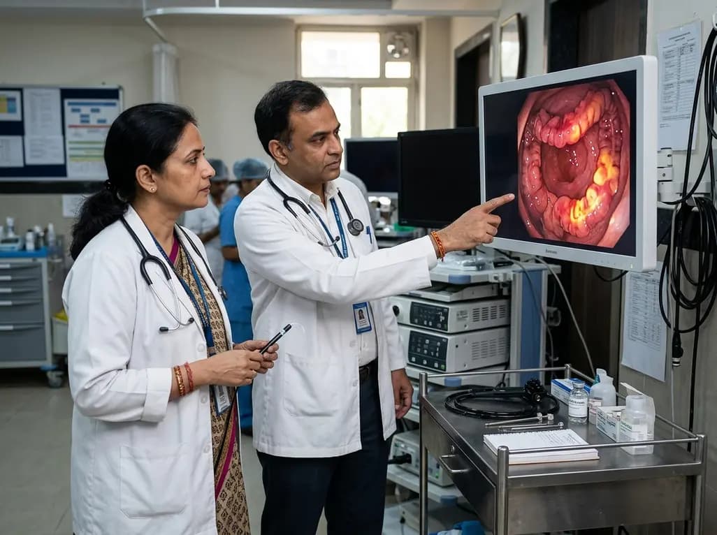
The novel chemiluminescent probe 2TPA-IsoL enables selective, deep-tissue monitoring of MPO-generated HOCl. This breakthrough offers real-time tracking of inflammatory bowel disease activity and therapeutic response.
Today

New evidence from Karolinska Institutet shows women vaccinated against HPV prior to conception have significantly lower rates of preterm birth and major perinatal complications. Clinicians in India can leverage these findings to strengthen adolescent immunisation and reproductive health advocacy.
Today

Cinematic rendering utilizes advanced global illumination to transform coronary CT angiography datasets. This imaging technique provides lifelike depth perception, helping cardiologists and surgeons precisely evaluate anomalous coronary artery origins, high-risk interarterial courses, and post-surgical anatomy.
Today
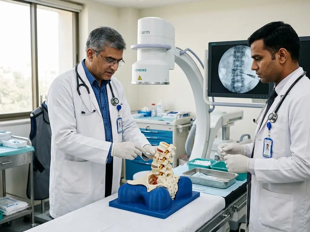
A novel 3D-printed lumbar spine simulator with tunable radiopacity provides realistic tactile and radiographic feedback for fluoroscopy-guided intervertebral disc puncture training, offering a low-cost, ethical alternative to cadaver models.
Today

A breakthrough study reveals that loss of SELENOT in hypothalamic POMC neurons triggers premature cellular senescence, ATF6α stress activation, and ER calcium depletion, linking neuroendocrine decay to metabolic disease.
Today
