

Loading, please wait...
Loading, please wait...
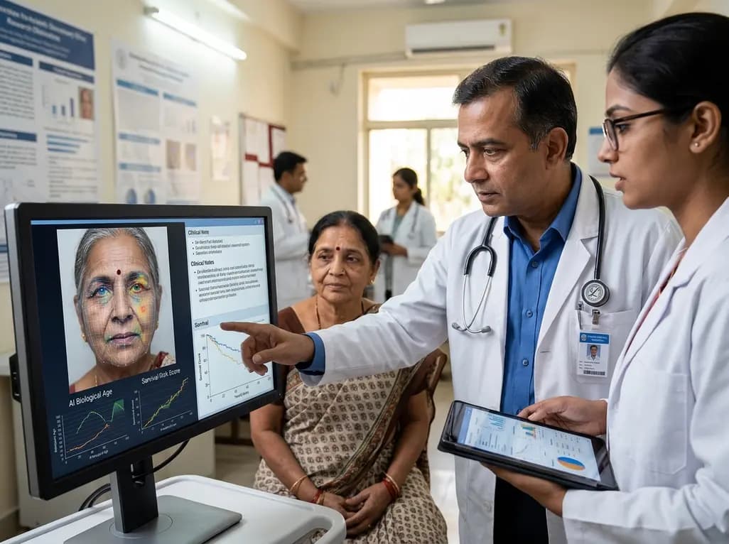
Modern oncologists continually seek non-invasive methods to evaluate baseline physiological reserve and patient frailty before initiating aggressive therapies. Recently, researchers introduced an advanced cancer survival prediction AI framework that extracts physiological insights directly from standard facial portraits. Investigators at Mass General Brigham published this breakthrough in The Lancet Digital Health, demonstrating how deep learning quantifies systemic health. Clinicians often observe that a patient's physical appearance reveals crucial clues regarding biological resilience and fitness. Now, automated algorithms can quantify these subtle visual indicators with reproducible mathematical precision. Consequently, this breakthrough presents significant implications for personalized cancer therapeutics, multidisciplinary decision-making, and risk stratification globally.
To establish this diagnostic tool, bioinformaticians created the Foundation Artificial Intelligence for Health Recognition-Face system, known as FAHR-Face. Developers initially pre-trained this neural network on more than forty million diverse facial photographs using self-supervised masked autoencoders. Consequently, the architecture learned complex, high-dimensional facial geometry across varied ages, ethnicities, and imaging environments. Subsequently, investigators developed two specialized downstream tools from this foundational base to address critical prognostic questions in clinical oncology.
The first tool, FAHR-FaceAge, estimates a patient's biological age by quantifying structural dermatological markers. In contrast, chronological age merely tracks calendar years without measuring functional physical wear. When researchers tested FAHR-FaceAge against public datasets, the algorithm calculated biological age with an average error of about five years. Furthermore, the second tool, FAHR-FaceSurvival, was trained on photographs of 34,363 cancer patients. Notably, FAHR-FaceSurvival bypasses age estimation entirely and predicts survival probabilities directly from facial pixels. Additionally, these two distinct algorithms capture complementary phenotypic signals from the same anatomical portrait. Thus, combining both algorithms provides clinicians with a multifaceted appraisal of baseline physiological reserve.
During multi-cohort validation, the cancer survival prediction AI revealed robust correlations with patient mortality across distinct oncological settings. Investigators analyzed survival records from 38,183 cancer patients treated across six Harvard-affiliated medical institutions in the United States. Remarkably, patients exhibiting a biological age significantly higher than their chronological age experienced poorer overall survival outcomes. For example, individuals who appeared biologically older faced substantially elevated mortality risks during curative radiation therapy.
Furthermore, the dedicated survival model demonstrated remarkable discriminatory power across diverse malignancies. When investigators stratified 3,820 validation patients, the highest-risk decile faced more than three times higher mortality than the lowest-risk group. Strikingly, approximately half of the patients in the highest-risk decile died within ninety days of assessment. In contrast, only two percent of patients in the lowest-risk cohort succumbed within that same window. These stark prognostic differences persisted after researchers adjusted for tumor stage, primary cancer site, and performance status. Therefore, facial imaging biomarkers reflect unmeasured physiological vulnerabilities that routine clinical assessments frequently overlook.
Interestingly, explainable artificial intelligence techniques revealed that the two algorithms inspect completely different regions of the human face. This divergence underscores how distinct biological processes imprint upon facial anatomy over time. Specifically, FAHR-FaceAge focuses predominantly on the forehead, the temporal regions, and the nasolabial folds running beside the mouth. These anatomical areas prominently reflect connective tissue remodeling, sun damage, and cumulative chronic lifestyle exposures. Indeed, clinical data show that cigarette smoking, heavy alcohol consumption, and chronic metabolic stress accelerate aging across these specific facial zones.
In contrast, FAHR-FaceSurvival evaluates entirely different anatomical territories, primarily inspecting the infraorbital hollows beneath the eyes and the nasal bridge. Clinicians find this discovery fascinating because these zones closely mirror ancient bedside descriptions. For instance, Hippocrates famously observed sunken eyes and hollowed temples as heralds of terminal illness over two millennia ago. Contemporary oncology now captures this phenomenon computationally as deep neural networks identify subtle manifestations of cachexia and microvascular decline. Additionally, these anatomical regions signal micro-nutritional deficiencies and systemic catabolism before gross muscular wasting becomes clinically obvious. Thus, algorithmic evaluation revives classical bedside semiotics through scalable computer vision.
The emergence of rapid facial phenotyping offers transformative possibilities for clinical oncology across India. Currently, Indian oncologists manage immense patient volumes with diverse socio-economic backgrounds and variable access to expensive molecular diagnostics. Dr. Mandeep Singh Malhotra, director of surgical oncology at the CK Birla Hospital in Delhi, highlighted the practical value of these algorithmic tools. Specifically, he noted that facial biomarkers could complement precision oncology when combined with genomic sequencing, liquid biopsy, and metabolic imaging.
Moreover, incorporating non-invasive visual algorithms into outpatient clinics could streamline oncology triage. A simple digital tablet could capture facial images during initial intake, providing immediate estimates of physiological frailty before clinicians prescribe aggressive therapy. Consequently, surgical teams could identify vulnerable individuals requiring prehabilitation, nutritional optimization, or gentler treatment regimens. Additionally, serial facial analyses could assist oncologists in monitoring therapy-induced physical decline during post-treatment surveillance. As precision oncology evolves in India, integrating facial artificial intelligence with circulating tumor DNA could build a truly comprehensive, multimodal framework for cancer care.
Despite these compelling results, clinicians must acknowledge significant technical and demographic limitations before adopting facial algorithms into everyday oncology workflows. First, investigators derived and validated these models primarily using cohorts from the United States and the Netherlands. Consequently, researchers have not yet established model reliability in Indian populations, which exhibit distinct genetic backgrounds, diverse facial morphologies, and unique epidemiological profiles. Dr. Malhotra emphasized that extensive domestic validation is mandatory to eliminate algorithmic biases across diverse South Asian demographics.
Furthermore, environmental factors and imaging conditions introduce potential confounding artifacts into visual predictive algorithms. Variations in ambient clinic lighting, camera angles, patient head orientation, and spontaneous facial expressions can alter pixel values. Similarly, cosmetic procedures, facial reconstructive surgery, severe facial palsy, or corticosteroid-induced facial changes might mislead algorithmic feature extraction. Most importantly, physicians must remember that FAHR-Face does not diagnose malignancies or calculate an absolute lifespan. Instead, it serves solely as an adjunctive decision-support biomarker. Therefore, oncologists must continuously combine computational estimates with comprehensive clinical examinations and laboratory investigations.
Q1: How does the FAHR-Face artificial intelligence model estimate biological age from a photograph?
The FAHR-Face model utilizes deep convolutional neural networks pre-trained on forty million facial photographs to analyze micro-structural facial features. Specifically, the FAHR-FaceAge sub-model evaluates tissue laxity, skin texture, and deep wrinkles across the forehead, temporal areas, and nasolabial folds. By comparing these subtle phenotypic indicators against vast population databases, the system generates an objective biological age estimate reflecting physiological resilience rather than chronological time.
Q2: Can facial AI models replace existing cancer diagnostic tools and genomic profiling?
No, facial artificial intelligence tools cannot replace tissue biopsy, histological grading, or comprehensive genomic sequencing in clinical oncology. Instead, researchers designed these tools to function as complementary biomarkers that gauge patient frailty, physiological reserve, and short-term mortality risks. Therefore, clinicians should incorporate facial analysis alongside established diagnostics like liquid biopsy and molecular profiling to personalize treatment intensities and improve multidisciplinary therapy planning.
Q3: Why is regional validation essential before deploying facial survival algorithms in India?
Domestic validation is crucial because the initial FAHR-Face studies evaluated patient populations exclusively in Western healthcare centers. Indian populations possess distinct craniofacial features, varied skin phototypes, diverse genetic backgrounds, and unique disease etiologies that algorithms must learn. Consequently, Indian medical researchers must validate and calibrate these foundation models on local cancer cohorts to guarantee diagnostic accuracy, prevent racial algorithmic bias, and ensure equitable clinical applicability.
Disclaimer: This content is for informational and educational purposes only. It does not constitute medical advice or replace professional judgment. Refer to the latest local and national guidelines for clinical practice.
References

Read summarized clinical updates, watch expert medical content, and earn CME certifications right from your smartphone.
Open your smartphone camera to scan and install.





A landmark Lancet Digital Health study introduces FAHR-Face, an artificial intelligence system assessing facial photos to estimate biological age and cancer mortality risk. We explore its clinical mechanics, validation data, implications for precision oncology, and essential limitations across diverse populations.
Today

A KGMU study reveals over half of women with breast abnormalities fail to undergo diagnostic confirmation, leading to advanced-stage detection. Discover the socio-cultural barriers, clinical risks of incomplete resection, and community-level solutions needed to bridge the critical screening-to-diagnosis gap in India.
Today

A comparative non-contact study reveals that high-power radiofrequency delivery with the QDOT MICRO catheter induces an artifactual contact force rise. Electrophysiologists must understand this power-dependent thermal and electrical phenomenon to avoid inadvertent catheter withdrawal and ineffective ablation.
Today
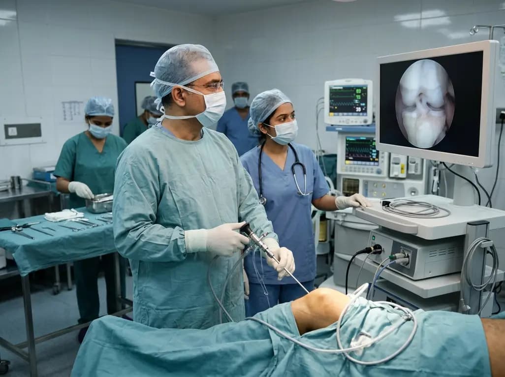
Healthium Medtech has transitioned its arthroscopy vertical into SIRONIX Orthopedics, aiming to capture key segments of the global joint care market. The strategic shift introduces dedicated portfolios in sports medicine, joint regeneration, surgical reconstruction, and digital operating technologies.
Today
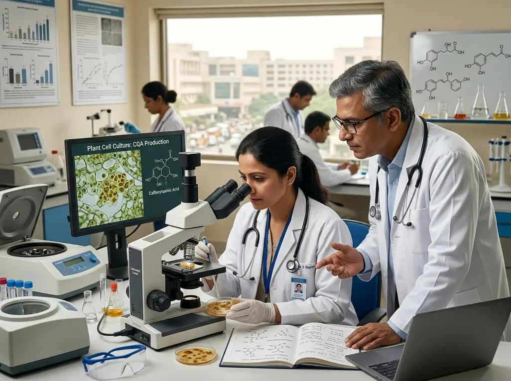
Caffeoylquinic acids, such as chlorogenic acid, offer broad antioxidant, antidiabetic, and anti-inflammatory properties. Advanced plant cell and organ culture methods now enable scalable, sustainable, and pure production of these high-value therapeutic metabolites for pharmaceutical and clinical applications.
Today
