.webp&w=384&q=75)

Loading, please wait...
Loading, please wait...

Rising rates of surgical deliveries worldwide have led to a noticeable increase in rare ectopic implantations. Among these clinical conditions, caesarean scar pregnancy represents an exceptionally hazardous obstetric presentation. In this disorder, the conceptus implants directly into fibrous scar tissue from a prior hysterotomy. Consequently, clinicians must establish an accurate diagnosis early to prevent catastrophic hemorrhage and preserve maternal fertility. Recent prospective investigations highlight the benefit of combining transvaginal sonography with maternal serum biomarkers. Furthermore, this multi-marker strategy provides critical prognostic insights regarding intraoperative bleeding risks during curettage.
A caesarean scar pregnancy develops when a blastocyst embeds within an anterior myometrial defect created by previous surgery. Because fibrous scar tissue lacks normal vascular remodeling and robust decidualization, trophoblastic invasion extends aggressively into the uterine wall. Therefore, affected patients face substantial risks of uterine rupture, severe hemoperitoneum, and emergency hysterectomy.
Clinically, early presentation often mimics ordinary miscarriage or typical early pregnancy complaints. For instance, many patients report painless vaginal spotting or mild lower abdominal discomfort. However, unguided interventions in unrecognized scar implantations provoke rapid, torrential bleeding from non-contractile fibrous niches. In addition, deep trophoblastic invasion frequently causes placenta accreta spectrum disorders if pregnancy advances unnoticed. Consequently, prompt differentiation from cervical ectopic pregnancy or intrauterine miscarriage remains vital. Preoperative risk stratification enables surgical teams to select appropriate interventions, assemble multidisciplinary support, and safeguard maternal outcomes effectively.
High-resolution transvaginal ultrasonography serves as the primary diagnostic modality for lower uterine segment evaluation. Specifically, sonographers focus on two vital measurements: myometrial thickness at the scar and gestational sac diameter. In patients with scar pregnancies, the muscular layer over the gestational sac measures significantly thinner than in normal controls. Often, this remaining myometrium measures less than three millimeters.
Furthermore, sonographers evaluate the average diameter and morphological configuration of the gestational sac or mass. Color Doppler imaging frequently reveals rich peritrophoblastic vascular flow surrounding the scar defect. Moreover, clinicians must carefully inspect the boundary between the gestational sac and maternal bladder wall. A marked loss of hyperechoic myometrial tissue indicates extensive trophoblastic invasion toward surrounding pelvic organs. Therefore, precise sonographic mapping defines anatomical distortion and alerts surgeons to fragile structural margins before any surgical attempt.
Biochemical monitoring provides crucial functional information that complements anatomical ultrasound imaging. In particular, clinicians assess serum beta-human chorionic gonadotropin and estradiol levels during initial diagnostic workups. Research indicates that patients with scar pregnancies display significantly lower concentrations of both hormones compared to healthy controls. This biochemical disparity reflects impaired trophoblastic development within poorly vascularized, fibrous scar tissue.
Consequently, diminished syncytiotrophoblastic proliferation produces lower quantities of human chorionic gonadotropin and steroid hormones. However, clinicians cannot rely on hormone levels alone for definitive diagnosis because individual ranges overlap considerably. In contrast, combining endocrine profiles with ultrasound parameters markedly improves diagnostic sensitivity and specificity. Furthermore, monitoring these hormonal parameters guides post-treatment surveillance by confirming complete trophoblastic demise. As a result, dual hormonal testing strengthens clinical diagnostic algorithms and helps clinicians verify therapeutic success.
Uterine curettage remains a standard management option, but it carries substantial risks of massive intraoperative hemorrhage. Fortunately, combined ultrasound parameters and endocrine markers reliably predict intraoperative blood loss volume during evacuation. Specifically, residual myometrial thickness at the scar correlates inversely with surgical bleeding. Because thinned fibrous tissue cannot contract to compress severed vessels, curettage often induces persistent local bleeding.
Additionally, larger gestational sac diameters and elevated beta-hCG values signal extensive local vascular recruitment. Therefore, patients exhibiting thin scar musculature alongside large gestational masses face the highest hemorrhagic risks. Conversely, patients with preserved myometrial thickness and lower trophoblastic volumes typically experience minimal blood loss. Accordingly, surgical teams can utilize these predictive markers to design targeted preoperative strategies. For example, clinicians can prepare intraoperative tamponade balloons, cross-match packed red blood cells, and secure interventional radiology support in advance.
Safe management of scar implantations requires structured clinical protocols and coordinated multidisciplinary collaboration. When predictive markers identify high bleeding risk, clinicians should avoid unguided suction curettage entirely. Instead, surgical teams often utilize preoperative uterine artery embolization to diminish local perfusion before surgical intervention. Furthermore, performing hysteroscopic resection under direct laparoscopic or ultrasound guidance significantly improves operative safety.
Direct visualization allows precise removal of gestational products while coagulating active feeding vessels under magnified control. In addition, inserting a cervical Foley catheter into the lower uterine segment provides immediate balloon tamponade if bleeding occurs. In settings like India, where cesarean birth rates continue to rise, implementing standardized diagnostic algorithms is essential. Moreover, early risk stratification prevents unnecessary emergency hysterectomies and preserves future reproductive potential for young women. Therefore, integrating hormonal testing with detailed sonographic metrics ensures safer interventions and superior clinical outcomes.
Obstetric practice in India increasingly encounters scar pregnancies due to rising cesarean section numbers in both urban and rural centers. However, diagnostic delays occur frequently when community healthcare facilities lack high-resolution transvaginal sonography equipment. Consequently, patients sometimes present with severe abdominal pain or advanced bleeding after incomplete medical terminations. Establishing robust referral pathways between primary health centers and tertiary hospitals is therefore paramount.
Additionally, combining routine serum hormone tests with standard ultrasound scans offers a cost-effective protocol for Indian tertiary institutions. Because maternal mortality and fertility preservation remain top public health concerns, minimizing curettage-related hemorrhage is crucial. Furthermore, clinicians must provide comprehensive preconception counseling before subsequent pregnancies, recommending detailed scar evaluations prior to conception. Ultimately, adopting standardized multi-marker protocols helps healthcare systems deliver equitable, life-saving obstetric care across diverse clinical settings.
Early identification of this condition prevents catastrophic maternal complications, including uncontained uterine rupture, massive pelvic hemoperitoneum, and emergency hysterectomy. When detected during initial first-trimester weeks, the gestational sac remains small and myometrial invasion remains limited. Consequently, gynecologists can implement conservative medical therapy or guided surgical interventions safely. In contrast, delayed recognition significantly elevates procedural failure rates, increases transfusion requirements, and threatens future reproductive potential in affected women.
Residual myometrial thickness directly reflects the degree of trophoblastic invasion and structural wall integrity at the hysterotomy site. When the muscular layer measures under three millimeters, the attenuated uterine wall cannot contract effectively to compress severed uteroplacental vessels. Consequently, surgical curettage induces rapid intraoperative hemorrhage and substantially elevates uterine perforation risks. Recognizing severe myometrial thinning preoperatively prompts surgical teams to prepare hemostatic balloons and arrange interventional radiology support.
Evaluating serum estradiol alongside beta-hCG assesses embryonic viability and syncytiotrophoblast metabolic activity within the lower uterine segment. In scar pregnancies, both hormonal concentrations are generally lower than in healthy intrauterine pregnancies due to poor scar vascularity. However, higher relative hormone levels correlate with increased tissue vascularity and larger gestational mass volumes. Therefore, combining both biomarkers with ultrasound parameters optimizes diagnostic precision and predicts surgical blood loss during curettage.
Disclaimer: This content is for informational and educational purposes only... Refer to the latest local and national guidelines for clinical practice.
References

Read summarized clinical updates, watch expert medical content, and earn CME certifications right from your smartphone.
Open your smartphone camera to scan and install.





Evaluating ultrasound markers alongside beta-hCG and estradiol improves early diagnosis of caesarean scar pregnancy. This multi-marker strategy also predicts intraoperative bleeding during curettage, guiding timely surgical planning and fertility-preserving clinical interventions.
Today
A network meta-analysis of 10 RCTs comprising 37,923 patients reveals that SGLT2 inhibitors offer the broadest cardiorenal protection in diabetic kidney disease. Dapagliflozin reduced kidney outcomes more than semaglutide, while finerenone increased hyperkalaemia and treatment discontinuation rates.
Today
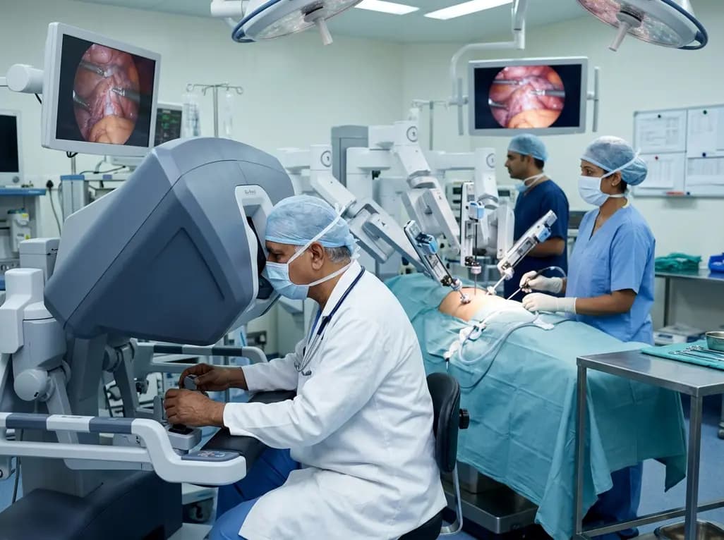
Surgical robotics is redefining modern operating theaters by replacing extensive open incisions with micro-dexterity and stereoscopic 3D vision. Multi-specialty surgeons highlight how robotic platforms mitigate tissue trauma, enhance functional organ preservation, and shorten postoperative hospital recovery.
Today
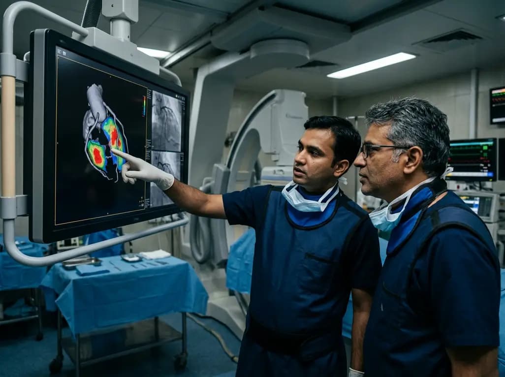
A groundbreaking study demonstrates that coronary angiography-derived radial wall strain reflects plaque morphology, predicting future rupture and erosion.
Today

The Delhi Cabinet has cleared an advanced seven-storey critical care expansion at Guru Tegh Bahadur Hospital under PMSSY Phase-IV. Costing Rs 244.87 crore, the new wing features 250 beds, specialized intensive care units, modern operation theatres, and round-the-clock emergency diagnostics for East Delhi.
Today
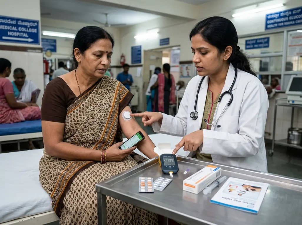
A landmark case demonstrates that favorable continuous glucose monitoring metrics cannot rule out SGLT2 inhibitor-associated euglycemic diabetic ketoacidosis. Clinicians must educate patients on symptom-based sick-day rules, uninterrupted basal insulin therapy, and proactive blood ketone testing.
Today
