.webp&w=384&q=75)

Loading, please wait...
Loading, please wait...
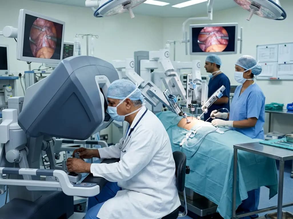
Robotic-assisted surgery is rapidly transforming surgical operating rooms across India and redefining operative standards. Traditional surgical paradigms relied heavily on wide incisions to secure adequate anatomical exposure. However, progressive technological innovation has shifted clinical practice toward high-precision, tissue-preserving approaches. Modern surgical platforms offer articulated endo-wrist instrumentation and high-magnification stereoscopic visualization. Consequently, surgical teams can now perform complex dissections within anatomical spaces once considered inaccessible. Furthermore, clinical evidence indicates that this technological leap improves clinical outcomes and significantly reduces patient recovery times. Surgeons across multiple surgical specialties now employ robotic technology as a standard tool rather than an experimental modality. Therefore, modern surgical disciplines are transitioning from conventional laparotomy toward refined precision surgery.
Surgeons historically faced physical constraints when maneuvering rigid instruments during traditional laparoscopic interventions. In contrast, modern robotic platforms integrate multi-jointed wristed instruments that replicate and enhance human wrist mobility. These articulated instruments provide seven degrees of freedom, which greatly expands surgical dexterity within narrow operative corridors. Moreover, advanced computerized filters actively eliminate physiological tremors from the clinician's fingertips. As a result, operators execute microscopic dissections and complex intracorporeal sutures with steady, unmatched precision. Additionally, the robotic optical console delivers magnified, high-definition three-dimensional stereoscopic imagery directly to the operating surgeon. Unlike standard laparoscopy where assistants manually steady the laparoscope, the robotic camera platform remains rock solid throughout long operations. Therefore, surgeons maintain stable operative views during tedious multi-hour reconstructions. Furthermore, this elevated visual depth perception allows accurate identification of critical tissue planes, fragile microvasculature, and delicate somatic nerves. Surgeons consequently minimize accidental tissue trauma and collateral thermal injury during vital dissections. Ultimately, the integration of superior optics with tremor-free dexterity elevates patient safety across diverse specialties.
General surgeons and surgical gastroenterologists are rapidly adopting robotic platforms for complex reconstructive abdominal procedures. For instance, abdominal wall reconstruction and complex ventral hernia repairs demand meticulous dissection and secure tension-free mesh fixation. Laparoscopic approaches often present mechanical ergonomic challenges in such extensive reconstructions. In comparison, robotic assistance allows clinicians to navigate difficult fascial planes and place durable sutures effortlessly. Similarly, transplant surgeons are successfully applying robotic instrumentation to kidney transplantation. Patients with end-stage renal disease frequently suffer from obesity, diabetes, and multiple cardiovascular comorbidities. Consequently, open renal transplant incisions carry significant risks of wound dehiscence, lymphoceles, and surgical site infections. Robotic kidney transplantation elegantly solves these clinical challenges by placing the donor graft through a tiny periumbilical incision. Subsequently, surgeons complete delicate vascular and ureteral anastomoses using wristed micro-instruments under high visual magnification. Furthermore, this minimally invasive method protects the immunocompromised graft recipient from major wound complications. Early clinical data confirm that patients experience reduced postoperative pain and accelerated graft reperfusion. As a result, hospitalized recipients mobilize earlier and return to routine physical activities much faster.
Cardiac and thoracic interventions traditionally required aggressive median sternotomy or extensive rib-spreading thoracotomies. These traumatic access routes often inflicted severe postoperative pain and required protracted rehabilitation periods. However, robotic cardiac surgery has emerged as a reliable solution for delicate intracardiac and coronary operations. Specifically, cardiac surgeons perform complex mitral valve repairs and coronary artery bypass procedures through minute intercostal ports. Additionally, clinicians harvest internal mammary artery conduits with exceptional safety while preserving chest wall integrity. Patients avoid the deep physical trauma and psychological fear associated with splitting the sternum. Consequently, individuals recover within days rather than languishing through months of debilitating musculoskeletal healing. Similarly, thoracic specialists utilize robotic platforms to address challenging mediastinal tumors, diaphragmatic anomalies, and complex pulmonary malignancies. For example, surgeons in eastern India now resect complex lung lesions and tuberculosis complications with minimal blood loss. The magnified three-dimensional optical system enables meticulous lymph node clearance around major pulmonary vessels. Therefore, robotic thoracic resections maintain rigorous oncological clearance without inflicting debilitating chest trauma on medically fragile patients.
The deep confines of the bony pelvis present severe anatomical obstacles for open and laparoscopic surgery. Fortunately, robotic instrumentation overcomes these natural geometric constraints through superior visualization and flexible angulation. In uro-oncology, robot-assisted radical prostatectomy represents the clinical benchmark for organ-confined prostate cancer management. The wristed instruments allow precise nerve-sparing dissection along the prostatic capsule. Thus, surgeons maximize oncological resection margins while actively preserving neurovascular bundles responsible for urinary continence and erectile potency. In addition, robotic partial nephrectomy enables urologists to resect renal tumors cleanly while sparing healthy nephron parenchyma. Gynecological oncologists similarly capitalize on robotic advantages when treating complex endometrial, cervical, and ovarian malignancies. For example, robotic platforms support near-infrared fluorescence imaging with Firefly technology for precise sentinel lymph node mapping. This optical technology pinpoints metastatic lymphatic drainage pathways without requiring extensive pelvic lymphadenectomy. Consequently, patients avoid severe postoperative lower-limb lymphedema and prolonged hospital stays. Medically fragile women with severe obesity and cardiac comorbidities tolerate robotic gynecological interventions with significantly fewer perioperative complications.
Despite remarkable clinical achievements, common misconceptions regarding surgical robotics persist among patients and healthcare providers. Many people mistakenly assume that autonomous artificial intelligence or automated robotic arms perform the surgical operation independently. However, medical specialists emphasize that the robotic console never acts autonomously. In reality, the surgeon retains complete real-time manual control over every single movement and incision. The platform simply functions as an advanced master-slave digital interface that mirrors human intent with mechanical precision. Furthermore, surgical leaders emphasize that advanced technology cannot replace sound oncological and surgical principles. Operating teams must always adhere to established resection margins and meticulous tissue handling techniques. Moreover, appropriate patient selection remains an absolute prerequisite for successful clinical outcomes. Certain clinical presentations, such as extensive intraperitoneal adhesions or severe cardiopulmonary intolerance to pneumoperitoneum, may preclude robotic approaches. Therefore, surgical institutions must invest in rigorous clinical credentialing, structured simulation training, and standardized proctoring. By maintaining strict clinical protocols, surgical departments ensure that robotic innovations translate directly into consistent patient safety.
Q1: Does the surgical robot perform operations independently without direct human intervention?
No, surgical robots cannot perform operations independently. The robotic console operates strictly as a master-slave interface entirely guided by the operating surgeon. Every single articulation, tissue cut, and suture reflects the direct, real-time hand movements of the clinician. Additionally, sophisticated onboard computer systems filter out hand tremors and scale motions for safety. The system features built-in fail-safes and safety locks that immediately halt all instrument movement if the surgeon looks away.
Q2: How does robotic-assisted surgery benefit oncological cancer clearance compared to open surgery?
Robotic-assisted surgery enhances oncological clearance by providing surgeons with magnified three-dimensional visualization and articulated wristed dexterity. Consequently, clinicians can clearly delineate tumor margins from adjacent healthy tissue, vital nerves, and delicate blood vessels within narrow anatomical cavities. Furthermore, integrated optical modalities like near-infrared fluorescence allow real-time identification of sentinel lymph nodes. These technological capabilities ensure complete, oncologically sound tumor extirpation while minimizing collateral tissue damage, intraoperative hemorrhage, and long-term functional morbidity for cancer patients.
Q3: Why is robotic surgery especially advantageous for high-risk and obese surgical candidates?
Robotic surgery significantly benefits high-risk and obese patients by eliminating massive incisions that carry elevated risks of infection, dehiscence, and hematomas. Instead, surgeons maneuver articulated instruments through keyhole ports without exerting excessive abdominal wall torque. In addition, stereoscopic 3D magnification enables rapid vascular control and precise tissue planes through dense adipose layers. Consequently, patients experience substantially lower blood loss, reduced postoperative pulmonary complications, less opioid dependence, and accelerated functional recovery following major operative interventions.
Disclaimer: This content is for informational and educational purposes only. It does not constitute medical advice or replace professional judgment. Refer to the latest local and national guidelines for clinical practice.
References

Read summarized clinical updates, watch expert medical content, and earn CME certifications right from your smartphone.
Open your smartphone camera to scan and install.





Surgical robotics is redefining modern operating theaters by replacing extensive open incisions with micro-dexterity and stereoscopic 3D vision. Multi-specialty surgeons highlight how robotic platforms mitigate tissue trauma, enhance functional organ preservation, and shorten postoperative hospital recovery.
Today

A network meta-analysis of 10 RCTs comprising 37,923 patients reveals that SGLT2 inhibitors offer the broadest cardiorenal protection in diabetic kidney disease. Dapagliflozin reduced kidney outcomes more than semaglutide, while finerenone increased hyperkalaemia and treatment discontinuation rates.
Today
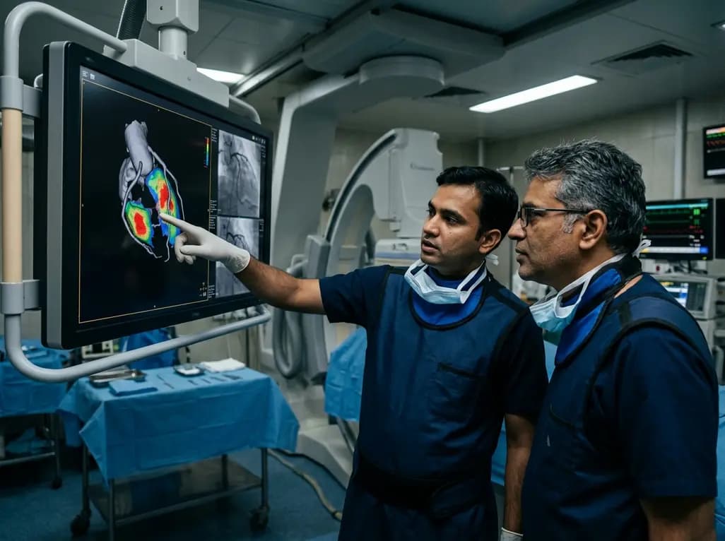
A groundbreaking study demonstrates that coronary angiography-derived radial wall strain reflects plaque morphology, predicting future rupture and erosion.
Today

The Delhi Cabinet has cleared an advanced seven-storey critical care expansion at Guru Tegh Bahadur Hospital under PMSSY Phase-IV. Costing Rs 244.87 crore, the new wing features 250 beds, specialized intensive care units, modern operation theatres, and round-the-clock emergency diagnostics for East Delhi.
Today

A landmark case demonstrates that favorable continuous glucose monitoring metrics cannot rule out SGLT2 inhibitor-associated euglycemic diabetic ketoacidosis. Clinicians must educate patients on symptom-based sick-day rules, uninterrupted basal insulin therapy, and proactive blood ketone testing.
Today
