

Loading, please wait...
Loading, please wait...
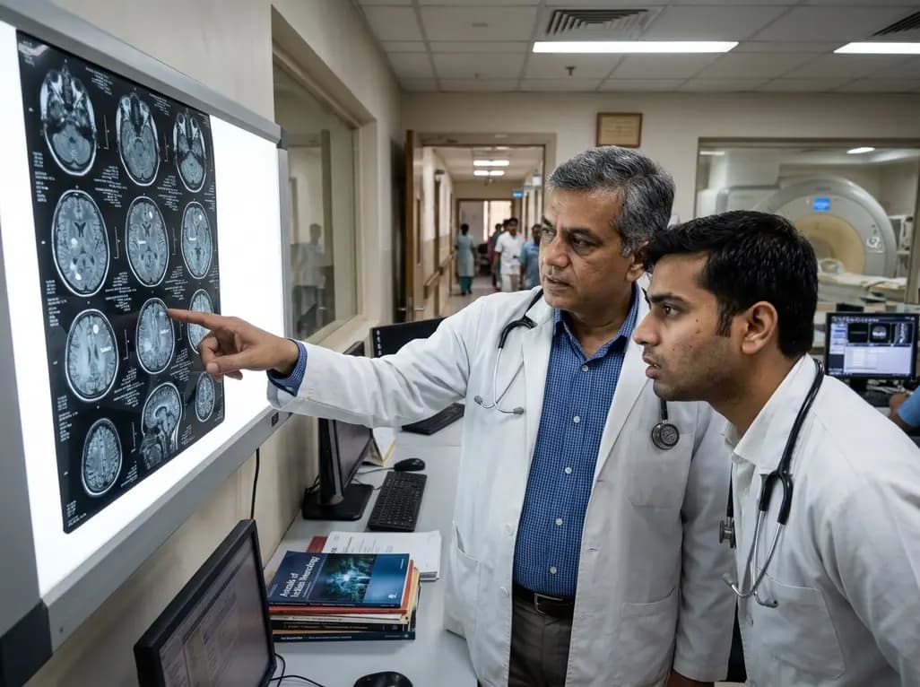
Central nervous system tuberculosis represents a devastating clinical manifestation of mycobacterial disease across India. When addressing intracranial tuberculomas, initiating prompt and effective brain tuberculosis treatment remains vital to avert neurological deficits. However, determining the precise therapeutic end point continues to provoke intense debate among physicians. Recent multicenter evidence reveals that many recovered patients take prolonged pharmacotherapy solely due to persistent MRI abnormalities. This lingering radiological enhancement frequently misleads clinicians into prescribing unnecessary medications.
Clinicians regularly evaluate therapeutic response through follow-up neuroimaging. However, neuroimaging abnormalities often outlast active mycobacterial proliferation. Specifically, a recent multicenter study in Lucknow evaluated 201 patients presenting with central nervous system tuberculomas. Investigators from King George's Medical University, SGPGI, and Era's Lucknow Medical College led this clinical evaluation. The researchers discovered that 41% of these individuals retained visible lesions on MRI after completing treatment. In contrast, only 35% of the cohort showed complete radiological clearance of their lesions. Consequently, many physicians mistakenly interpret stable parenchymal enhancements as persistent active disease. Therefore, practitioners frequently extend therapeutic regimens beyond twelve or even twenty-four months.
Prof Ravindra Kumar Garg and his colleagues emphasized that residual lesions usually represent inactive pathology. Non-viable tissue, reactive gliosis, localized edema, and dystrophic calcification frequently account for these ongoing imaging artifacts. Thus, persisting contrast enhancement alone does not confirm bacterial survival or therapeutic failure. Clinicians must recognize that anatomical recovery often lags months or years behind microbiological cure. As a result, relying solely on scan clearance drives substantial pharmaceutical toxicity and elevates economic burdens. Patients who are already cured endure prolonged therapeutic regimens without clinical gain.
During intracranial mycobacterial therapy, practitioners must frequently anticipate paradoxical immune responses. The Lucknow study documented that nearly 47% of patients developed paradoxical reactions during treatment. Under these circumstances, pre-existing tuberculomas enlarge or entirely new nodular lesions appear despite effective bactericidal activity. Mechanistically, this phenomenon arises from robust delayed-type hypersensitivity triggered by mycobacterial antigens released from dying bacilli. Consequently, unaccustomed clinicians often misidentify this immune-mediated deterioration as drug resistance or treatment failure.
Moreover, standard structural MRI sequences cannot reliably differentiate sterilizing granulomatous inflammation from living bacilli. While advanced spectroscopic sequences offer physiological detail, conventional scanners remain standard in clinical practice. For example, fibrous capsules, vascular remodeling, and localized mineral deposits retain paramagnetic contrast agents long after bacillary death. When clinicians lack this crucial understanding, they frequently add toxic second-line antitubercular medications or needlessly prolong first-line agents. Therefore, healthcare providers must interpret paradoxical reactions as signs of dynamic host immunity rather than therapeutic failure. Clinicians should confidently manage persistent cerebral swelling with targeted corticosteroids while maintaining baseline anti-tubercular therapy according to standard timelines.
Fortunately, overall long-term clinical prognosis remains positive when patients complete standard first-line regimens. In the multicenter investigation, approximately 49% of the cohort achieved complete recovery. This group experienced total symptom resolution accompanied by scan clearance. Furthermore, another 37% experienced marked neurological improvement despite retaining non-progressive structural lesions or minor complications. Most importantly, disease relapse emerged in only 3.5% of the participants across prolonged follow-up periods. This remarkably low relapse rate confirms that microbiological sterilization typically occurs within standard treatment durations.
Nevertheless, fear of disease recrudescence drives healthcare practitioners to overtreat asymptomatic individuals who harbor quiet parenchymal shadows. Unnecessary extensions of multidrug therapy carry substantial iatrogenic risks for the patient. For instance, prolonged use of isoniazid, rifampicin, and pyrazinamide heightens the risk of drug-induced hepatotoxicity. Additionally, extended therapy induces peripheral neuropathy, gastrointestinal distress, and profound fatigue. Excessive medication regimens also create hefty financial distress for families. Hence, specialists emphasize that physicians must treat the patient rather than chasing radiological perfection. When patients demonstrate robust neurological recovery and normal inflammatory markers, residual MRI lesions rarely signify viable organisms requiring protracted chemotherapy.
Establishing a definitive conclusion to intracranial antitubercular therapy requires a comprehensive clinical evaluation. The investigative team, including Dr Neeraj Kumar, Dr Imran Rizvi, and Dr Sanjay Singhal, underscored this message. They highlighted that clinicians should never base termination decisions exclusively on MRI findings. Instead, physicians must assess systemic wellness, resolution of fever, cessation of headaches, and stabilization of neurological signs. In addition, practitioners should evaluate serial imaging to verify that residual tuberculomas remain stable or progressively calcify over time. Clinicians must also examine systemic inflammatory indicators, such as erythrocyte sedimentation rates and C-reactive protein, to ensure quiet biochemistry.
However, physicians must exercise caution before applying rapid discontinuation protocols uniformly across all clinical presentations. Patients exhibiting progressive neurological deterioration, expanding mass effect, or suspected multidrug-resistant tuberculosis demand intensive microbiological scrutiny. Similarly, individuals with underlying immunocompromising conditions, such as HIV coinfection, require careful long-term surveillance. Practitioners must explicitly advise patients never to alter dosing schedules independently without physician guidance. Ultimately, adopting a holistic clinical strategy shields fully recovered individuals from chronic pharmacotherapy while safeguarding complex patients against premature cessation.
The findings published in the American Journal of Tropical Medicine and Hygiene present immediate clinical implications. Current national protocols frequently advocate 9 to 12 months of therapy for neurotuberculosis. However, ambiguous radiological criteria permit arbitrary treatment extensions lasting several years. Therefore, expert committees must formulate standardized radiological endpoints that distinguish active tuberculomas from quiescent cerebral scars. Furthermore, researchers advocate for the broader integration of advanced imaging biomarkers within clinical research trials.
Specialized modalities like magnetic resonance spectroscopy and diffusion-weighted MRI help confirm biological quiescence when conventional scans remain ambiguous. In addition, large prospective randomized controlled trials are urgently required to establish precise, safe cessation parameters for asymptomatic individuals. Such systematic evidence will provide frontline neurologists and general practitioners with the clinical confidence needed to halt therapy safely. Consequently, modern healthcare delivery will minimize adverse pharmaceutical reactions and curb needless healthcare expenditures across India. By shifting focus toward clinical recovery rather than cosmetic scan perfection, clinicians can protect recovering patients from the avoidable hazards of therapeutic inertia.
Q1: Why do MRI scans show abnormalities after brain tuberculosis treatment?
Magnetic resonance imaging frequently visualizes persistent tissue alterations rather than living mycobacteria. Following successful treatment, brain tuberculomas often resolve into quiescent fibrous scar tissue, localized gliosis, or dystrophic calcification. Contrast enhancement can persist across these healing tissues for months or years due to remodeled local vasculature. Therefore, residual imaging findings primarily represent sterile, structural remodeling rather than viable bacterial replication, meaning asymptomatic patients do not require prolonged chemotherapy.
Q2: How does a paradoxical reaction differ from treatment failure in CNS tuberculosis?
A paradoxical reaction represents an exaggerated host immune response occurring during effective antitubercular therapy. As bactericidal drugs destroy mycobacteria, released bacterial antigens stimulate intense localized delayed-type hypersensitivity. This causes existing brain tuberculomas to expand or new lesions to emerge on neuroimaging. Conversely, true therapeutic failure involves microbiological resistance accompanied by clinical deterioration. Paradoxical inflammation improves with adjunctive corticosteroids while clinicians maintain standard anti-TB medications without prematurely declaring regimen failure.
Q3: When can clinicians safely stop therapy if brain lesions persist on imaging?
Clinicians can safely discontinue medication when the patient achieves sustained clinical recovery and completes standard therapy duration. Safe discontinuation criteria require complete symptom resolution, absence of fever, stable neurological status, and normalized inflammatory markers. Furthermore, serial neuroimaging must confirm that residual tuberculomas remain stable or progressively calcify without active surrounding vasogenic edema. Decisions must rely on holistic medical evaluation rather than solely seeking complete scan clearance.
Disclaimer: This content is for informational and educational purposes only. It does not constitute medical advice or replace professional judgment. Refer to the latest local and national guidelines for clinical practice.
References

Read summarized clinical updates, watch expert medical content, and earn CME certifications right from your smartphone.
Open your smartphone camera to scan and install.





New clinical research shows that four in ten central nervous system tuberculosis patients continue anti-TB medications despite achieving full recovery. Persistent non-viable lesions on cranial MRI often mislead clinicians into prescribing unnecessary therapy, underscoring the urgent need for clinical-first decisions.
Today
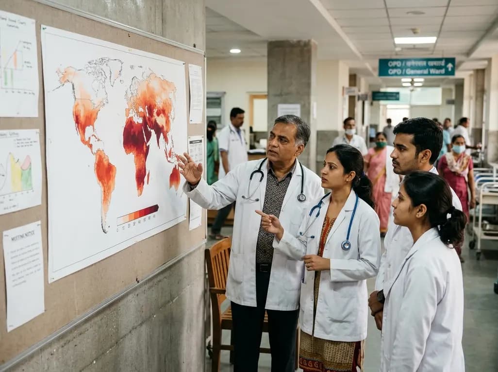
A landmark Lancet report reveals that heat claimed three million lives between 2016 and 2023, with over seventy percent linked to human activity. As healthcare systems face accelerating climate impacts, international experts urge unified policy action, resilient infrastructure, and targeted research investments.
Today

A KGMU study reveals over half of women with breast abnormalities fail to undergo diagnostic confirmation, leading to advanced-stage detection. Discover the socio-cultural barriers, clinical risks of incomplete resection, and community-level solutions needed to bridge the critical screening-to-diagnosis gap in India.
2 days back
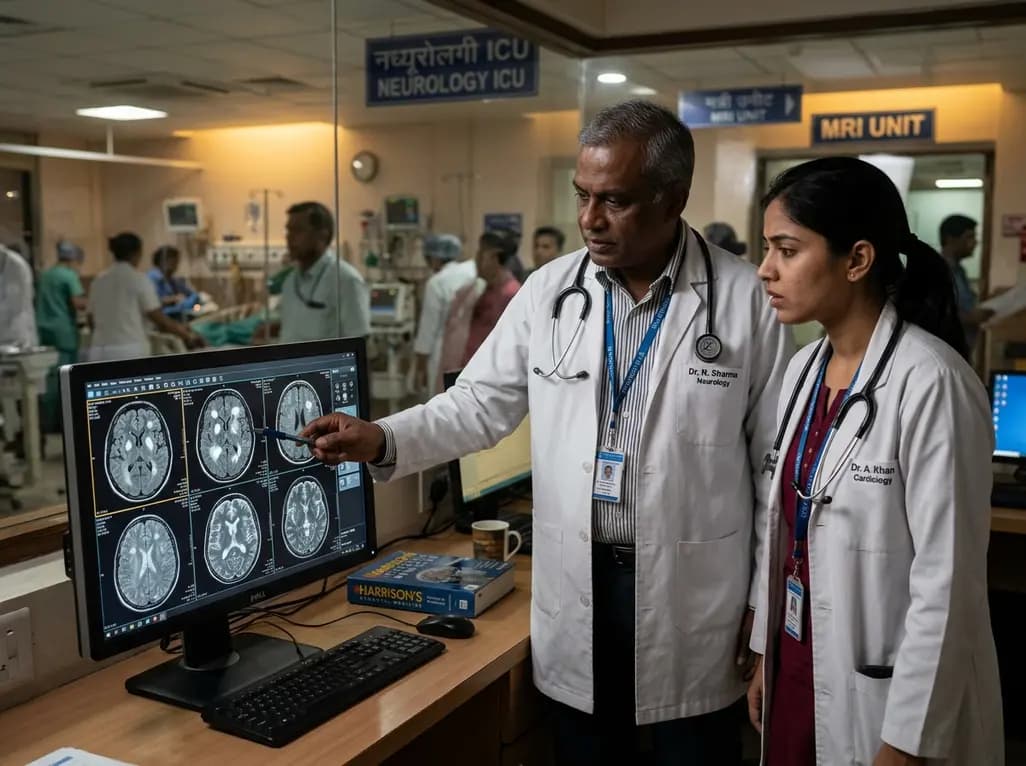
Recent findings from the VESUVE study demonstrate that brain MRI detects intracranial lesions in over 76% of patients with infective endocarditis. Acute ischemic stroke and subarachnoid hemorrhage independently elevate six-month mortality, underscoring the critical prognostic utility of neuroimaging.
Yesterday
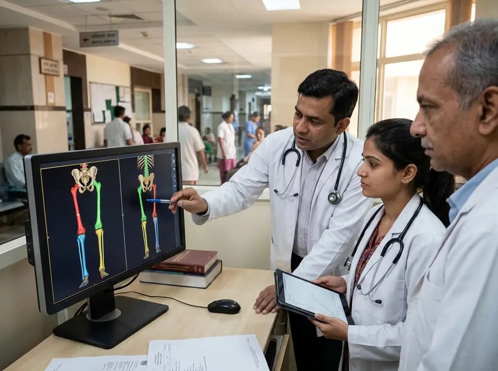
Modern 3D anthropometry reveals that pediatric leg growth ceases years before spinal elongation, exposing the clinical illusion of the classical Anderson-Green model. Learn how segmental growth dissociation improves surgical timing for epiphysiodesis in adolescent leg length discrepancy.
Yesterday

A meta-analysis of 18 studies reveals that bariatric surgery does not significantly alter circulating homocysteine levels, regardless of follow-up duration. Clinicians must maintain vigilance over post-surgical micronutrient supplementation to prevent hyperhomocysteinemia.
Yesterday
