

Loading, please wait...
Loading, please wait...
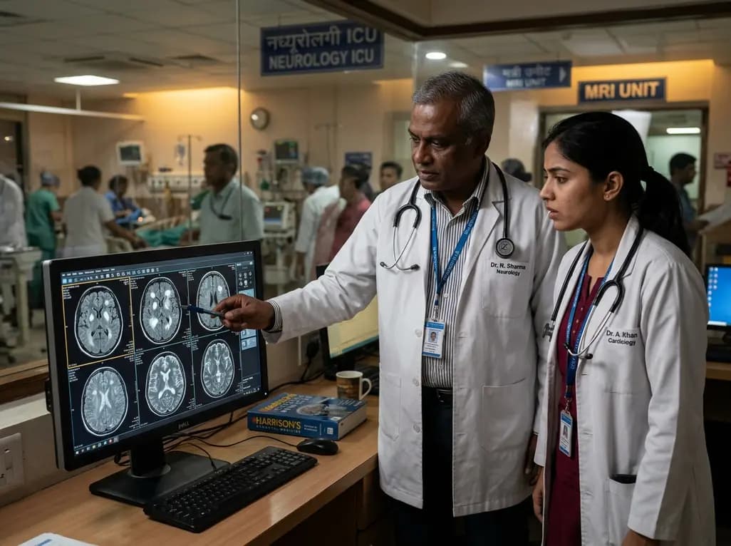
Infective endocarditis remains a formidable cardiovascular challenge associated with substantial in-hospital morbidity and mortality. Clinicians frequently encounter secondary central nervous system embolization during the disease trajectory. Therefore, early identification of intracranial pathologies represents a paramount diagnostic goal. Historically, physicians relied primarily on computed tomography to detect macrovascular strokes or gross hemorrhage. However, conventional computed tomography fails to identify subtle microstructural damage. Consequently, performing a brain MRI in infective endocarditis has gained remarkable momentum across tertiary care centers. Recent evidence from the VESUVE multicenter registry sheds new light on cerebral involvement. In this study, investigators systematically evaluated 212 patients with definite endocarditis using standardized magnetic resonance imaging protocols. The findings revealed that more than 76% of all hospitalized patients exhibited distinct intracranial abnormalities. Furthermore, a senior neuroradiologist reviewed all neuroimaging scans, ensuring diagnostic consistency across the cohort. Because cerebral emboli frequently occur prior to antibiotic initiation, rapid neuroimaging provides pivotal insight into systemic disease severity. Clinicians can thereby establish an accurate baseline before complications escalate. Ultimately, routine magnetic resonance screening shifts clinical practice toward proactive neurovascular risk assessment.
The spectrum of intracranial damage uncovered by magnetic resonance imaging extends well beyond overt focal deficits. Indeed, silent neurological events occur with striking frequency in endocarditis patients. Within the VESUVE study cohort, acute ischemic stroke was detected in over 60% of patients. In addition, cerebral microbleeds emerged as the single most prevalent radiological finding, affecting more than 62% of individuals. These microbleeds signify subclinical petechial extravasation from microemboli or localized vasculitis. Moreover, investigators observed hemorrhagic transformation in nearly 20% of patients, demonstrating the fragile nature of ischemic brain tissue. Subarachnoid hemorrhage developed in over 12% of subjects, whereas intracranial mycotic aneurysms appeared in 5%. Clinicians also detected sulcal microaneurysms in nearly 24% of scans. Importantly, true brain abscesses occurred infrequently, developing in only 1.5% of cases. These diverse findings prove that clinically silent emboli continuously seed the cerebral vasculature during active bacteremia. Therefore, bedside neurological examinations substantially underestimate tissue injury. Incorporating advanced diffusion-weighted imaging and susceptibility-weighted sequences allows clinicians to detect occult embolic cascades early. Consequently, imaging clarifies diagnostic categorization under contemporary criteria.
Neurological complications decisively dictate overall survival in infective endocarditis. In the VESUVE cohort, the overall six-month mortality rate reached 19%. However, mortality differed dramatically based on specific intracranial findings observed on neuroimaging. Most notably, the presence of acute ischemic stroke increased six-month mortality to nearly 27%. Statistical regression confirmed that acute ischemic stroke independently multiplied the odds of death more than fivefold. Even more strikingly, patients presenting with subarachnoid hemorrhage faced a devastating six-month mortality rate of 44%. Multivariable analysis demonstrated that subarachnoid hemorrhage increased mortality risk more than fourfold. In contrast, isolated cerebral microbleeds and small asymptomatic sulcal aneurysms did not independently predict premature death. This crucial distinction helps physicians stratify clinical risk with superior precision. When clinicians identify either acute ischemic infarction or subarachnoid hemorrhage, they must recognize an exceptionally high-risk clinical phenotype. Consequently, these radiological biomarkers alert the medical team to imminent hemodynamic or neurological decompensation. Early risk stratification therefore empowers clinicians to intensify supportive monitoring. Thus, magnetic resonance imaging provides objective prognostic intelligence that transcends traditional clinical scores alone.
Managing surgical indications in endocarditis patients with intracranial lesions requires nuanced clinical judgment. Urgent cardiac valve surgery frequently offers the only definitive cure for severe valvular destruction or refractory heart failure. Nevertheless, operating during active cerebral ischemia raises substantial safety concerns. Cardiopulmonary bypass and mandatory systemic intraoperative anticoagulation can trigger catastrophic intracranial hemorrhage within freshly infarcted brain parenchyma. Conversely, unnecessary surgical delays expose the patient to persistent septic embolization and irreversible cardiogenic shock. Modern cardiology guidelines suggest proceeding promptly with urgent valve repair when neurological damage remains limited and stable. However, if neuroimaging identifies extensive parenchymal hemorrhage or extensive cerebral infarction, postponing surgery for several weeks remains prudent. Magnetic resonance imaging precisely differentiates bland microinfarcts from high-risk cortical hemorrhages and vascular aneurysms. Furthermore, identifying unruptured mycotic aneurysms prompts early endovascular coiling prior to valve replacement. Therefore, routine magnetic resonance scanning resolves critical diagnostic dilemmas regarding surgical timing. Consequently, tailored neuroimaging ensures optimal operative timing without compromising cerebral safety.
Optimal outcomes in infective endocarditis require seamless collaboration across medical subspecialties. Specialized endocarditis teams must unite cardiologists, infectious disease physicians, neurologists, cardiac surgeons, and neuroradiologists. Because intracranial lesions occur silently in most patients, institutional protocols should establish standardized access to advanced neuroimaging. Magnetic resonance imaging should include diffusion-weighted, susceptibility-weighted, and fluid-attenuated inversion recovery sequences. In addition, contrast-enhanced vascular imaging remains vital to detect developing mycotic aneurysms before rupture occurs. When ischemic stroke occurs, intravenous thrombolysis remains strictly contraindicated due to prohibitive hemorrhagic transformation risks. Instead, clinicians should consider mechanical thrombectomy for emergent large-vessel occlusions when technically feasible. Simultaneously, physicians must administer targeted bactericidal intravenous antibiotics to sterilize vegetations and eliminate ongoing embolic threats. Repeat neuroimaging remains essential if the patient exhibits new headache, mental status changes, or neurological deficits. In conclusion, systematic brain imaging provides invaluable prognostic clarity and directly shapes therapeutic decision-making. Through coordinated multidisciplinary vigilance, healthcare teams can successfully mitigate neurological catastrophe and dramatically improve patient survival.
Clinicians should consider brain MRI because more than seventy percent of endocarditis patients harbor occult intracranial lesions. Standard neurological examinations frequently miss silent microinfarctions, microbleeds, and asymptomatic mycotic aneurysms. Detecting these subclinical vascular events fulfills major and minor diagnostic criteria. Furthermore, identifying silent cerebral injury stratifies long-term mortality risk and prevents catastrophic perioperative hemorrhage if urgent valve surgery becomes necessary. Consequently, early imaging guides critical medical and surgical decisions.
According to recent clinical investigations, acute ischemic stroke and subarachnoid hemorrhage represent the strongest independent predictors of six-month mortality. Acute ischemic stroke increases the odds of death fivefold, whereas subarachnoid hemorrhage carries an alarming forty-four percent mortality rate. Conversely, incidental cerebral microbleeds and asymptomatic sulcal microaneurysms do not independently worsen survival. Therefore, identifying acute ischemia or subarachnoid blood signals an aggressive disease course requiring intensive multidisciplinary management and hemodynamic stabilization.
Finding an acute ischemic stroke on neuroimaging requires careful multidisciplinary evaluation rather than an automatic surgical cancellation. If the stroke is small, non-hemorrhagic, and the patient suffers from severe valve dysfunction or heart failure, clinicians should proceed with urgent cardiac surgery without unnecessary delay. However, if the brain scan demonstrates extensive territorial infarction or secondary hemorrhagic transformation, guidelines recommend delaying surgery for at least two to four weeks to minimize lethal intraoperative intracranial bleeding.
Disclaimer: This content is for informational and educational purposes only... Refer to the latest local and national guidelines for clinical practice.
References
Clottes P et al. Prevalence and prognostic significance of intracranial lesions assessed with MRI in infective endocarditis patients: Data from the Vesuve study. Rev Neurol (Paris). 2026 Oct 26. doi: undefined. PMID: 42934033.
Delgado V, Ajmone Marsan N, de Waha S, et al. 2023 ESC Guidelines for the management of endocarditis. Eur Heart J. 2023;44(39):3948-4042.
Ahn Y, Joo L, Suh CH, et al. Impact of Brain MRI on the Diagnosis of Infective Endocarditis and Treatment Decisions: Systematic Review and Meta-Analysis. AJR Am J Roentgenol. 2022;218(6):958-968.

Read summarized clinical updates, watch expert medical content, and earn CME certifications right from your smartphone.
Open your smartphone camera to scan and install.





Recent findings from the VESUVE study demonstrate that brain MRI detects intracranial lesions in over 76% of patients with infective endocarditis. Acute ischemic stroke and subarachnoid hemorrhage independently elevate six-month mortality, underscoring the critical prognostic utility of neuroimaging.
Today

A KGMU study reveals over half of women with breast abnormalities fail to undergo diagnostic confirmation, leading to advanced-stage detection. Discover the socio-cultural barriers, clinical risks of incomplete resection, and community-level solutions needed to bridge the critical screening-to-diagnosis gap in India.
Yesterday

A controlled evaluation reveals that AI raters in clinical LLM benchmarking disagree significantly due to divergent scoring models, challenging automated healthcare AI validation.
Today

A phase I/II trial evaluated E. coli-derived rhBMP-2 combined with beta-tricalcium phosphate for long-bone nonunion. The combination demonstrated high radiographic union, functional limb recovery, undetectable systemic absorption, and transient antibody formation without clinical adverse events.
Today

A meta-analysis of 18 studies reveals that bariatric surgery does not significantly alter circulating homocysteine levels, regardless of follow-up duration. Clinicians must maintain vigilance over post-surgical micronutrient supplementation to prevent hyperhomocysteinemia.
Today
