.webp&w=384&q=75)

Loading, please wait...
Loading, please wait...

Decompressive craniectomy remains a life-saving intervention for severe intracranial hypertension following traumatic injuries or major cerebrovascular events. Consequently, reconstruction of the cranial vault through autologous cranioplasty is essential to restore cerebral protection and normalize cerebrospinal fluid dynamics. However, autologous bone grafts carry a notable clinical complication termed bone flap resorption. Bone flap resorption represents the gradual aseptic osteolysis and structural degradation of the reimplanted calvarial bone. Therefore, neurosurgeons frequently witness progressive thinning, structural instability, and cosmetic deformity across long-term follow-up.
Recent clinical investigations reveal that resorption rates vary widely across neurosurgical cohorts, spanning from low single digits to extensive destruction. Furthermore, storing bone flaps in specialized bone tissue banks helps reduce graft contamination. Tissue banking protocols provide strict sterilization and cold storage to preserve bone architecture. Nevertheless, cryopreservation does not completely prevent biological degradation once surgeons reimplant the graft into vascularized host tissues. Thus, understanding the pathophysiological trajectory of aseptic resorption is vital for timely surgical decision-making. Clinicians must identify high-risk individuals early to optimize post-cranioplasty outcomes and prevent unnecessary secondary operations.
Accurate assessment of osteolytic progression requires standardized and reproducible radiological classification criteria. Therefore, clinicians increasingly rely on validated tools such as the Oulu Resorption Scale to quantify cranial defect remodeling. This system systematically categorizes patients into four distinct structural tiers based on computed tomography findings. Specifically, these categories encompass no bone flap resorption, mild resorption, intermediate resorption, and severe resorption. Mild resorption typically involves superficial cortical thinning without compromising overall mechanical stability. In contrast, intermediate resorption displays distinct areas of full-thickness bone erosion that challenge cranial architecture.
Severe resorption exhibits extensive osteolysis exceeding fifty percent of the original graft thickness and surface area. Consequently, severe cases frequently generate mobile cranial segments, profound cosmetic irregularities, and painful sunken flaps. Longitudinal observations demonstrate that severe resorption develops rapidly, occurring predominantly within the first postoperative year. In fact, severe structural degradation manifests at a mean interval of approximately three hundred days after cranioplasty. Conversely, resorption occurring after the initial twelve months usually remains mild or intermediate. Hence, systematic classification ensures precise monitoring and guides surgical planning for potential reconstructive revisions.
Identifying predisposing variables enables surgical teams to predict which patients face heightened osteolysis risks. Recent clinical analyses show that primary pathology plays a foundational role in subsequent graft survival. Specifically, patients who undergo decompressive craniectomy for traumatic brain injury experience significantly higher resorption rates than those treated for stroke. Traumatic injuries provoke massive systemic inflammatory surges that recruit circulating immune cells to the healing bone. Moreover, elevated pro-inflammatory cytokines stimulate intense osteoclast differentiation while actively suppressing local osteoblast activity. Trauma patients also frequently sustain microscopic skull fractures during the initial injury, which impairs microvascular revascularization.
In addition, active cigarette smoking emerges as another potent independent risk factor for graft failure. Nicotine and toxic tobacco metabolites induce profound peripheral vasoconstriction and impair capillary sprouting into reimplanted autografts. Furthermore, chronic hypoxia prevents proper osteocyte integration and accelerates avascular necrosis of the stored bone matrix. Consequently, clinicians must emphasize strict smoking cessation before and after reconstruction to protect graft viability. Addressing these modifiable and biological hazards helps safeguard the cranial reconstruction against early graft breakdown.
Beyond patient-related variables, surgical anatomy and mechanical characteristics directly influence the rate of bone degradation. Retrospective multivariate modeling demonstrates that a cranial defect surface area exceeding ninety-two square centimeters significantly increases resorption likelihood. Larger calvarial gaps demand extensive neovascularization from surrounding dural and scalp capillary beds. However, revascularizing extensive bone surfaces requires substantial metabolic support that the host bed cannot always provide. Consequently, the central zones of large autologous grafts often remain ischemic and undergo progressive osteoclastic resorption.
Furthermore, the physical integrity of the cranial bone flap strongly governs postoperative longevity. Having a single intact flap serves as a significant protective factor against pathological osteolysis. Conversely, fragmented grafts comprising multiple separate pieces markedly elevate resorption rates. When surgeons piece together fragmented flaps, the exposed cancellous bone surface area expands dramatically. This expanded surface area exposes abundant necrotic bone marrow elements and triggers extensive foreign body reactions. Additionally, fibrous scar tissue quickly invades inter-fragmentary spaces, preventing osteoblast bridging and hastening structural collapse. Thus, single-piece cranial grafts provide vastly superior durability compared to fragmented reconstructions.
Standardized bone tissue banking represents a sophisticated storage method designed to overcome traditional preservation pitfalls. Historically, subcutaneous abdominal pockets or unmonitored hospital freezers produced elevated surgical site infection rates. In contrast, certified bone tissue banks utilize controlled-rate freezing, strict microbiological screening, and liquid nitrogen storage protocols. Clinical data confirm that tissue bank-preserved bone flaps achieve remarkably low infection rates of approximately 1.6 percent. Nevertheless, low infection rates do not eliminate the non-infectious cellular processes that drive aseptic graft resorption.
Therefore, neurosurgical teams must establish proactive, long-term surveillance protocols for all autologous cranioplasty recipients. High-resolution computed tomography scans at six, twelve, and twenty-four months allow early detection of asymptomatic osteolysis. Surveillance becomes particularly crucial during the first postoperative year when severe structural collapse most frequently occurs. If progressive bone loss destabilizes the construct, surgeons should plan an elective revision cranioplasty promptly. Modern synthetic alternatives, such as titanium mesh or polyetheretherketone implants, provide excellent structural durability for revision cases. Ultimately, structured surveillance safeguards neurological recovery and prevents sudden cosmetic or functional reconstruction failures.
Severe bone flap resorption involves extensive osteolysis and marked loss of cranial graft structural integrity. Clinicians identify it when osteolysis exceeds fifty percent of the original graft thickness or volume on imaging. This severe structural compromise frequently destabilizes the construct, resulting in contour deformities and necessitating surgical revision with alloplastic materials.
Traumatic brain injury elevates resorption risk through profound systemic and local inflammatory cascades. Circulating inflammatory cytokines actively drive osteoclast differentiation and osteolysis. In addition, trauma patients frequently sustain microfractures within the graft and exhibit altered osteogenesis. Furthermore, these patients are often younger, which independently accelerates metabolic bone remodeling and graft degradation.
Bone tissue banking offers strict sterilization, controlled freezing, and regulated storage protocols that dramatically suppress microbial contamination. Recent clinical investigations demonstrate postoperative surgical site infection rates as low as 1.6 percent. However, cryopreservation preserves structural proteins while non-viable cells undergo osteoclastic turnover, necessitating careful monitoring for aseptic osteolysis over time.
Disclaimer: This content is for informational and educational purposes only... Refer to the latest local and national guidelines for clinical practice.
References

Read summarized clinical updates, watch expert medical content, and earn CME certifications right from your smartphone.
Open your smartphone camera to scan and install.





A clinical analysis of autologous cranioplasty with tissue bank-stored grafts highlights key risk factors for bone flap resorption, including smoking, defect size, and traumatic brain injury, alongside low infection rates and critical first-year surveillance windows.
Today
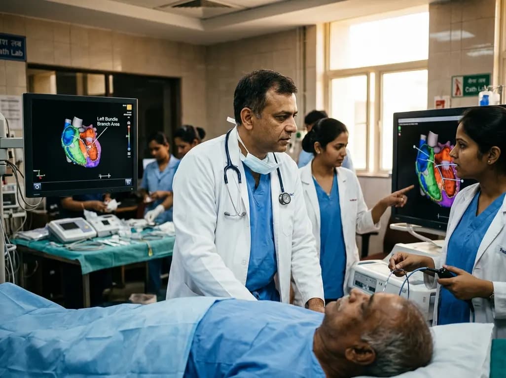
Left bundle branch pacing is an emerging physiological resynchronization strategy for heart failure. While observational data and some trials show benefits over biventricular pacing, divergent trial outcomes highlight the importance of confirmed conduction capture and specialized operator technique.
Today

A retrospective study of 86,012 pregnancies demonstrates that size-selective cfDNA enrichment boosts mean fetal fraction to 18.88% and achieves a 59.20% PPV for fetal copy number variants. However, 85.83% of CNVs showed size discrepancies, underscoring the mandatory role of confirmatory microarray testing.
Today

Radiofrequency catheter ablation of the cavotricuspid isthmus successfully terminates typical atrial flutter. This article reviews the mechanisms driving coronary sinus electrogram changes, wavefront activation pathways, and clinical criteria to verify bidirectional block during electrophysiology studies.
Today

A recent investigation examined load-dependent kinetic and kinematic profiles during two-way ballistic exercise across 30% to 80% 1-RM. Findings highlight load effects on velocity, impulse, and power, offering sports physicians objective guidance for neuromuscular conditioning and rehabilitation.
Today
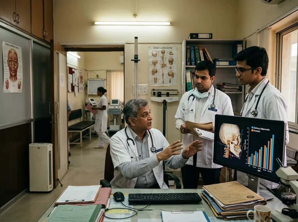
A cross-sectional study demonstrates that increased BMI in adolescents significantly enhances mandibular length and alters salivary adipokines like adiponectin and resistin, providing novel insights into craniofacial growth and orthodontic planning.
Today
