.webp&w=384&q=75)

Loading, please wait...
Loading, please wait...
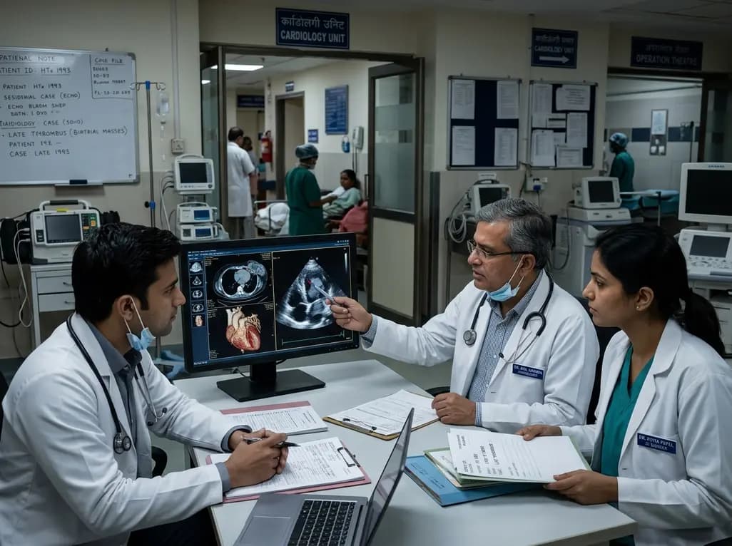
Orthotopic heart transplantation remains the definitive therapy for selected patients facing end-stage heart failure. However, clinicians occasionally encounter severe late complications decades after the initial procedure. The development of a post-transplant atrial thrombus represents an exceptionally rare yet perilous clinical entity, particularly in long-term transplant survivors. Historically, surgeons frequently utilized the Shumway biatrial anastomosis technique to reconstruct the cardiac chambers. Although this traditional surgical method offered technical simplicity and reduced operative ischemic time, it inherently disrupted native atrial architecture. Specifically, the biatrial approach retains substantial portions of recipient atrial tissue attached to donor atrial cuffs. Consequently, this surgical configuration creates enlarged, distorted atrial cavities characterized by abnormal geometry. Over subsequent decades, these altered chambers develop progressive electromechanical dissociation, asynchronous contraction, and persistent blood stasis. In this notable clinical case, a 63-year-old male presented three decades after heart transplantation with massive bilateral intracardiac clot formation. Therefore, clinicians must recognize that surgical anatomy established thirty years prior can continue to generate profound hemodynamic vulnerabilities. Modern cardiologists and transplant teams must maintain vigilance regarding late anatomical deterioration in this aging cohort.
Patients harboring extensive intracardiac masses frequently present with progressive dyspnea, exercise intolerance, or sudden hemodynamic compromise. In this clinical scenario, the patient developed worsening exertional dyspnea that prompted urgent diagnostic evaluation. Multimodality cardiovascular imaging plays a central diagnostic role when evaluating suspected intracardiac pathology. Transthoracic echocardiography typically provides the initial bedside evaluation, revealing large intracavitary echogenic structures. However, transesophageal echocardiography offers markedly superior resolution for visualizing the atrial appendages and posterior atrial walls. Furthermore, cardiac magnetic resonance imaging and contrast-enhanced computed tomography allow precise tissue characterization. These advanced imaging modalities help clinicians differentiate avascular organized thrombus from hypervascular neoplastic tissue or mobile infective vegetations. In this case, multimodality imaging confirmed extensive, mobile masses occupying significant portions of both atria. Consequently, severe mechanical obstruction across the atrioventricular valves produced acute diastolic filling impairment and progressive pulmonary congestion. Clinicians must immediately recognize that subtle respiratory complaints in transplant recipients may reflect severe structural obstruction rather than simple respiratory infection.
When physicians encounter an intracardiac mass in a cardiac transplant recipient, they must systematically evaluate several potential etiologies. Intracardiac thrombus remains a critical diagnostic consideration, but several other pathological entities present identically on initial scans. First, benign neoplasms such as cardiac myxomas or fibroelastomas can originate along the endocardial surfaces. Second, immunosuppressed recipients harbor an elevated lifetime risk of post-transplant lymphoproliferative disorder, which can manifest as infiltrative intracavitary cardiac tumors. Third, opportunistic infective endocarditis with large friable vegetations must remain high on the differential list. Fourth, structural pseudomasses, such as prominent suture lines or retained native atrial remnants, frequently mimic pathological lesions. Distinguishing among these diverse clinical conditions requires careful correlation of clinical history, laboratory biomarkers, and tissue imaging characteristics. In the reported patient, urgent surgical exploration and histopathological examination confirmed extensive organized thrombi without malignant cells or microorganisms. The largest extracted clot measured an astonishing 88 by 47 by 37 millimeters. Therefore, clinicians must combine tissue pathology with high-resolution imaging to confirm the definitive diagnosis and guide subsequent therapy.
Multiple interrelated pathophysiologic factors drive intra-atrial clot propagation in long-term heart transplant survivors. Virchow's classic triad—endothelial injury, circulatory stasis, and hypercoagulability—operates aggressively within this specialized population. First, the surgical suture lines along the anastomotic junctions create permanent endocardial irregularities that serve as persistent thrombogenic niduses. Second, chronic atrial dilation combined with loss of coordinated atrial kick promotes profound blood stasis. Third, cardiac allograft vasculopathy induces chronic microvascular ischemia, contributing to progressive myocardial fibrosis and regional wall motion abnormalities. In addition, long-term immunosuppressive regimens, systemic inflammatory states, and metabolic comorbidities can generate a hypercoagulable milieu. In many instances, the temporary interruption or subtherapeutic dosing of systemic anticoagulation triggers rapid thrombus expansion over pre-existing structural niduses. Consequently, even a brief lapse in antithrombotic coverage can allow massive clots to organize and occlude critical inflow tracts. Clinicians must rigorously evaluate each patient's individual prothrombotic risks to deliver timely preventive therapy.
The acute management of massive biatrial thrombi demands rapid multidisciplinary coordination between cardiac surgeons, intensivists, and cardiologists. When clots produce critical hemodynamic obstruction or demonstrate high embolic potential, surgical thrombectomy represents the definitive treatment of choice. In this challenging case, surgeons successfully performed an urgent bilateral thrombectomy to restore unobstructed intracardiac blood flow. However, surgical manipulation and underlying coronary pathology can precipitate severe perioperative crises. Postoperatively, this patient developed refractory cardiogenic shock secondary to an acute thromboembolic occlusion of the left anterior descending coronary artery. Consequently, the clinical team immediately instituted venoarterial extracorporeal membrane oxygenation to provide crucial mechanical circulatory support. Simultaneously, interventional cardiologists performed emergency percutaneous coronary intervention to restore epicardial myocardial perfusion. Through intensive hemodynamic monitoring, careful inotropic titration, and structured critical care protocols, the team successfully stabilized cardiac function. Eventually, the patient was successfully decannulated from extracorporeal membrane oxygenation support, demonstrating the life-saving potential of timely mechanical intervention.
Following surgical stabilization, long-term therapeutic planning requires meticulous secondary prevention and structured multidisciplinary surveillance. Clinicians must initiate guideline-directed medical therapy for heart failure to support allograft recovery and optimize ventricular loading conditions. Furthermore, uninterrupted therapeutic anticoagulation is vital to prevent recurrent intra-atrial thrombosis, particularly when underlying anatomical distortions persist. Physicians must carefully balance thromboembolic risks against potential bleeding complications, considering drug-drug interactions with maintenance immunosuppressive agents. In addition, long-term care plans should incorporate routine transthoracic echocardiography to detect silent clot reaccumulation or progressive allograft vasculopathy. When patients exhibit complex comorbidities or limited physiological reserve, structured palliative care teams provide essential holistic support. In this case, following complete clinical stabilization and initiation of heart failure therapies, the patient transitioned to a structured palliative and outpatient follow-up framework. Therefore, successful long-term management requires continuous collaboration across transplant cardiology, hematology, cardiac surgery, and primary care specialties.
The Shumway biatrial technique joins donor atrial cuffs directly to preserved recipient atria, creating abnormally enlarged and geometrically distorted chambers. Over decades, this surgical design causes profound electromechanical dysfunction, severe asynchronous wall motion, and localized blood stasis. Furthermore, permanent intra-atrial suture lines serve as persistent thrombogenic niduses. When combined with systemic hypercoagulability or interrupted anticoagulation, these anatomical factors significantly accelerate clot formation.
Clinicians utilize multimodality cardiovascular imaging, including transesophageal echocardiography, contrast-enhanced computed tomography, and cardiac magnetic resonance imaging, to characterize intracardiac masses. Thrombi typically appear avascular, display low contrast enhancement, and attach near suture lines or stagnant blood zones. Conversely, neoplasms like myxomas or lymphomas demonstrate active vascularity, contrast uptake, and tissue infiltration. Definitive differentiation often requires histopathological evaluation following surgical excision.
Late cardiac transplant survivors require regular clinical monitoring by a multidisciplinary team to detect silent structural and vascular complications. Recommended protocols include annual or biannual transthoracic echocardiography, regular coronary evaluation for cardiac allograft vasculopathy, and periodic screening for hypercoagulability. Clinicians must also monitor anticoagulation adherence closely, review drug-drug interactions with immunosuppressants, and utilize transesophageal imaging promptly if patients report unexplained dyspnea.
Disclaimer: This content is for informational and educational purposes only. It is not intended to substitute professional medical advice, diagnosis, or treatment. Always consult a qualified healthcare provider with any questions regarding medical conditions or clinical decisions. Refer to the latest local and national guidelines for clinical practice.
References

Read summarized clinical updates, watch expert medical content, and earn CME certifications right from your smartphone.
Open your smartphone camera to scan and install.





A rare case of massive biatrial thrombus 3 decades post-heart transplant highlights the long-term anatomical risks of the Shumway technique, surgical thrombectomy, ECMO rescue, and vital anticoagulation.
last month
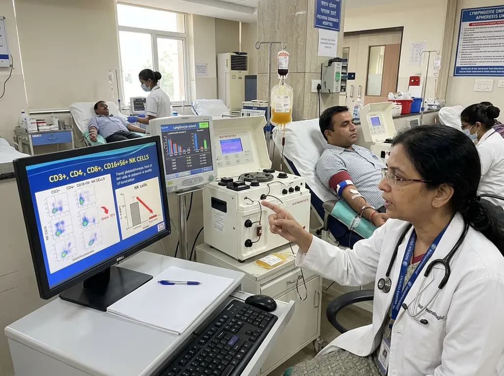
A landmark study assesses how frequent plateletpheresis alters peripheral immune profiles. Regular donors demonstrate notable reductions in circulating natural killer cells, emphasizing the critical need for routine lymphocyte subset surveillance in long-term blood donor safety programs.
Today
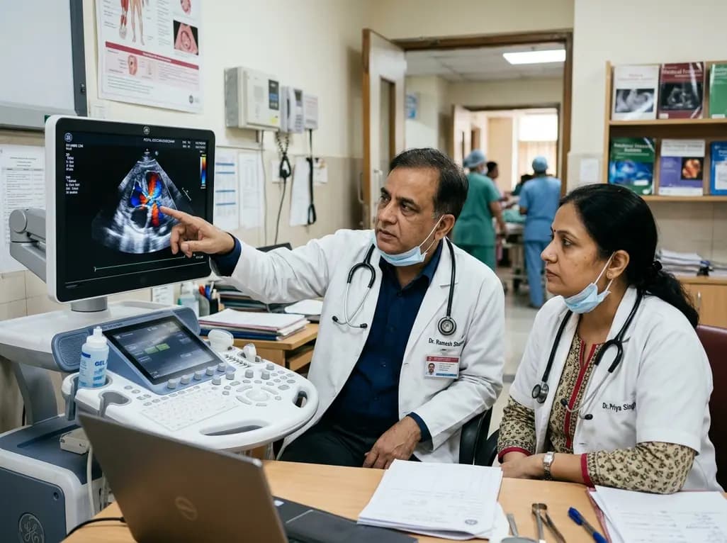
Fetal foramen ovale restriction typically emerges in the third trimester with right heart enlargement. Evidence demonstrates it does not independently mandate cesarean delivery, and over 93% of postnatal cardiac findings resolve spontaneously within one year, supporting conservative obstetric management.
Today
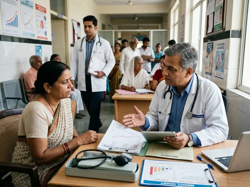
The latest WHO Global Health Estimates highlight India's life expectancy reaching 71 years alongside a healthy lifespan of 60.6 years. While longevity rebounds post-pandemic, non-communicable conditions now account for three-quarters of top mortalities, underscoring urgent clinical shifts in routine primary care.
Today
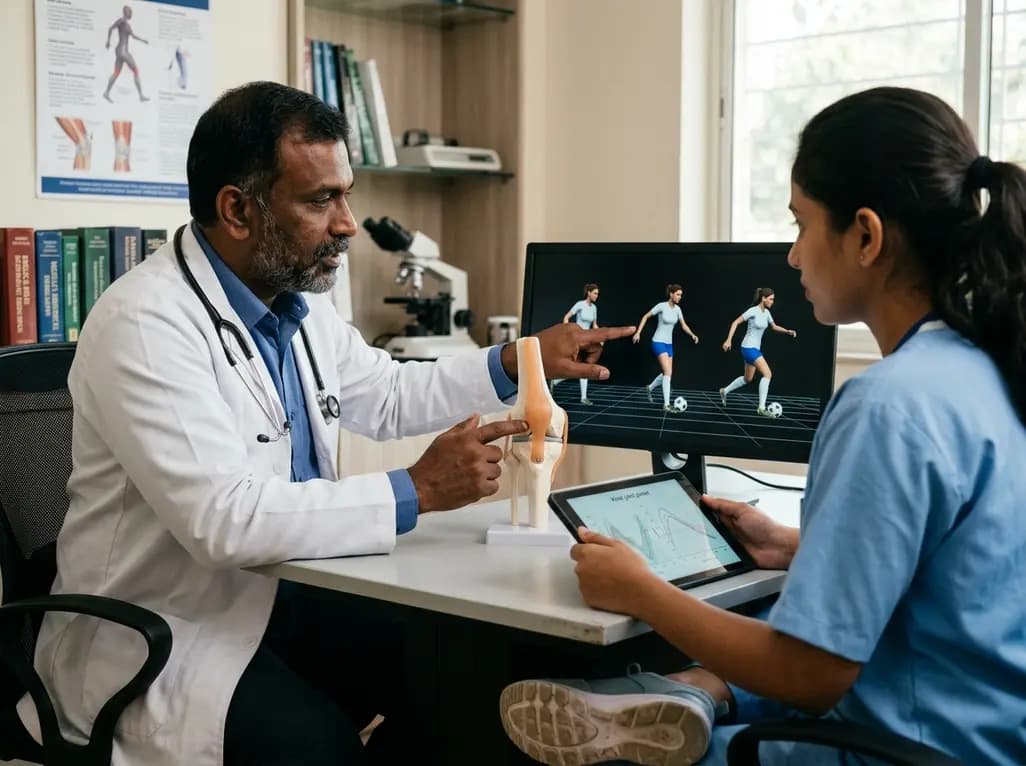
New biomechanical research reveals that foot-strike angle significantly influences knee energy absorption during cutting tasks in female soccer players. Understanding these kinematic patterns offers clinicians valuable pathways for targeted ACL injury prevention and athletic retraining.
Today

A comprehensive review analyzes periconceptional GLP-1 receptor agonist exposure, revealing reassuring data on congenital anomalies and adverse perinatal outcomes while emphasizing the need for cautious patient counseling.
Today
