

Loading, please wait...
Loading, please wait...

Modern oral rehabilitation increasingly relies on computer-assisted protocols to optimize fixture positioning and protect vital anatomic structures. In contemporary implantology, clinicians frequently deliberate between dynamic optical tracking and static physical guides. A recent benchmark investigation evaluated the precision of augmented reality implant surgery relative to conventional template-guided workflows across standardized anatomical models. This comparative evaluation delivers valuable technical benchmarks for dental surgeons who seek predictable prosthetically driven outcomes.
Digital dentistry continues to transform restorative care through rapid technological innovation. Historically, static surgical guides represented the standard benchmark for computer-aided implantology. These resin templates faithfully transfer virtual cone-beam computed tomography plans to the surgical field via mechanical sleeves. However, physical templates possess notable limitations. For instance, they frequently restrict surgical irrigation, obscure direct optical inspection, and limit interocclusal clearance in posterior regions. Consequently, researchers developed dynamic computer-assisted navigation systems to overcome these physical constraints.
More recently, head-mounted displays such as the Microsoft HoloLens 2 have introduced wearable mixed reality into oral surgery. Through augmented reality implant surgery, clinicians project three-dimensional anatomical reconstructions and planned drilling trajectories directly over the patient's maxilla. Therefore, surgeons maintain an uninterrupted line of sight without shifting their gaze toward distant external monitors. Moreover, this optical integration preserves vital tactile sensitivity during osteotomy preparation. Although wearable technology presents immense ergonomic benefits, operators must thoroughly validate whether virtual overlays provide geometric precision comparable to rigid mechanical guides. Determining these accuracy parameters remains essential before adopting wearable headsets into routine restorative workflows.
To establish definitive accuracy benchmarks, researchers conducted a controlled in-vitro comparative study utilizing twenty standardized maxillary bone models. In total, the operators executed eighty implant placements across identical anatomical conditions. Specifically, each maxillary model received four osteotomies to balance experimental variables and eliminate structural bias. Two implants per model utilized wearable head-mounted guidance via the Microsoft HoloLens 2 platform. Conversely, the remaining two fixtures utilized static template-guided insertion planned through advanced SMOP software.
Furthermore, the investigators implemented a rigorous digital evaluation workflow to quantify spatial deviations. Prior to drilling, technicians generated comprehensive cone-beam computed tomographic scans and optical surface registrations to formulate standardized prosthetically driven plans. Following implant insertion, investigators acquired postoperative volumetric scans of each maxillary model. Subsequently, advanced three-dimensional superimposition algorithms aligned postoperative datasets against the original presurgical plans. This robust digital registration protocol facilitated precise geometric analysis. The researchers designated three-dimensional angular deviation as their primary outcome parameter. Concurrently, they evaluated depth deviation and global displacement as secondary outcomes to establish a comprehensive spatial profile for each surgical method.
Quantitative assessment revealed critical biomechanical performance distinctions between optical dynamic guidance and static mechanical templates. Regarding the primary outcome parameter, the comparative analysis demonstrated a statistically significant difference in angular deviation between the two modalities. Specifically, the template-guided approach exhibited superior angular control compared to the augmented reality system. Because static acrylic templates physically confine the surgical drill within metal sleeves, they effectively restrict lateral trajectory deviations during cortical bone penetration.
In contrast, augmented reality navigation relies entirely on the operator's manual motor control to align the handpiece with floating virtual vectors. Consequently, physiological hand tremor and visual parallax slightly elevate angular variance during freehand dynamic execution. However, evaluation of the secondary outcome parameter presented a markedly different scenario. Depth deviation measurements showed comparable performance between augmented reality navigation and template guidance. Through clear visual depth cues and real-time vertical stops, the HoloLens 2 system enabled operators to achieve longitudinal seating accuracy equivalent to physical drill keys. Thus, while mechanical guides dominate trajectory alignment, augmented reality ensures dependable control over vertical apical placement.
These experimental findings provide critical guidance for practicing oral surgeons and restorative dentists. In maxillary rehabilitation, anatomical complexity and variable trabecular bone density constantly challenge surgical precision. Close proximity to the maxillary sinus and adjacent root apices demands exceptionally tight margins of error. Therefore, static surgical guides remain the safest option when placing fixtures near critical anatomic boundaries or in narrow interdental spaces. Their rigid physical boundaries prevent inadvertent drill slippage and trajectory errors.
Nevertheless, augmented reality systems offer distinct advantages in expansive edentulous spans and complex full-arch reconstructions. For example, wearable navigation eliminates bulky acrylic appliances that might otherwise compromise cooling saline irrigation during osteotomy drilling. As a result, clinicians minimize the danger of thermal osteonecrosis in vulnerable maxillary bone. Furthermore, surgeons retain direct visual assessment of soft-tissue contours and alveolar architecture throughout the entire operative procedure. When treating patients with severe trismus or reduced posterior vertical clearance, augmented reality overcomes the physical limitations imposed by tall guide sleeves and extended drills. Consequently, clinicians can tailor their digital guidance strategy according to individual anatomical constraints.
Although augmented reality navigation demonstrates remarkable clinical potential, several technical barriers currently restrict its widespread commercial implementation. First, optical tracking systems depend heavily on direct line-of-sight between fiducial tracking markers and headset sensors. If surgical instruments or operative lighting inadvertently obstruct these optical markers, real-time spatial calibration momentarily degrades. Therefore, engineers must develop multi-camera tracking arrays and robust optical algorithms that resist intermittent visual occlusions.
Additionally, hardware ergonomics and software calibration demand ongoing refinement. Current head-mounted displays can induce operator fatigue during lengthy surgical procedures due to headset weight and visual focal convergence. Furthermore, intraoral registration protocols must become more automated to streamline chairside efficiency. In regions like India, where high-volume dental practices prioritize cost-effectiveness and rapid patient turnover, system affordability and operational simplicity represent decisive adoption factors. As artificial intelligence algorithms merge with spatial computing, future iterations will likely automate landmark recognition and dynamic drill tracking. In conclusion, while static templates currently provide superior mechanical angular precision, augmented reality represents the definitive future of non-restrictive computer-guided implant dentistry.
Augmented reality provides direct optical superimposition of presurgical plans onto the surgical field without restricting physical access. In contrast, static surgical templates guide drills through physical sleeves, ensuring mechanical stability. Studies demonstrate that both techniques achieve acceptable clinical precision. However, template-guided methods typically provide superior angular control, whereas augmented reality navigation offers continuous real-time visualization, preserves natural tactile feedback, and eliminates the laboratory fabrication delays inherent to physical stereolithographic guides.
Several clinical factors introduce deviations during computer-assisted implant placement. First, cumulative registration inaccuracies between cone-beam computed tomographic scans and optical tracking systems alter virtual projections. Second, mechanical drill chatter and bone density variances cause drill deflection during osteotomy preparation. Additionally, patient movement, optical marker displacement, and visual latency within head-mounted displays like the HoloLens can reduce placement accuracy, requiring clinicians to maintain rigorous calibration and continuous intraoperative verification.
Adopting augmented reality systems in routine clinical practice requires dedicated capital investment and technical training. Although wearable displays offer ergonomic benefits and eliminate line-of-sight distractions, practitioners face a steep learning curve during registration and depth assessment. Furthermore, high equipment acquisition costs and software maintenance currently restrict widespread adoption. As tracking fiducials and head-mounted hardware improve, dynamic augmented navigation will become increasingly viable for private clinics and dental hospitals.
Disclaimer: This content is for informational and educational purposes only. It is not intended to be a substitute for professional medical advice, diagnosis, or treatment. Always seek the advice of your physician or other qualified health provider with any questions you may have regarding a medical condition. Never disregard professional medical advice or delay in seeking it because of something you have read here. Refer to the latest local and national guidelines for clinical practice.
References

Read summarized clinical updates, watch expert medical content, and earn CME certifications right from your smartphone.
Open your smartphone camera to scan and install.





An in-vitro study compared augmented reality navigation using HoloLens 2 against conventional template-guided implant placement across 80 maxillary sites. Static templates achieved superior angular precision, whereas both modalities demonstrated comparable depth control, highlighting key clinical considerations.
Today
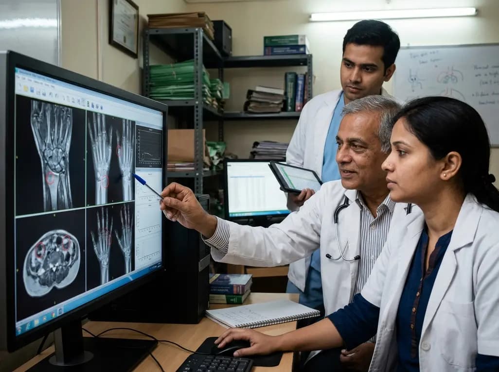
This methodological viewpoint establishes ten rules to constrain noise and enhance precision across image scoring, database handling, and data analysis in rheumatology research.
Today

A study in JAMIA Open shows lightweight open-source reasoning LLMs achieve up to 95% sensitivity and accelerate evidence synthesis screening by nearly twentyfold, presenting a cost-effective, scalable AI solution for biomedical research and systematic reviews.
Yesterday
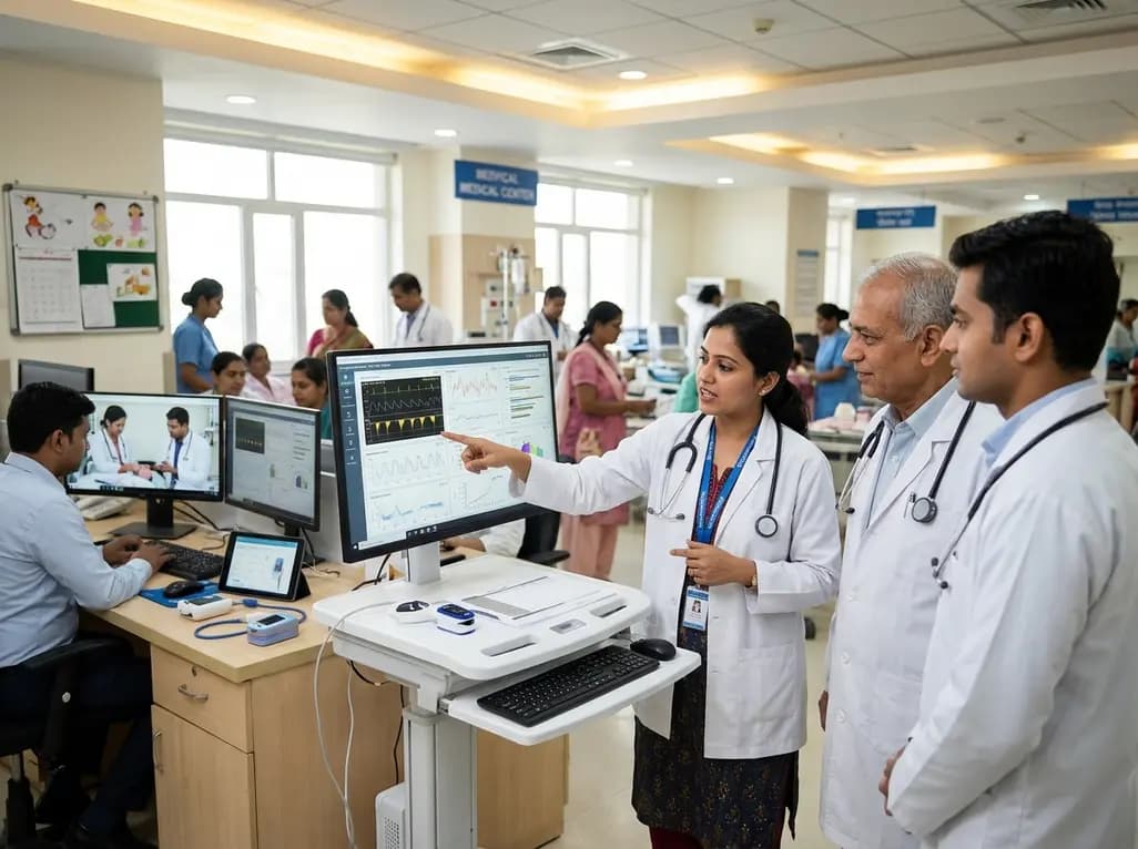
A landmark study details how an enterprise pediatric remote patient management model expanded from a single cardiology pilot to 16 diverse specialty programs by combining centralized digital operations with specialty clinical leadership.
Yesterday
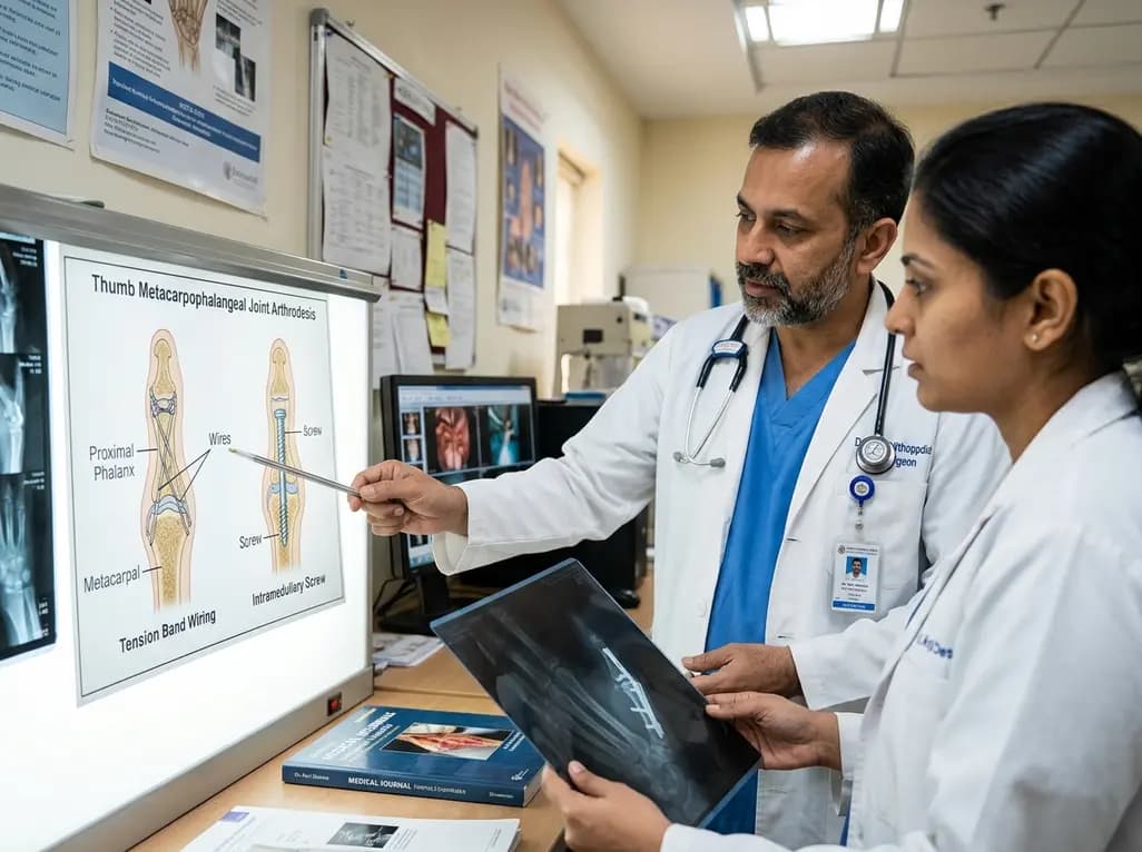
A comparative review of tension band wiring and intramedullary screw fixation in thumb metacarpophalangeal arthrodesis, evaluating union rates, hardware complications, and clinical outcomes.
Yesterday
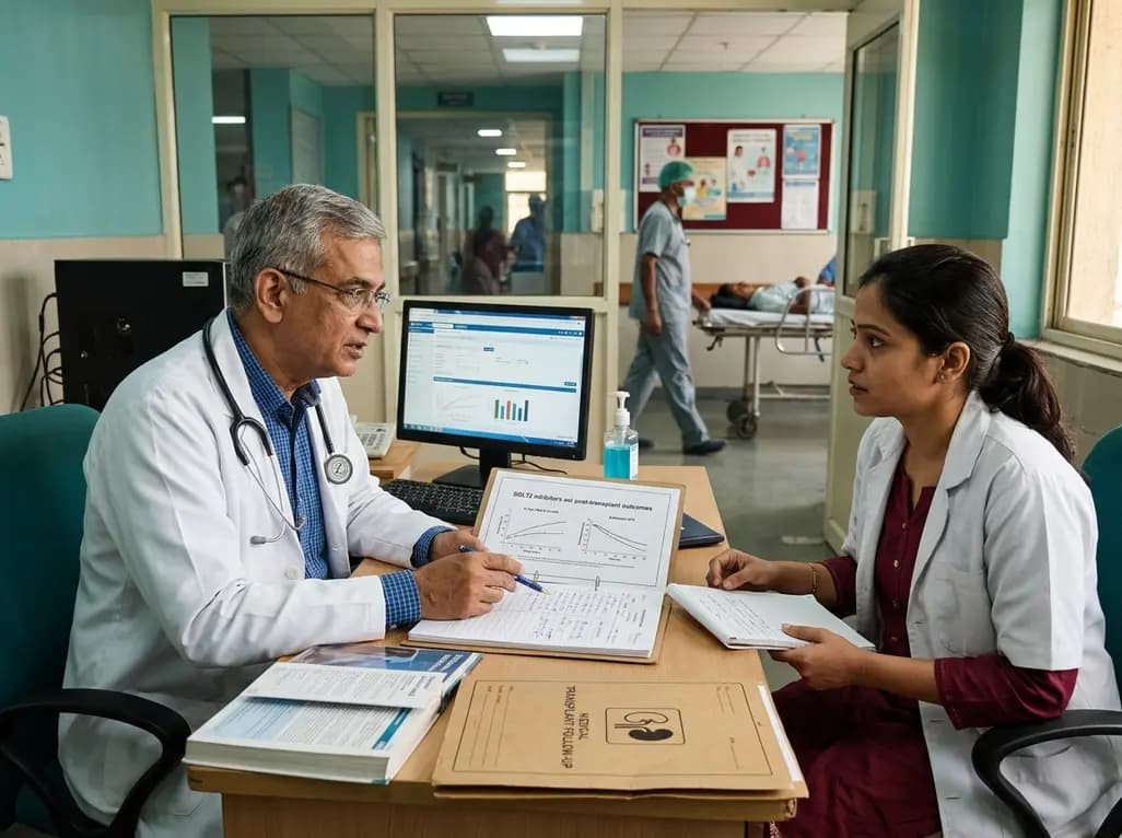
A nationwide observational study from Norway investigates whether SGLT2 inhibitor therapy improves 5-year metabolic parameters in kidney transplant recipients with post-transplantation diabetes mellitus (PTDM), finding comparable glycemic, renal, and blood pressure profiles between user and non-user cohorts.
Today
