.webp&w=384&q=75)

Loading, please wait...
Loading, please wait...
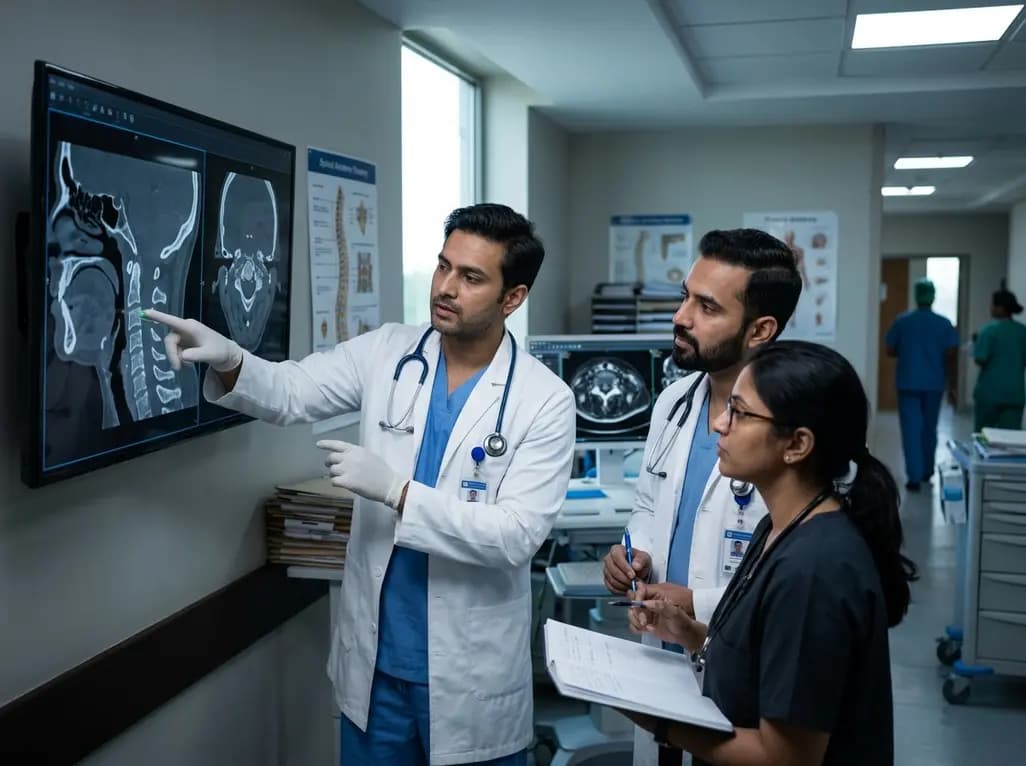
Managing craniovertebral junction instability represents one of the most intricate challenges in modern spine surgery. Specifically, atlantoaxial dislocation produces significant biomechanical instability that can compromise cervical spinal cord integrity and cranial nerve function. Over the past decade, surgical techniques have advanced from aggressive anterior decompressions toward posterior reduction and fixation. A comprehensive 10-year multicenter study across eight tertiary centers now presents an updated treatment algorithm for this complex condition. By analyzing 2,354 surgical cases with mid-to-long-term follow-up, researchers have validated a refined strategy that minimizes transoral procedures while maximizing anatomical realignment.
Surgical teams historically classified craniovertebral instability using isolated institutional experience. Consequently, therapeutic strategies varied widely across global centers, leading to inconsistent outcomes and higher complication rates. In 2013, spine specialists introduced a four-tier classification system that systematically categorized dislocations based on reducibility and structural pathology. However, rapid innovations in instrumentation and facet manipulation necessitated an objective update based on broader real-world evidence.
Therefore, investigators initiated a multicenter evaluation spanning eight tertiary centers across China between 2011 and 2021. The updated classification categorizes patients into four distinct pathological tiers. Type I dislocations demonstrate dynamic reducibility on extension radiographs, whereas Type II lesions achieve reduction under supervised skeletal traction. Furthermore, Type III lesions represent irreducible dislocations without bony fusion, and Type IV captures complex cases with osseous malformation or spontaneous fusion.
Importantly, the multicenter cohort demonstrated that Type I and Type II dislocations comprise 76.0% of all clinical presentations. Because the vast majority of patients exhibit reducible patterns, surgeons can safely avoid aggressive anterior releases in most cases. Instead, clinicians prioritize definitive posterior stabilization while maintaining safe neurological margins throughout surgery.
For patients diagnosed with Type I or Type II instability, the surgical algorithm prioritizes direct posterior reduction and rigid internal fixation. Because these lesions reduce either spontaneously or under axial traction, surgeons achieve anatomical alignment without extensive soft-tissue release. Consequently, operative teams can perform posterior instrumentation safely using modern C1 lateral mass and C2 pedicle or pars screws.
Furthermore, this posterior-only approach eliminates the severe morbidity historically associated with anterior transoral exposures. In earlier surgical eras, clinicians frequently performed transoral soft-tissue dissection or anterior release even for borderline reducible instability. However, modern spinal navigation and rigid screw-rod constructs allow surgeons to achieve powerful intraoperative reduction maneuvers directly from behind.
Additionally, rigid internal fixation establishes an optimal biomechanical environment for bone healing. Surgeons place autologous bone grafts or structural spacers along decorticated posterior elements to promote robust arthrodesis. Overall, this standardized approach produces exceptional long-term stability with minimal blood loss and brief hospital stays. Therefore, posterior screw fixation remains the benchmark therapy for reducible craniovertebral dislocation, offering predictable decompression and preservation of neurological function.
Historically, clinicians considered irreducible lesions, classified as Type III atlantoaxial dislocation, an absolute indication for transoral anterior release. Anterior approaches carry substantial risks, including pharyngeal wound breakdown, retropharyngeal infection, and prolonged ventilatory dependence. Therefore, the updated algorithm introduces a pivotal shift toward posterior intra-articular facet release as the initial intervention.
Specifically, surgeons approach the C1-C2 facet joints posteriorly, releasing contracted joint capsules and clearing fibrous tissue. This intra-articular maneuver unlocks the lateral masses, thereby converting the rigid deformity into a reducible Type II dislocation. In the multicenter study of 523 Type III patients, posterior intra-articular release successfully converted 40.5% of cases without requiring any anterior surgery.
Nevertheless, when posterior release fails to mobilize the joint, surgeons promptly convert to an anterior transoral release. In the reported cohort, 59.5% of Type III patients required transoral soft-tissue release to achieve mobilization. Consequently, surgeons do not discard anterior approaches entirely; rather, they reserve them strictly for cases that resist posterior facet mobilization. This sequential strategy dramatically reduces overall surgical trauma while preserving high rates of anatomical restoration.
Type IV dislocations involve congenital osseous fusion, malformed bony bridges, or post-traumatic heterotopic ossification across the craniovertebral junction. Consequently, simple soft-tissue release cannot unlock these structural deformities. To manage these formidable challenges, the updated algorithm advocates targeted osteotomy to restore anatomical mobility.
Remarkably, surgical teams successfully converted 88.1% of Type IV patients to reducible alignments using either posterior or transoral osteotomy. By carefully resecting obstructing bone bridges, surgeons avoided destructive neural decompression and restored physiological spinal axis alignment. Furthermore, this reconstructive philosophy protects the structural column and optimizes long-term load bearing across the occipitocervical junction.
In contrast, transoral or transnasal odontoidectomy served as an absolute last resort. Surgeons performed odontoidectomy in only 11.9% of Type IV patients when severe bony dysplasia completely prohibited reduction despite extensive osteotomy. Historically, surgeons widely applied odontoidectomy as standard therapy for irreducible compressive myelopathy. However, this updated evidence proves that modern osteotomy techniques and reduction maneuvers make odontoidectomy necessary in only a minor fraction of cases.
The multicenter investigation followed 2,354 patients for an average duration of 5.3 years, providing robust validation for this updated algorithm. Over this mid-to-long-term period, 85.9% of patients achieved complete anatomical reduction of their atlantoaxial misalignment. Furthermore, an impressive 98.8% of participants demonstrated clinical and radiological signs of solid bony fusion at final follow-up.
Moreover, these high realignment rates translated into meaningful neurological recovery for individuals presenting with cervical myelopathy. Restoring canal diameter through anatomical reduction relieved spinal cord compression without requiring extensive neural unroofing. In addition, complication rates declined substantially compared to historical series that relied heavily on primary transoral odontoidectomy.
Accordingly, these multicenter findings establish an evidence-based framework for neurosurgeons and orthopedic spine specialists worldwide. In regions like India, where craniovertebral anomalies, congenital basilar invagination, and post-traumatic dislocations occur frequently, this algorithm offers clear clinical guidance. By emphasizing sequential posterior release before anterior exposure, spine surgeons can improve patient safety, achieve predictable fusion, and consistently restore spinal alignment.
Type I dislocations reduce spontaneously during dynamic cervical extension radiographs, indicating preserved mobility. In contrast, Type II dislocations remain unreduced on dynamic views but achieve complete anatomical reduction under supervised skeletal traction. Both types permit definitive posterior-only stabilization and fusion without necessitating anterior soft-tissue or bony release.
Posterior intra-articular release permits direct joint mobilization and screw fixation through a single posterior incision. Consequently, it circumvents transoral exposure, which carries elevated risks of pharyngeal infection, dehiscence, swallowing difficulty, and prolonged intubation. The updated multicenter data show that posterior release successfully mobilizes over forty percent of irreducible cases.
Transoral or transnasal odontoidectomy remains an absolute last-resort intervention reserved strictly for persistent ventral spinal cord compression. Clinicians utilize it only when comprehensive posterior intra-articular releases and targeted osteotomies fail to achieve anatomical reduction. In contemporary multicenter cohorts, fewer than twelve percent of complex bony dislocations required this aggressive resection.
Disclaimer: This content is for informational and educational purposes only. It is not intended to provide medical advice or to be a substitute for professional medical advice, diagnosis, or treatment. Always seek the advice of your physician or other qualified health provider with any questions you may have regarding a medical condition. Refer to the latest local and national guidelines for clinical practice.
References

Read summarized clinical updates, watch expert medical content, and earn CME certifications right from your smartphone.
Open your smartphone camera to scan and install.





A 10-year multicenter study of 2,354 patients updates the atlantoaxial dislocation surgical algorithm, demonstrating high reduction and fusion rates while minimizing transoral release and odontoidectomy.
last month

As monsoon seasons elevate arboviral disease threats, Tamil Nadu has initiated an ICMR-monitored evaluation of the tetravalent dengue vaccine in Tenkasi district. Clinicians emphasize clinical monitoring, public prevention, and high-quality early pediatric care to reduce disease morbidity across endemic regions.
Today

Leucine Rich Bio has introduced South Asia's first comprehensive vaginal microbiome test, BugSpeaks. By profiling microbial dysbiosis, pathogens, and antimicrobial resistance, this next-generation diagnostic platform provides clinicians with deeper insights into recurrent infections, fertility, and reproductive health.
Today

Review how tailored exercise programs benefit frail older patients with heart failure. Explore pathophysiological mechanisms, individualized training prescriptions, and strategies to overcome adherence barriers in clinical practice.
Today

This comparative study evaluates posterior labral tears with and without glenohumeral instability, highlighting distinct demographic patterns, MRI underestimation in stable shoulders, and successful arthroscopic repair outcomes.
Today

A clinical study demonstrates that early intervention with tocilizumab, either as monotherapy or bridged with corticosteroids, achieves significant proptosis reduction and resolves active inflammation in treatment-naive thyroid eye disease.
Today
