.webp&w=384&q=75)

Loading, please wait...
Loading, please wait...
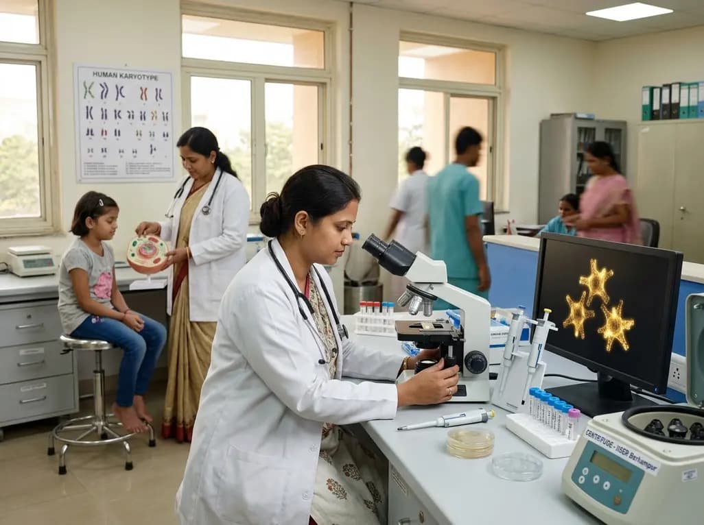
Turner syndrome affects approximately one in every 2,000 female live births globally. Consequently, early clinical recognition remains critical for managing associated cardiovascular, endocrine, and developmental morbidities. Researchers at the Indian Institute of Science Education and Research (IISER), Berhampur, have introduced a breakthrough platform to accelerate Turner syndrome detection. By harnessing functional nanomaterials, this diagnostic method identifies disease-specific chromosomal anomalies within hours rather than weeks. This advancement addresses substantial diagnostic bottlenecks in public health systems, offering timely diagnostic confirmation for pediatricians and endocrinologists across diverse healthcare environments.
Turner syndrome arises from the complete or partial monosomy of the X chromosome. Clinicians frequently encounter classical 45,X karyotypes, as well as complex mosaic variants. However, standard diagnosis relies predominantly on conventional chromosomal karyotyping of peripheral blood lymphocytes. This cytogenetic workflow requires viable cell culturing, metaphase arrest, and skilled microscopic evaluation. As a result, processing times routinely exceed three to four weeks from initial sample collection. Furthermore, specialized cytogenetic facilities remain concentrated in tertiary academic centers and metropolitan hospitals. Families living in semi-urban and rural areas often face significant diagnostic delays or high out-of-pocket testing expenses. These operational hurdles prevent timely therapeutic action during crucial developmental windows. Delayed confirmation impedes the optimal timing for growth hormone therapy and pubertal induction. In addition, unrecognized cardiovascular malformations, such as aortic coarctation, pose silent life-threatening risks. Therefore, clinicians urgently require faster, cost-effective molecular alternatives that streamline diagnostic pipelines without compromising analytical precision. Developing rapid point-of-care assays ensures immediate risk stratification and reduces long waiting periods for vulnerable families. Such technological improvements would democratize genetic screening across resource-limited clinical settings.
To overcome traditional diagnostic delays, investigators engineered an innovative biosensing platform combining advanced nanostructures. Assistant Professor Parikshit Moitra from the Department of Chemical Sciences at IISER Berhampur spearheaded this study. Specifically, the technology integrates a nucleotide-guided covalent organic framework with plasmonic gold nanostars. Covalent organic frameworks provide highly ordered, porous architectures that display intense intrinsic luminescence. Meanwhile, gold nanostars offer unique optical properties and exceptional electromagnetic field enhancements at their sharp tips. The research team functionalized the framework with gene-specific oligonucleotide probes designed to target characteristic X-chromosome sequences. Consequently, hybridization between the target genomic DNA and the complementary probes triggers a measurable fluorescence modulation. This coordinated molecular recognition system operates with remarkable sensitivity, detecting minute quantities of unamplified genetic material. Moreover, the assay eliminates lengthy cell-culturing steps entirely. The entire analytical workflow takes place rapidly in a straightforward liquid phase. Thus, the nanomaterial-based architecture transforms complex cytogenetics into a streamlined, high-throughput biosensing readout suitable for point-of-care clinical deployment. Laboratory evaluations demonstrated that this hybrid nano-assembly maintains robust structural stability under varied physiological buffers. Furthermore, the platform successfully distinguishes intact genomic targets from mismatched sequences with high fidelity. Because the signal generation occurs via direct optical changes, laboratory technicians do not require cumbersome isotope labeling or intricate fluorescence microscopy tools.
Translating nanotechnological concepts into reliable clinical tools demands rigorous validation against genuine patient samples. Therefore, the IISER Berhampur team partnered with leading clinical endocrinologists, including Dr. Sunetra Mondal and Dr. Satinath Mukhopadhyay. The collaborating physicians obtained clinical blood specimens from individuals presenting with suspected Turner phenotypes at a specialized hospital in West Bengal. Subsequently, the researchers tested the biosensor directly against these blinded patient samples to benchmark diagnostic reliability. Notably, the nano-assembly demonstrated extraordinary concordance with conventional karyotyping results. The assay accurately differentiated samples exhibiting monosomy X from healthy female and male controls. Furthermore, the platform preserved high sensitivity even when analyzing degraded genomic extracts or low sample volumes. This high analytic resilience represents a crucial advantage over standard polymer-based assays that often suffer from enzyme inhibition. In addition, the assay achieved near-perfect specificity, generating negligible false-positive signals in cross-reactivity experiments. Consequently, the clinical findings confirm that synthetic nanomaterials can reliably decode human chromosomal aneuploidies. This successful translational effort establishes a sturdy foundation for multicentric trials across Indian healthcare institutions. Clinicians emphasized that the rapid turnaround time significantly minimized parental anxiety during diagnostic evaluations. Moreover, the simple workflow ensures reproducible performance across diverse laboratory conditions.
Prompt identification of Turner syndrome dramatically improves long-term health outcomes for affected girls. Typically, growth failure begins in early childhood, leading to severe short stature if left untreated. When clinicians detect the condition early, pediatric endocrinologists can initiate recombinant human growth hormone therapy promptly. Consequently, patients gain substantial height velocity and reach near-normal adult stature before epiphyseal plates fuse. Furthermore, timely diagnosis allows structured surveillance for congenital cardiovascular defects. Clinicians can immediately order echocardiograms and cardiac magnetic resonance imaging to assess aortic root dimensions and bicuspid valve architecture. Early cardiovascular monitoring prevents devastating aortic dissection in early adulthood. In addition, endocrinologists can carefully plan hormone replacement therapy to induce secondary sexual characteristics at a physiologically appropriate age. Early estrogen replacement promotes normal breast development, optimizes uterine growth, and supports bone mineral density accumulation. Moreover, routine screening for autoimmune conditions, including Hashimoto thyroiditis and celiac disease, can commence during early childhood. Thus, rapid diagnostic confirmation transforms clinical care from reactive treatment into proactive, preventive healthcare management. Physicians can also offer timely reproductive counseling and psychological support to adolescent girls and their parents. Ultimately, expedited diagnostic pathways protect metabolic health, cardiovascular function, and emotional well-being throughout the patient lifespan.
The breakthrough reported by IISER Berhampur represents a transformative paradigm for clinical genetics and precision medicine. Beyond Turner syndrome, the modular nature of covalent organic frameworks offers extensive versatility. Researchers can readily modify the immobilized oligonucleotide probes to target other sex-chromosome aneuploidies, such as Klinefelter syndrome. Similarly, the platform holds immense promise for diagnosing autosomal trisomies, including Down syndrome and Edwards syndrome. However, broad routine implementation requires prospective multicenter clinical trials to validate diagnostic parameters across larger populations. Scientists must also establish standardized manufacturing protocols to guarantee batch-to-batch consistency of synthesized gold nanostars. In addition, developing portable microfluidic integration could soon enable decentralized point-of-care testing in district hospitals and rural primary health centers. Such decentralization would drastically lower health disparities and reduce testing turnaround times to under two hours. Prof Ashok Kumar Ganguli emphasized that continued institutional support will foster innovative diagnostic technologies for national health priorities. Consequently, this nanotechnological framework highlights how domestic biomedical engineering can address pressing unmet clinical needs. In summary, rapid molecular platforms will soon redefine genetic screening paradigms throughout India. Furthermore, affordable diagnostic cartridges will empower frontline community clinicians to detect rare genetic conditions without relying on metropolitan laboratories. Ultimately, this scalable approach promises equitable access to state-of-the-art cytogenomics across global public health systems.
Q1: What makes the IISER Berhampur test faster than standard karyotyping?
Standard chromosomal karyotyping requires peripheral blood lymphocyte culturing and metaphase chromosome analysis, taking three to four weeks. In contrast, the IISER Berhampur biosensor directly captures target genomic sequences using nucleotide-guided covalent organic frameworks and gold nanostars. Consequently, the system bypasses living cell culture entirely, delivering sensitive optical results within hours. Therefore, this streamlined molecular recognition saves crucial clinical time and facilitates rapid medical decision-making for endocrinologists.
Q2: How does the covalent organic framework and gold nanostar system work?
The biosensing platform utilizes a luminescent covalent organic framework functionalized with sequence-specific oligonucleotide probes. When these probes bind matching Turner syndrome DNA signatures, they assemble hierarchically with plasmonic gold nanostars. Consequently, this proximity-driven interaction modulates optical emissions, generating a distinct fluorescent signal. Furthermore, the sharp nanoscale tips of the gold nanostars enhance electromagnetic fields, enabling the detection of minute quantities of genetic material without complex amplification.
Q3: When should pediatricians suspect Turner syndrome in clinical practice?
Pediatricians should suspect Turner syndrome in any female infant or child presenting with unexplained growth failure, short stature, or delayed pubertal development. Furthermore, physical signs such as a webbed neck, low posterior hairline, cubitus valgus, recurrent otitis media, and lymphedema warrant prompt genetic evaluation. Early identification ensures immediate screening for congenital cardiac malformations and enables the timely initiation of growth hormone therapy to optimize final adult stature.
Disclaimer: This content is for informational and educational purposes only. It does not constitute medical advice or replace professional judgment. Refer to the latest local and national guidelines for clinical practice.
References

Read summarized clinical updates, watch expert medical content, and earn CME certifications right from your smartphone.
Open your smartphone camera to scan and install.





A research team from IISER Berhampur has engineered an advanced biosensor combining covalent organic frameworks and gold nanostars. This platform detects Turner syndrome genetic signatures rapidly, providing a scalable alternative to month-long karyotyping and enabling timely clinical interventions.
last month

Review how tailored exercise programs benefit frail older patients with heart failure. Explore pathophysiological mechanisms, individualized training prescriptions, and strategies to overcome adherence barriers in clinical practice.
Today

Leucine Rich Bio has introduced South Asia's first comprehensive vaginal microbiome test, BugSpeaks. By profiling microbial dysbiosis, pathogens, and antimicrobial resistance, this next-generation diagnostic platform provides clinicians with deeper insights into recurrent infections, fertility, and reproductive health.
Today

In high-risk pulmonary embolism complicated by severe anemia and active uterine bleeding, systemic thrombolysis is contraindicated. Large-bore aspiration thrombectomy provides rapid hemodynamic rescue without fibrinolysis, supported by inferior vena cava filter placement and multidisciplinary critical care.
Today

This comparative study evaluates posterior labral tears with and without glenohumeral instability, highlighting distinct demographic patterns, MRI underestimation in stable shoulders, and successful arthroscopic repair outcomes.
Today

A clinical study demonstrates that early intervention with tocilizumab, either as monotherapy or bridged with corticosteroids, achieves significant proptosis reduction and resolves active inflammation in treatment-naive thyroid eye disease.
Today
