.webp&w=384&q=75)

Loading, please wait...
Loading, please wait...
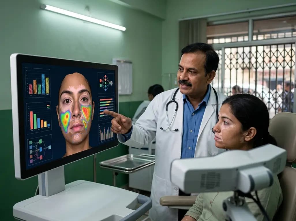
Vitiligo is a chronic autoimmune condition that presents a significant challenge for dermatologists due to the subjective nature of traditional assessment tools. Historically, clinicians have relied on visual inspection and 2D photography to estimate the extent of depigmentation. However, these methods often fail to account for the complex contours of the human face, leading to inconsistencies in disease monitoring. Recent advancements in facial vitiligo quantification have introduced artificial intelligence and 3D reconstruction as potential solutions. A pivotal study by Lee S et al. has demonstrated that integrating deep learning models with 3D mapping can significantly enhance the accuracy and reliability of lesion measurement. This technological leap addresses the long-standing need for standardized diagnostic support in busy clinical settings.
For years, the Vitiligo Area Scoring Index (VASI) has been the gold standard for clinical evaluation. While useful, it remains prone to interobserver variability and relies heavily on the experience of the practitioner. In many cases, subtle changes in repigmentation or lesion expansion are difficult to detect through 2D analysis alone because flat images cannot accurately represent the surface area of curved anatomical regions like the nose or jawline. This lack of precision often complicates long-term treatment planning and the evaluation of therapeutic efficacy. By shifting toward an automated 3D approach, dermatologists can bypass these subjective hurdles. The study highlights that manual assessments often result in a higher mean absolute error, emphasizing the necessity for more robust, data-driven tools in routine practice.
The researchers developed a sophisticated system that utilizes standardized clinical facial photographs to create a virtual 3D model of the patient’s face. A deep learning segmentation model was trained specifically to identify depigmented lesions with high precision. Once identified, these lesions were mapped onto the 3D surface, which allowed the system to account for facial curvature during the calculation of the lesion area. This process ensures that facial vitiligo quantification is not just a visual estimate but a precise mathematical measurement. The study included a retrospective analysis of over 300 patients, ensuring a robust dataset for validation. By incorporating surface mapping, the system provides a more realistic representation of the total affected area compared to traditional methods.
The performance of the AI segmentation model was remarkable, achieving an F1 score of 0.98 for boundary delineation. When dermatologists used the AI-guided system, the mean absolute error in lesion quantification dropped from 6.71 to 3.13. This indicates that AI assistance nearly doubles the accuracy of human assessment alone. Furthermore, the Pearson correlation coefficient between AI measurements and reference lesion areas was significantly higher than that of manual measurements. Bland-Altman analysis also showed a marked reduction in systematic bias, suggesting that the AI provides a level of consistency that is difficult to achieve manually across multiple sessions. These findings suggest that AI-driven tools can effectively standardize outcomes in both clinical trials and daily dermatology practice.
The ability to precisely track lesion changes over time is crucial for managing vitiligo. With facial vitiligo quantification becoming more objective, clinicians can provide patients with clearer evidence of treatment progress, which significantly improves treatment adherence and patient satisfaction. This system is particularly valuable for evaluating the response to newer therapies, such as JAK inhibitors, where subtle repigmentation might be a key indicator of success. Moreover, the reduction in interobserver variability means that a patient can see different doctors within the same clinic and still receive consistent, comparable data regarding their condition. This reliability is a cornerstone of modern personalized medicine and evidence-based dermatological care.
Integrating AI-powered 3D analysis into routine dermatological workflows represents a significant step toward digital health transformation. While the current study focuses on facial lesions, the underlying technology could potentially be adapted for other body regions or different pigmentary disorders. The high speed of automated inference makes it a practical choice for real-time clinical use without disrupting the physician-patient interaction. As these tools become more accessible, they will likely become an essential component of standardized outcome reporting. The transition from subjective estimation to objective quantification ensures that every patient receives a diagnosis and monitoring plan based on the highest level of technical precision available today.
Traditional 2D photography often underestimates the true surface area of lesions because it cannot account for the natural curves and contours of the face. 3D analysis uses surface mapping to provide a more accurate and mathematically sound measurement of depigmented patches. This ensures that the quantification of facial vitiligo is standardized and precise, allowing for better detection of even minor changes in the size of the lesion over time.
AI-assisted tools are designed to augment, not replace, clinical judgment. While the AI provides highly accurate data and reduces manual errors, the dermatologist still plays a critical role in interpreting the results and making treatment decisions. The primary benefit of the AI is to remove the subjectivity and variability associated with manual measurements, providing a reliable baseline and tracking system that supports the physician's overall diagnostic and therapeutic strategy.
The main benefits include increased consistency, higher accuracy, and reduced diagnostic time. AI models can delineate lesion boundaries with a high level of precision, often achieving F1 scores near 0.98. This level of detail minimizes the mean absolute error in quantification. For the clinician, this means less time spent on manual tracing and more time focusing on patient care, while ensuring that the data used for monitoring is reproducible across different sessions.
Disclaimer: This content is for informational and educational purposes only and does not constitute medical advice. It is intended for healthcare professionals. Always consult with a qualified specialist for diagnosis and treatment. Refer to the latest local and national guidelines for clinical practice.
References
Lee S et al. Objective and Quantitative Assessment of Facial Vitiligo via AI-powered 3D Analysis for Diagnostic Support. Br J Dermatol. 2026 Jul 20. doi: undefined. PMID: 42473859.
Parikh M et al. Technological advances in vitiligo management: perspectives on AI, mobile tools, and clinical utility. Frontiers in medicine. 2025. doi: 10.3389/fmed.2025.145678.
Ganesan A et al. Three-dimensional imaging for facial vitiligo: Results from a phase 2 randomized controlled trial investigating upadacitinib in patients with vitiligo. J Invest Dermatol. 2026. doi: 10.1016/j.jid.2025.12.036.

Read summarized clinical updates, watch expert medical content, and earn CME certifications right from your smartphone.
Open your smartphone camera to scan and install.





A new study reveals that AI-assisted 3D reconstruction provides superior objective quantification of facial vitiligo lesions compared to traditional 2D photography and manual assessment, significantly reducing diagnostic error and interobserver variability.
3 months ago

Review how tailored exercise programs benefit frail older patients with heart failure. Explore pathophysiological mechanisms, individualized training prescriptions, and strategies to overcome adherence barriers in clinical practice.
Today

Leucine Rich Bio has introduced South Asia's first comprehensive vaginal microbiome test, BugSpeaks. By profiling microbial dysbiosis, pathogens, and antimicrobial resistance, this next-generation diagnostic platform provides clinicians with deeper insights into recurrent infections, fertility, and reproductive health.
Today

In high-risk pulmonary embolism complicated by severe anemia and active uterine bleeding, systemic thrombolysis is contraindicated. Large-bore aspiration thrombectomy provides rapid hemodynamic rescue without fibrinolysis, supported by inferior vena cava filter placement and multidisciplinary critical care.
Today

This comparative study evaluates posterior labral tears with and without glenohumeral instability, highlighting distinct demographic patterns, MRI underestimation in stable shoulders, and successful arthroscopic repair outcomes.
Today

A clinical study demonstrates that early intervention with tocilizumab, either as monotherapy or bridged with corticosteroids, achieves significant proptosis reduction and resolves active inflammation in treatment-naive thyroid eye disease.
Today
