

Loading, please wait...
Loading, please wait...

Posterior shoulder pain creates a challenging diagnostic scenario in sports medicine and orthopedic practice. Clinicians frequently encounter active patients who report deep, persistent shoulder discomfort without any sensation of subluxation. In recent years, orthopedic surgeons have recognized that posterior labral tears do not always produce gross glenohumeral instability. In fact, many individuals experience debilitating mechanical pain while maintaining full joint stability during physical examinations. A retrospective study by Seo and Shin provides essential insights into these distinct clinical presentations. The authors analyzed eighty-five patients undergoing arthroscopic repair, comparing those with instability against those without instability. Their findings demonstrate that patients without laxity exhibit unique demographic traits, insidious mechanisms, and subtle radiological findings. Consequently, recognizing these differences ensures timely diagnosis and optimal operative intervention.
Patients with posterior labral tears display distinct clinical characteristics based on joint stability status. In this comparative investigation, patients without instability presented at an older average age than unstable patients. Specifically, stable individuals averaged over thirty-one years of age, whereas unstable individuals presented significantly younger. Furthermore, the duration of symptoms differed remarkably between the two study cohorts. Patients without mechanical instability endured chronic discomfort for an average of thirty months prior to surgery. In contrast, unstable patients sought surgical intervention much sooner after symptom onset. Additionally, an antecedent traumatic injury occurred far less frequently in the non-instability cohort. Only sixteen patients without instability recalled a discrete traumatic injury. In addition, these patients frequently report that deep posterior aching worsens when carrying heavy objects or sitting with arms extended forward. Instead, chronic mechanical stress predominated in this group. Conversely, patients with instability consistently associated their pain with sudden athletic impacts or falls. Therefore, clinicians evaluating mature, active individuals with prolonged shoulder discomfort must consider labral pathology, even without a traumatic history or frank joint subluxation.
Accurate preoperative imaging remains notoriously elusive in patients who lack joint laxity. Standard magnetic resonance imaging frequently underestimates the structural severity of these posterior capsulolabral lesions. In the comparative analysis, magnetic resonance scans failed to detect incomplete posterior tears significantly more often in non-instability patients than in unstable patients. Moreover, surgeons identified occult posterior labral tears during arthroscopy in ten non-instability patients, compared to only two unstable patients. Consequently, routine magnetic resonance scans often mislead clinicians when posterior subluxation is absent. The lack of capsular enlargement or joint effusion frequently conceals subtle chondrolabral detachment. Furthermore, radiologists should note that fluid tracking along the posterior glenoid rim provides a critical clue to underlying labral pathology. Because standard axial views may show preserved labral morphology, internal fissuring and partial detachment easily escape detection. Thus, orthopedists should not exclude labral injury solely based on normal imaging findings. In ambiguous cases, clinicians should consider magnetic resonance arthrography to enhance visualization. Ultimately, diagnostic arthroscopy provides the definitive assessment when persistent localized symptoms resist conservative management.
The biomechanical forces responsible for posterior labral pathology differ considerably across athletic disciplines. The study demonstrated that competitive sports participation occurred far more frequently among patients with frank instability. These high-demand athletes regularly experience sudden collision forces, forced adduction, and rapid internal rotation that avulse the labrum. Conversely, patients without instability engaged much more heavily in repetitive resistance training. Specifically, over half of the stable cohort regularly performed intensive weightlifting, including heavy bench press and overhead barbell routines. During heavy bench pressing, posteriorly directed axial loads transfer directly through the flexed arm against the posterior glenoid rim. This repetitive microtrauma gradually shears the posterior labrum from the glenoid cartilage without stretching the joint capsule. Therefore, weightlifters develop localized tissue breakdown rather than generalized capsular laxity. Identifying these contrasting activity profiles helps sports physicians pinpoint vulnerable fitness enthusiasts who report posterior pain during upper-body pressing movements.
Fortunately, arthroscopic repair achieves excellent subjective and objective functional recovery regardless of stability status. The researchers assessed patient outcomes after a mean follow-up of thirty-one months using validated shoulder scoring systems. Following arthroscopic posterior labral refixation, both patient cohorts demonstrated statistically significant improvements across all functional measures. Visual analog scale pain scores dropped dramatically, while American Shoulder and Elbow Surgeons scores improved substantially. Importantly, postoperative evaluations demonstrated equivalent tissue healing rates between the two groups. Incomplete healing appeared in seven unstable patients and five stable patients, showing no statistically significant difference. In contrast to anterior repairs, isolated posterior repairs typically allow earlier rehabilitation because the posterior capsule tolerates passive forward elevation well. Furthermore, patients without instability required simple labral re-anchoring without extensive capsular plication, which preserved postoperative joint range of motion. However, achieving successful outcomes requires meticulous operative technique. Surgeons must thoroughly debride degenerative tissue edges and secure stable anchor fixation on the glenoid rim. Consequently, timely surgical intervention reliably eliminates pain and prevents progressive joint deterioration.
These clinical insights offer valuable practical guidance for orthopedic surgeons, sports physicians, and radiologists. As gym-based fitness and resistance training become increasingly popular, clinicians encounter more active adults with unexplained posterior shoulder pain. Practitioners must recognize that intact joint stability does not eliminate the possibility of a labral tear. Therefore, clinicians should routinely perform targeted provocative tests, including the Kim test and jerk test, during physical evaluations. In addition, radiologists and surgeons must maintain active interdisciplinary communication. When reviewing shoulder scans in weightlifters, radiologists should specifically inspect the posteroinferior chondrolabral junction for subtle tearing or concealed avulsions. If structured conservative therapy fails after several months, surgical evaluation becomes warranted. Arthroscopic labral repair offers reliable symptom relief and durable restoration of function. By adopting this heightened clinical awareness, practitioners can avoid prolonged diagnostic delays and facilitate an efficient return to full physical activity.
Patients without instability typically present with insidious, chronic posterior shoulder pain rather than sudden traumatic subluxation. They frequently report localized aching during heavy bench pressing, overhead resistance exercises, or prolonged internal rotation. On clinical examination, these patients show localized posteroinferior joint line tenderness and positive provocative maneuvers like the Kim or jerk test, but they maintain normal glenohumeral translation without apprehension or gross joint laxity.
Conventional magnetic resonance imaging frequently underestimates these lesions because the detached labrum may remain anatomically aligned without displacement or extensive joint effusion. Furthermore, occult tears often involve the articular surface or deep chondrolabral junction while leaving the superficial capsular attachment intact. Without joint distension, imaging cannot clearly delineate these subtle incomplete avulsions. Consequently, magnetic resonance arthrography or diagnostic arthroscopy is often necessary to confirm the diagnosis.
Arthroscopic posterior labral repair delivers excellent clinical outcomes, producing significant pain relief and functional restoration. Standardized measures, including visual analog scale pain scores and American Shoulder and Elbow Surgeons scores, improve markedly within two years after surgery. Most patients achieve durable labral healing, regain full range of motion, and successfully return to recreational exercise or competitive sports without persistent posterior shoulder discomfort or joint instability.
Disclaimer: This content is for informational and educational purposes only and should not be considered medical advice. Healthcare professionals should exercise their independent clinical judgment. Refer to the latest local and national guidelines for clinical practice.
References

Read summarized clinical updates, watch expert medical content, and earn CME certifications right from your smartphone.
Open your smartphone camera to scan and install.





This comparative study evaluates posterior labral tears with and without glenohumeral instability, highlighting distinct demographic patterns, MRI underestimation in stable shoulders, and successful arthroscopic repair outcomes.
Today
A recent breakthrough reveals that capsaicin mitigates microglial inflammation through a novel TRPV1-SSR4 protein-protein interaction independent of classic calcium influx. This discovery highlights fresh therapeutic avenues for managing neurodegenerative diseases like Alzheimer's.
Today
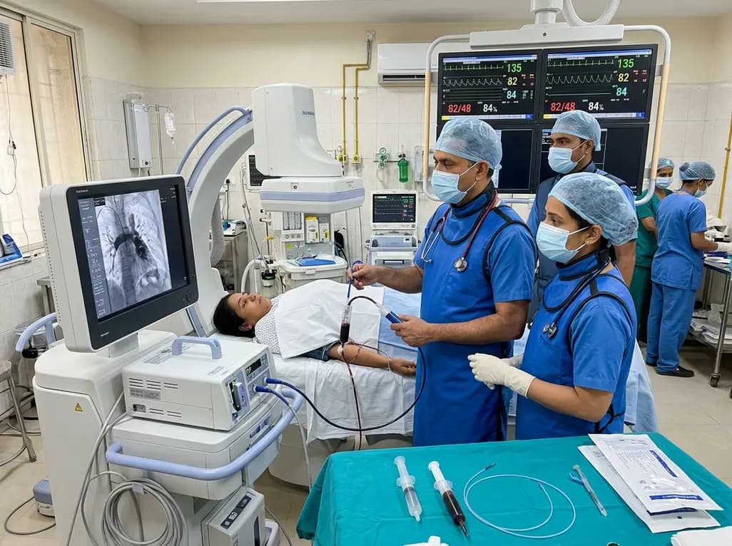
In high-risk pulmonary embolism complicated by severe anemia and active uterine bleeding, systemic thrombolysis is contraindicated. Large-bore aspiration thrombectomy provides rapid hemodynamic rescue without fibrinolysis, supported by inferior vena cava filter placement and multidisciplinary critical care.
Today

A cross-sectional study in Zanzibar reveals that 46.5% of adults with type 2 diabetes experience multidomain autonomic dysfunction on the COMPASS-31 tool, primarily orthostatic intolerance, while 45.9% exhibit cardiovascular autonomic neuropathy, highlighting the urgent need for structured clinical screening.
Today
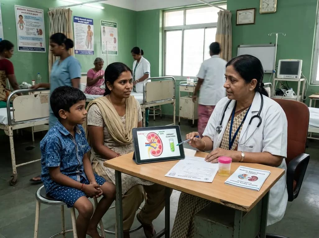
A recent case-control study reveals that urinary clusterin provides superior diagnostic performance for early diabetic kidney disease detection in pediatric type 1 diabetes mellitus, identifying tubular damage before conventional microalbuminuria occurs.
Today
